Pituitary Gland
Historical document, translated for reference. It reflects medical knowledge of the 1920s–30s and is not medical advice.
Summary
This article from the first edition of the Great Medical Encyclopedia (1928–1936) provides a detailed anatomical and physiological overview of the pituitary gland. It describes the gland's structure, including the neurohypophysis and the glandular anterior lobe, as well as its location within the sella turcica.
Encyclopedia article (1928–1936)
192 Normal and pathological physiology.
194 The pituitary gland (hypophysis cerebri, brain appendage; syn. gl. pituitaria, phlegm gland; old names: caput rosae, colatorium, sentina cerebri) is an incretory organ located in the depression of the sella turcica of the sphenoid bone and connected to the tissue of the base of the brain by means of the latter's processus infundibuli. The shape of the organ is rounded, flattened in the dorso-ventral and cranio-caudal directions. Average dimensions in an adult male: sagittal 11.9 mm, transverse 14.4 mm, and vertical 5.5 mm. Average weight 61.2 mg (in nulliparous women of corresponding age 61.8 mg) (Erdheim and Stumme). Structure of the pituitary gland. Macroscopically, the following are distinguished in the pituitary gland: the posterior lobe, or neurohypophysis (brain lobe, pars nervosa), which represents the terminal thickening of the infundibular process of the diencephalon, and the anterior lobe, which is genetically independent of it, the so-called glandular lobe (the pituitary gland proper). (In carnivores, the neurohypophysis is enveloped by glandular tissue over its entire surface, being pressed into the anterior lobe like a button into its socket.) In turn, the glandular part of the pituitary gland is divided into: 1) the intermediate part (p. intermedia, p. paranervosa, p. juxtanervosa infundibularis, epithelial rim), which directly covers the brain lobe; 2) the main (principal) lobe, separated from the previous one by a narrow slit-like cavity (pituitary cavity); and 3) glandular masses, which macroscopically represent a kind of anterior continuation of the main lobe and cover the pituitary stalk more or less completely—the prehypophysis (lob. bifurcatus, p. juxtanervosa tuberalis, covering part, tongue-shaped process, lob. peduncularis), separate, more sharply defined sections of which have received the names lob. chiasmaticus, lob. praemamillaris, etc. (see separate table, fig. 1). The human pituitary gland in the embryonic state fully corresponds to this structural scheme, but in extrauterine life it deviates from it due to the disappearance of the pituitary cavity, the significant regression of the intermediate part, which turns into a group of cystic formations wedged between the main and posterior lobes, and the weak expression of the prehypophysis (see separate table, fig. 2). The entire organ is covered by a fibrous capsule, which is a derivative of the dura mater, forming a circular fold on its cranial surface—the diaphragm of the sella turcica. Being a direct continuation of the tissue of the tuber cinereum of the diencephalon, the neurohypophysis retains the basic structural features of the supporting tissue of the central nervous system, while its microscopic picture is complicated due to the rather abundant penetration of the organ by connective tissue elements accompanying blood vessels, on the one hand, and the penetration into it of epithelial elements from the intermediate part of the pituitary gland, on the other. Thus, the following are found in the composition of the nerve lobe: 1) glial elements with typical glial fibers, 2) connective tissue fibers and cells, and 3) individual epithelial cells, usually in various stages of degeneration. (In the case of the presence of the recessus infundibuli, the latter is lined with characteristic ependyma.) The constant presence of colloid droplets in the tissue spaces is characteristic.
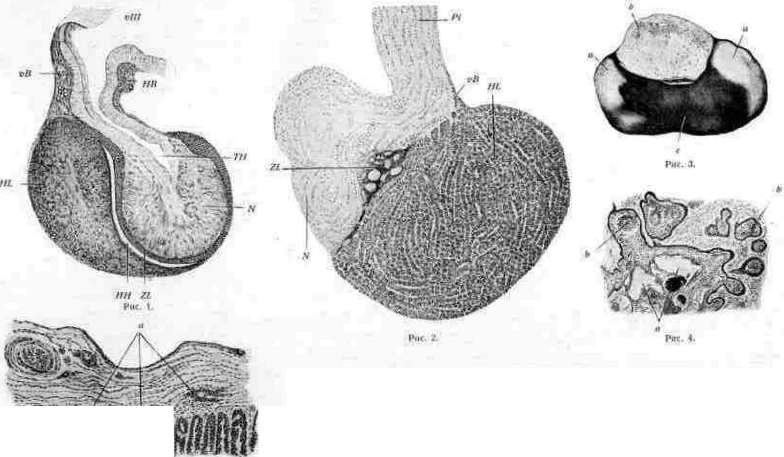
The presence of pigment is observed, predominantly in the elements of the glia; more or less abundant lipoid inclusions are also encountered in them. The intermediate part (pars intermedia), connected with the previous part by a layer of delicate connective tissue (a remnant of the primary dura mater), in humans is rudimentary, reduced to a small area permeated with colloid cysts; in most mammals, it appears as an epithelial layer, limiting the cavity of the pituitary gland from behind with its free surface and covering the neurohypophysis to a greater or lesser extent. In its developed state, this layer is built of several rows of polygonal cells with weak basophilic protoplasm; it often contains, between typical epithelial elements, elements of the glia originating from the posterior lobe. A few capillaries penetrate from the border of the same lobe. Individual colloid cysts are characteristic, increasing in number with age. Wrapping around the edges of the pituitary cavity, the tissue of the pars intermedia passes without a sharp boundary into the tissue of the main lobe. The most voluminous part of the organ, the main (principal) lobe, is built of anastomosing epithelial cords, separated from each other by lacunar capillaries and appearing in cross-sections as rounded cell groups. Since the time of Dostoevsky and Flesch, two main types of cellular elements have been distinguished in the composition of the glandular parenchyma represented by these cords: 1) larger chromophiles, characterized by the specific granularity of their protoplasm, and 2) chromophobes (chief cells), which do not have such granularity. According to their reaction to common microscopic stains, the former in turn fall into acidophiles (eosinophiles) and basophiles (cyanophiles) [see color plate (pp. 187-188), Fig. 2]. The so-called "clusters of naked nuclei" described by a number of researchers are the result of post-mortem changes in the chief cells, which undergo cadaveric decomposition extremely rapidly. In terms of the size of individual elements, basophiles stand in first place, followed by acidophiles, and finally chromophobes. The morphological significance of the described types is controversial; the most well-founded hypothesis is one that accepts chromophiles as functionally active elements of the parenchyma, developing from chief cells in two different directions. In the thickness of the cellular cords, drops of colloid are encountered, small droplets of which are also observed intracellularly, predominantly in chromophobes. Lipoid inclusions have also been described in the cells (predominantly in chromophiles), and the number of these inclusions increases with age. The stroma of the lobule is represented by delicate connective tissue, the few fibers of which are located along the blood vessels. The prehypophysis (lobus bifurcatus) is a system of epithelial cords embedded in loose, blood-vessel-rich connective tissue, consisting of small cells that, in their external appearance and staining reactions, resemble the chromophobes of the previous lobe. Along the course of the cords (due to the formation of colloid clusters), swellings are located that are extremely similar to the follicles of the thyroid gland. The arteries of the pituitary gland originate from the system of the internal carotid artery, partly branching off directly as it passes through the cavernous sinus (lower level), and partly in the form of branches of the Circle of Willis (Fuchs). A general arterial plexus forms in the capsule of the pituitary gland, from where small branches penetrate into the organ, breaking up into capillaries in its tissue. The exiting veins also form two levels, of which the upper one drains into the circular sinus of Ridley. In both the glandular and nervous lobes of the pituitary gland, abundant plexuses of lymph vessels have been described, both in the form of capillaries and in the form of lymphatic tissue spaces. The nerves of the pituitary gland, which apparently belong to the parasympathetic system, penetrate into the organ from the base of the brain through the pituitary stalk, branching in the neurohypophysis and proceeding from there into the glandular part, where they form abundant terminal plexuses in the cords of the main lobe, while some end in branches among the elements of the intermediate lobe. The specific metabolic products of the glandular parenchyma of the pituitary gland apparently enter the general economy of the organism in two ways: on the one hand, it is generally accepted that the secretion of the chromophiles of the main lobe is removed directly by its blood capillaries; on the other hand, a certain part of these products undoubtedly moves from the intermediate, as well as from the anterior lobe, through tissue spaces into the posterior lobe, in the direction of the infundibular recess, or, in the absence of the latter, through the pituitary stalk to the cavity of the third ventricle itself, which can be easily verified by observing the position of colloid droplets in the tissue of the nervous lobe. Embryology of the pituitary gland. By its origin, the pituitary gland is a heterogeneous formation: its posterior lobe develops directly from the wall of the diencephalon; the anterior, glandular part is a derivative of the epithelium of the embryonic oral pocket. The beginning of the embryonic primordium in humans is the 3rd week, in rabbits—the 6 mm stage. The epithelial primordium of the pituitary gland appears in the form of an unpaired dorsal invagination of the epithelium of the oral cavity immediately anterior to the remnants of the pharyngeal membrane. This invagination turns into a deep pocket (Rathke's pouch), which bulges toward the base of the diencephalon, which in turn gives rise to an outgrowth toward the described pocket (the future infundibular process). As Rathke's pouch grows, it expands at its distal end, forming an epithelial sac (pituitary sac), which subsequently detaches from its place of origin and applies its caudal wall to the wall of the hollow infundibular process. During the subsequent development of the body of the sphenoid bone, the pituitary sac, losing its connection with the epithelium of the oral cavity, closely adheres to the developing outgrowth of the cerebral funnel from the front, with its antero-inferior wall giving rise to the main lobe of the pituitary gland, and its postero-superior wall giving rise to its intermediate part, while the proliferation of the distal section of the infundibular process forms the posterior lobe. The prehypophysis has an independent primordium in the epithelium of the oral cavity, immediately in front of Rathke's pouch, in the form of a solid epithelial outgrowth, which subsequently also detaches and, as a solid mass of epithelial cells, applies itself to the pituitary primordium proper from the front, covering the cerebral funnel and giving rise to the lobus bifurcatus. The connection of the pituitary sac with the epithelium of the pharyngeal cavity is maintained for some time in the form of the so-called pituitary duct, represented in later stages as individual epithelial nests stretching from the spheno-vomerine fossa to the body of the sphenoid bone, continuing in the latter in the form of the craniopharyngeal canal, which in some cases persists into extrauterine life. A permanent remnant of the epithelial formations of the pituitary duct in humans is the so-called pharyngeal pituitary gland (hypophysis pharyngea, parahypophysis, German: Rachendachhypophyse), located in the form of an elongated cord, 5-6 mm long, 0.5-1 mm thick, under the mucous membrane of the pharynx, near the spheno-vomerine fossa, and close in its structure to the anterior lobe of the pituitary gland.
A.
Mislavsky. Pathological anatomy. Among developmental defects, the cases of congenital underdevelopment or complete absence of the anterior lobe of the pituitary gland, usually accompanied by various manifestations of hypopituitarism (for example, pituitary dwarfism), are of the greatest practical importance. Complete absence of development of the neurohypophysis is observed in anencephaly (see). Small accessory pituitary glands, located along the path of the pituitary duct, i.e., under the mucosa of the pharynx, in the body of the sphenoid bone, in the region of its sella turcica, and near the stalk of the pituitary gland, and consisting partly of groups of squamous epithelial cells and partly of cells resembling the cells of the anterior lobe of the pituitary gland; apart from casuistic interest, they are significant as sources for the subsequent development of tumors (see below). According to Erdheim, groups of squamous epithelial cells belonging to the remnants of the pituitary duct can be found near the stalk of the pituitary gland in 80% of all people. Congenital displacement, or dystopia of the pituitary gland, concerns only its posterior lobe and is expressed by the fact that the neurohypophysis turns out to be located in the stalk of the pituitary gland or in the region of the infundibulum (Priesel). Atrophic processes of the pituitary gland concern mainly its anterior lobe and are manifested by its shrinkage and induration. The microscope reveals a decrease in the volume of glandular acini, the disappearance of chromophilic (especially eosinophilic) cells, and the proliferation of interstitial connective tissue. Atrophy of the pituitary gland can be a particular expression of general senile atrophy, but it can also be observed in arteriosclerosis of the brain vessels, during starvation (Sedletsky), and in various other cachexias. In chronic hydrocephalus, atrophy of the pituitary gland usually develops due to its compression by the infundibulum and the floor of the III ventricle of the brain. Atrophy from pressure and various displacements of certain parts of the pituitary gland can be caused by tumorous growths next to the pituitary gland, as well as those developing within it itself (e.g., cysts of its intermediate part). Kraus speaks of atrophy of the lateral parts of the pituitary gland, which can develop due to pressure from sclerosed and aneurysmatically dilated internal carotid arteries. Besides these atrophies of clear origin, so-called idiopathic atrophy can occur in the pituitary gland, in which the origin of the atrophic changes of the parenchyma of the anterior lobe and its sclerosis remain unclear. Sometimes this sclerotic atrophy of the pituitary gland is combined with a similar change in other endocrine glands ("multiple Blutdrüsensklerose" - Falta). Atrophies of the pituitary gland, in case of their significance, can serve as the basis for the manifestation of symptoms of hypopituitarism. Degenerative changes in the pituitary gland most often affect the anterior lobe and are expressed in the form of parenchymatous and fatty degeneration of its epithelial cells, observed under the same conditions as similar changes in other parenchymatous organs. Particularly severe necrobiotic changes of the pituitary gland cells occur in diphtheria. Foci of necrosis, sometimes with hemorrhages, are observed in eclampsia. Deposition of amyloid in the walls of the vessels of the pituitary gland in general amyloidosis is a rare finding. Among the manifestations of pigmentation, the accumulation of brown pigment, partly giving reactions for iron (Lubarsch), in the neurohypophysis in progressive paralysis deserves mention. -Of the circulatory disorders in the pituitary gland, infarction has the most significant importance due to its consequences [see separate table (art. 183-184), fig. 3]. Infarction of the pituitary gland most often occurs as a result of embolism of an artery, whereby the source of the embolus can be endocarditis or thrombosis in one or another part of the vascular network of the organism. Less frequently, an infarct develops due to the closure of an arterial branch in atherosclerosis or syphilitic endarteritis, as well as in local thrombosis of an artery due to the spread of an inflammatory process or neoplasm from neighboring parts to it; infarcts, sometimes observed in eclampsia, are explained by arterial spasm. The site of formation of an infarct (sometimes several infarcts) is the anterior lobe of the pituitary gland, the arteries of which (according to Simmonds) belong to functionally end arteries; in contrast to this, in the middle and posterior lobes of the pituitary gland, in connection with the independence of their blood supply, infarcts are very rare. By their nature, infarcts of the pituitary gland belong to typical ischemic infarcts and represent wedge-shaped foci of ischemic necrosis, involving either only parts of the anterior lobe or its entirety. Subsequently (e.g., in embolic and arteriosclerotic infarcts
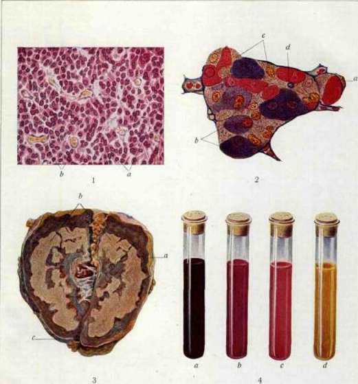
Fig. 1. Eosinophilic adenoma of the pituitary gland in acromegaly: a - groups of eosinophilic cells; b - erythrocytes. Figure 1. Section of the epithelial part of the pituitary gland (after Zhemchuzhnikov): a - adenoma; b - eosinophilic cells; c - connective tissue; d - remnants of the anterior lobe. Figure 3. Tuberculous hypertrophy in Addison's disease (longitudinal section); a - remnants of the adrenal cortex; b - caseous masses, replacing the medullary part of the organ; c - capsule (from the preparation of Muehlmann [Institute of Pathological Anatomy, Moscow State University]). Fig. 4. Monocellular pituitary adenoma: a - initial stage of growth and development; b - stage of development (eosinophilic adenoma); c - pituitary gland; d - capsule of the pituitary gland (after P. Bailey). Pituitary gland, hypophysis, pituitary body. The organization of the dead mass and the development of a scar at the site of the infarct occurs. Pituitary infarct is important because it can be the cause of the subsequent development of pituitary cachexia (see). Hemorrhages in the pituitary gland, which can have the most diverse etiology, are of lesser importance. Inflammation of the pituitary gland, hypophysitis (hypophysitis), most often develops due to the transition of the inflammatory process to the appendage from neighboring parts (meninges, cavernous sinus, sphenoid bone), while at first the inflammation in the form of edema and infiltrate is limited to the capsule of the pituitary gland (perihypophysitis) and partly to its stalk, and only later, involving the vessels, passes to the tissue of the appendage itself and causes the formation of necrotic-purulent foci in it. However, foci of hematogenous inflammation in the pituitary gland (as established by Simmonds) are not uncommon; it is precisely in septicopyemias of various origins that bacterial emboli can form in the anterior lobe of the pituitary gland with the subsequent development of abscesses. In typhus, typical granulomas occur in the neurohypophysis (Davydovsky). In generalized miliary tuberculosis, miliary tubercles can be found in the pituitary gland. In addition, tuberculosis can affect the pituitary gland in the form of the formation of large caseous foci in one part or another of it, which are hematogenous metastases of tuberculosis in their origin. Sometimes such a tuberculous focus destroys the entire pituitary gland completely. In congenital syphilis of newborns, a lesion of the pituitary gland is observed in 57% of cases (Schmidt), manifesting in interstitial proliferation of cell-rich connective tissue and the formation of miliary gummata. In acquired syphilis of adults, the formation of a gumma can sometimes occur in the pituitary gland. In addition, with syphilitic lesions of the meninges of the base of the brain, the process can involve the capsule of the pituitary gland, cause its thickening, the penetration of fibrous strands deep into the organ, and atrophy of its tissue. Most inflammatory changes in the pituitary gland, both non-specific and specific, destroy its tissue to one degree or another. In connection with this, both during the course of these changes and especially as a result of them, certain signs of pituitary insufficiency, i.e., hypopituitarism, may appear. Among hyperplastic processes in the pituitary gland, an increase in individual types of cells of its anterior lobe can be observed, without an increase in the latter in volume. Thus, Kraus found an increase in the number of basophils in progressive paralysis, eosinophils in diabetes, etc.; however, these data cannot yet be considered fully established. An increase in the anterior lobe of the appendage during pregnancy must be considered a constant physiological phenomenon. Microscopically, diffuse hyperplasia and an increase in the size of the main (chromophobic) cells are established, with their transformation into large lamellar elements ("pregnancy cells" - German "Schwangerschaftzellen"). An increase in the anterior lobe of the pituitary gland occurs after the loss of function of the gonads in men and women, as well as in animals (e.g., during castration or destruction of the gonads by a disease process), while in these cases some (Rossle) saw an increase in the number of eosinophilic cells, others (Okinchits) - of main cells. An increase in the anterior lobe (sometimes with the formation of colloid-containing follicles) is almost always observed with a decrease or loss of thyroid function, for example, in congenital underdevelopment of it, in acquired myxedema, in cretinism, in goiters. Focal hyperplasia of cells and small (sometimes multiple) adenomas of the anterior lobe of the pituitary gland are a not uncommon finding (approx. 10%) in subjects over 40 years of age. Less frequently, adenomas (struma pituitaria) are encountered, reaching larger sizes and, during their growth, compressing and atrophying the remaining parts of the pituitary gland, expanding the volume of the sella turcica of the sphenoid bone or bulging from the latter and compressing the corresponding region of the brain. Both focal hyperplasias and adenomas that are difficult to distinguish from them represent a proliferation of cells of the anterior lobe, as a rule, of only one type; therefore, adenomas are observed either from eosinophilic cells (eosinophilic adenomas) (see Figure 1 and color plate, Fig. 1) or from basophilic (basophilic adenomas); adenomas from main cells are rarely encountered. These tumors, although benign in their nature, can, with significant growth, destroy normal elements of the pituitary gland, erode bone, and compress the brain. In addition, sometimes an adenoma, along with the appearance of signs of cellular atypism in it, shows rapid infiltrative growth, which can be considered proof of the tumor's transition into a malignant adenoma or cancer; often in this case, their granularity (eosinophilic or basophilic) disappears in the tumor cells. In comparison with adenomas, fibromas, angiomas, sarcomas, and gliomas of the pituitary gland are much more rarely encountered. Sometimes cholesteatomas and teratomas are found in the region of the pituitary gland. Metastases of tumors (cancers, sarcomas, etc.) to the pituitary gland are not a particularly great rarity. As established by Erdheim, tumors can also originate not from the pituitary gland itself, but from the elements of the pituitary duct [see Figure 2 and separate plate (pp. 183-184), Fig. 4]. Most often, the source of the formation of such tumors (German - Hypophysengangsgeschwülste) are groups of squamous epithelial cells located in the region of the sella turcica and near the stalk of the pituitary gland. The tumor grows intracranially,
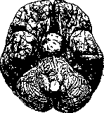
Figure 1. Pituitary adenoma.
can displace the pituitary gland, grow into the brain, and erode the bones of the skull base. Histologically, such a tumor has a peculiar structure, sometimes resembling the structure of an adamantinoma (see), sometimes a basalioma (see); the formation of cysts in the tumor in some cases gives it the character of
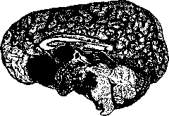
Figure 2. Tumor of the pituitary duct. (After Erdheim.)
a polycystoma. The course of the tumor is usually benign, but sometimes malignant growth of the tumor with metastases to the cervical lymph nodes is observed; histologically, malignant tumors of the pituitary duct most often have the appearance of squamous cell carcinoma without keratinization. In addition to these tumors, adenomas (basophilic and eosinophilic) characteristic of the anterior lobe of the pituitary gland can also arise from the elements of the pituitary duct along its entire length; in the same way, cysts with liquid or colloid content can also develop, sometimes with papillary growth on the inner surface. Cysts of the pituitary gland most often form in its intermediate part, representing an increase in the volume of small cysts that exist there normally; less often, cysts develop in the anterior lobe. These cysts are filled with either serous or thick colloid content. Reaching a significant volume, they compress and strongly atrophy both the anterior and posterior lobes of the appendage. Less common are cysts of embryonic origin, lined with tall ciliated epithelium. As for the influence of tumors and cysts of the pituitary gland on its function, it can be noted that all of them, reaching a significant volume, atrophying and destroying the tissue of the pituitary gland, usually lead to the loss of its function, i.e., to the manifestation of hypopituitarism, most often in the form of dystrophia adiposogenitalis; in particular, a basophilic adenoma, which does not manifest itself in any way when small in volume, gives the same result when it grows. The eosinophilic adenoma stands completely apart, the presence of which in the pituitary gland, as well as its development from the elements of the pituitary duct, manifests itself early with signs of hyperpituitarism in the form of the development of a picture of gigantism (see Giants, gigantism) or acromegaly (see). However, even here, in the case of significant growth of the eosinophilic adenoma and its destruction of the rest of the pituitary tissue, some signs related to hypopituitarism may be added.
A. Abrikosov. Chemistry of the pituitary gland. The study of the chemistry of the anterior lobe of the pituitary gland is in an embryonic state; more definite information is available regarding the posterior lobe. - Anterior lobe of the pituitary gland. The question of isolating the hormone of the anterior lobe of the pituitary gland has been occupied primarily by one author, who has devoted a series of works to this since 1915, namely Robertson. He isolated a lipoid-like substance, which he named "tethelin," the specific action of which is the enhancement of growth in young animals. To obtain tethelin, Robertson grinds 1 part of the anterior lobe of the bovine pituitary gland with 3 parts of a mixture of Na2SO4 and CaSO4 (this removes water from the organ); the mixture is dried and extracted with absolute alcohol. The evaporated extract is subjected to the action of ether; the active substance passes into the solution and is obtained by evaporation. This is an almost white powder, which begins to darken at 100°. It is soluble in water, alcohol, ether, and chloroform, contains 1.4% P and 2.6% S; upon boiling with baryta water, it yields inositol. For one atom of P, there are 4 atoms of N, which indicates an affinity with phosphatides. Tethelin contains 2 amino and 1 imino groups. In small doses (4 mg), tethelin stimulates the growth of young mice, the healing of wounds, and the growth of implanted cancerous tumors. Criticism of Robertson's data has for the time being cast doubt on the question of whether tethelin is a chemically individual, specific substance. - Further attempts to isolate the specific substance are proceeding along somewhat different paths. Evans and Long use an extract obtained by the action of a physiological saline solution. Upon applying this extract to young females, they obtained an increase in growth and a delay in estrus. According to their hypothesis, there are 2 high-molecular specific substances in the anterior lobe. B. Zondek and Aschheim induce the growth of ovaries and estrus in infantile and senile animals using the substance of the anterior lobe of the pituitary gland. Zondek calls the anterior lobe of the pituitary gland the "motor of sexuality." Zondek and Aschheim find this specific substance in large quantities in the urine of pregnant women. By means of dialysis and precipitation, they managed to isolate this water-soluble substance, which they named "prolan." Posterior lobe of the pituitary gland. In 1906, Dale first discovered the effect of the pituitary gland on the isolated uterus of guinea pigs, and with this, a biological method was provided for measuring the activity of preparations of this gland. After attempts by various authors to isolate the specific hormone, Fulmer in 1916 managed to achieve more definite and practically important results. Various patented methods for preparing hypophysin are based in general on the principle of this author. Weakly acidic substances of the posterior lobe of the pituitary gland are purified by dialysis, after which, under the action of heavy metal salts or substances that precipitate alkaloids, a specific product precipitates. After appropriate treatment of the precipitate, it is possible to separate 4 different substances from each other by fractional crystallization; 3 of them give a positive biuret reaction and Pauli reaction and rotate polarized light to the left. At the present time, there is reason to think that this method will not succeed in isolating all the active substances of the gland, because in the process of chemical treatment, molecular rearrangements occur, and the isolated substances easily decompose in the presence of alkalis, and can also be adsorbed by precipitates with a large surface area. The works of Dale (1919) came closer to the goal. He performed extraction using weak acetic acid, and precipitation with colloidal iron hydroxide. By boiling with chloroform, it was possible to separate the specific substance from histamine, which is insoluble in chloroform. Further attempts to purify the specific substance led Dudley (1919, 1923) to the conclusion that the pituitary gland contains no less than three different specifically active substances. He obtained a substance easily soluble in butyl alcohol, active with respect to the uterus, and a less soluble one, which has a pressor effect. Draper (1927), Dale, and others believe that in the posterior lobe of the pituitary gland there are 2 different substances: one acts on the uterus, the other has a pressor effect and an effect on diuresis, and also has a stimulating effect on the melanophores of frog skin, but the latter effect is hardly specific; it is obtained, among other things, with adrenaline as well. These data are also confirmed by clinical practice. Thus, according to the data of Gargley, Gilligon, and Blumgart (1928), "vasopressin" in four healthy individuals delayed the excretion of 1 liter of urine for 5-6 hours, and in two with diabetes insipidus, there was no thirst or polyuria; "oxytocin" (acts on the uterus) produced no effect here. The greatest successes in the question of the active substance of the pituitary gland were achieved by Abel (1919). He obtained a histamine-free preparation, which gives a pressor effect, influences diuresis, and also acts on the uterus (Trendelenburg and others object to such a unitary view). Thus, if previously 4, later 3 and 2 different hormones were isolated, Abel now speaks of a single one. Abel's method consists briefly in the following. The cooled and finely ground glands are treated quickly with hydrochloric acid and mercuric chloride, and then purified with phosphotungstic acid, tannin, picric and picrolonic acids. As a result, a compound of the crystalline hormone with tartaric acid is obtained. This precipitation is associated with adsorption by precipitates with a large surface area. The tartrates obtained in this way possess a threefold action: pressor, action on diuresis, and on the uterus. The tartrates are 1,000 times more active than the corresponding amount of histamine phosphate. These preparations become inactive upon boiling with a normal solution of NaOH or 1% HCl; they give a positive biuret reaction, which probably depends on unremoved impurities. In his latest works, Abel comes to the conclusion that even these tartrates are still not of a single nature, and he isolates 3 different fractions: A, B, and C, of which A is the specific tartrate, B possesses a depressor action, i.e., it is similar to histamine (which means the preparation is apparently not freed from the histamine impurity), and C is an albumose, which has no specific action. All attempts to isolate the hormone encounter a fundamental obstacle, namely the absence of a completely reliable test. The effect of the extract on the uterus does not provide absolute figures of action. At one time, histamine was used as a substance for comparison, but only as long as it was believed that the active substance of the pituitary gland was identical to histamine. Subsequently, as with insulin, they began to produce a standard preparation. In America, Voegtlin (1925) prepared such a dry acetone powder; other preparations are expressed in "Voegtlin units." By a Voegtlin unit is understood an extract of the posterior lobe of the pituitary gland, 1 cubic centimeter of which has the same effect as 0.005 mg of defatted, dried, and powdered posterior lobe of the pituitary gland of cats. Attempts to use other properties of the pituitary extract as a standard proved even less successful. - In conclusion, it must be said that, despite many contradictions, there is still sufficient reason to assume that the posterior lobe contains one specific substance. The fact that, upon inactivation of the preparation with alkali, all three effects are destroyed almost simultaneously, and that the process of inactivation upon heating proceeds according to the type of a monomolecular reaction, speaks in favor of this, among other things. The fact that the rates of diffusion through collodion filters are the same for all three fractions also speaks in favor of the unity of this substance. In any case, the active substance of the posterior lobe of the pituitary gland is a substance of complex structure. Guggenheim suggests that we are dealing with an acyl derivative of alkanolamine, while Leschke considers it a polypeptide. Both points of view harmonize with the ability of this substance to be precipitated by heavy metal salts and phosphotungstic acid and to be hydrolyzed by alkalis, acids, and enzymes. Salting-out ability and the ability to diffuse easily speak in favor of the albumose nature of the hormone. The attempt to synthetically prepare the active substance of the posterior lobe of the pituitary gland has not yet been crowned with success.
The normal and pathological physiology of the pituitary gland is based on data obtained from studying the effect on the organism of the removal of the cerebral appendage and the action of extracts from its anterior and posterior lobes, as well as on the clinical syndromes of hypopituitarism and hyperpituitarism and the pathological-anatomical changes of this organ discovered in them. The pharmacodynamic effect of an extract of an endocrine organ, as Gley emphasizes, does not always allow for a correct conclusion regarding its physiological significance. Nevertheless, the action of pituitary gland extracts on smooth muscle is still of great interest. In addition to the uterus, the gallbladder responds to the introduction of pituitary extract with energetic contractions. The posterior lobe extract, administered subcutaneously or intravenously, causes complete discharge of bile from the gallbladder (pituitary reflex of the gallbladder). Pituitary extract has a similar effect on the musculature of the intestine, sometimes after a short-term inhibition of its motor function. The urinary bladder shows less sensitivity. Blood pressure under the influence of the posterior lobe extract rises, sometimes very significantly—by 20-30 mm Hg. Repeated injections of the extract usually yield an increasingly smaller effect. Blood coagulation (in vitro) under the influence of the posterior lobe extract is accelerated; the anterior lobe extract, on the contrary, slows down coagulation. The posterior lobe extract has an inhibitory effect on urine secretion in humans. The decrease in urine secretion in humans occurs immediately after the injection of the extract, reaches its maximum in about four hours, and lasts about a day. The use of the posterior lobe extract of the pituitary gland for diabetes insipidus as a good means of symptomatic therapy is based on this effect. Its mechanism of action—vasomotor or directly on the renal epithelium—requires further research. The posterior lobe extract shows the same effect in dogs with experimental polyuria caused by a lesion of the tuber cinereum in the region of the infundibulum (Camus, Roussy). In normal dogs, however, the extract causes only a short-term decrease in urine secretion, which is then replaced by an increase in diuresis. In parallel with this, the renal vessels first constrict and then dilate (Houssay and Carrion). Dilation of the pupil under the influence of the posterior lobe extract occurs even after the transection of the cervical sympathetic nerve. The loss of the smooth muscle-stimulating influence of the internal secretion of the pituitary gland, which one would expect upon removal of the pituitary gland, does not, however, cause sharp functional changes in the corresponding organs. Experiments on its removal provide much more data for judging the physiological significance of the pituitary gland. The evaluation of the results of this operation, given by various authors, is, however, extremely contradictory. The reason for the disagreements is the close anatomical connection of the pituitary gland with the tuber cinereum, which contains vegetative nerve centers for various organs, very important for the regulation of metabolism and blood circulation, and conducting pathways to them from higher centers. The pathophysiological effect of hypophysectomy can therefore give rise to doubt as to whether it was caused by the removal of the pituitary gland or by damage to the tuber cinereum, and the absence of an effect may give rise to the assumption of the preservation of the pars tuberalis of the pituitary gland. Even the fundamental question—is the life of an animal possible without the pituitary gland—continues for this reason to cause disputes. The studies of Aschner, Cushing, Camus and Roussy, Arkhangelsky, and others have proven with certainty that the life of animals without a pituitary gland is quite possible and can last for years. The diverse pathological-anatomical changes that occur in hypophysectomized animals show that the pituitary gland is of very great importance for the normal growth and development of the organism and the regulation of metabolic processes within it. Removal of the pituitary gland, both complete and of only the anterior lobe, if the operation is performed at an early age, causes retardation in growth and sexual development and significant obesity with a characteristic localization of fat, especially in the region of the kidneys and pelvis. Despite the obesity, the weight of the operated animals several months after the operation is significantly, three to four times, less than the weight of control animals of the same litter. Short-term hyperglycemia and insignificant glycosuria, often observed in the first days after the operation, are apparently partly a consequence of anesthesia, and partly of irritation during the operation of the nerve centers and conducting pathways of the tuber cinereum. They are replaced by hypoglycemia and an increase in the threshold of carbohydrate assimilation, lasting for two or more months until the appearance of significant obesity. By this period, the blood sugar content becomes normal, and the threshold of carbohydrate assimilation decreases. Polyuria, described by many authors as a consequence of the removal of the pituitary gland, is by no means always observed after hypophysectomy. If there are no persistent changes in the tuber cinereum, this polyuria usually lasts from a few days to a few weeks after the operation and in most cases does not reach particularly large proportions. The same polyuria, and sometimes a very significant and prolonged one, can be obtained by damaging the diencephalon behind the pituitary gland without removing the latter. Sirotinin showed that the destruction of certain areas of the tissue of the tuber cinereum causes dilation of the renal vessels. Perhaps the so-called pituitary polyuria is also caused by a disturbance in the function of the renal vasomotor nerves, arising as a result of damage to the tuber cinereum during the hypophysectomy operation. Likewise, it is impossible to consider as proven the hypotheses attributing to the pituitary gland an important role in the regulation of water metabolism and water content in tissues. (See also Diuresis, Metabolism.) A persistent decrease in temperature in animals deprived of the pituitary gland is very characteristic, developing both after complete extirpation of this organ and after removal of only the anterior lobe. Throughout the entire life of the animal, the rectal temperature is invariably 1–2 degrees lower than that of the control animal. Retardation in growth of hypophysectomized animals ("pituitary dwarfism") is a constant consequence of the removal of the pituitary gland at an early age. Contrary to previous views, early ossification of the epiphyseal cartilages has been noted (Ginzburg). Experimentally obtained "pituitary dwarfs" usually retain normal proportions of body parts. Removal of the pituitary gland in tadpoles causes not only a cessation of growth but also the impossibility of metamorphosis (Smith, Atwell, Allen, and others). Removal of the pituitary gland causes retardation in the development of the sex glands, absence of spermatogenesis, and suppression of libido sexualis. Hypophysectomy thus allows for the experimental creation of the syndrome of dystrophia adiposogenitalis. Despite its obviousness, this fact continues to be disputed by proponents of the tuberal origin of this syndrome. The physical infantilism of hypophysectomized animals corresponds for a long time to the infantilism of the manifestations of their higher nervous activity. The increased excitability and emotivity characteristic of normal puppies persists in hypophysectomized dogs significantly longer than in control ones. However, in the manifestations of both emotive and intellectual reactions, animals without a pituitary gland do not show the defectiveness that is characteristic, for example, of experimental hypothyroidism. The study of the effect of hypophysectomy on the central nervous system does not provide any confirmation for the hypothesis about the pituitary gland and the "infundibulo-tuberal region" as the place from which the "regulation of psychic functions" originates, just as it does not for the now abandoned "pituitary theory of sleep." All of the above shows that the pituitary gland possesses a peculiar activating influence on the processes of intermediate metabolism, which is very important for the normal growth and development of the organism. Experimental data find confirmation in the clinical syndromes of hypo-, hyper-, and dyspituitarism. The mechanism of this influence and the nature of the substances of the internal secretion of the pituitary gland remain for the time being unexplained. Pituitary extracts cannot eliminate the vast majority of the consequences of physiological insufficiency of the cerebral appendage. By means of them, it is also impossible to induce the phenomena of hyperpituitarism—gigantism, acromegaly. Apparently, they do not contain the complete products of the internal secretory activity of the pituitary gland. It is therefore quite fair to point out that it is impossible to draw direct and categorical conclusions about the physiological action of the internal secretion of the pituitary gland based on the pharmacodynamic effect of these opotherapeutic preparations. A. Bogomolets.
Related articles
Mentioned in
Cite this page
“Pituitary Gland.” Soviet Medical Encyclopedia. English translation of Bolshaya Meditsinskaya Entsiklopediya, 1st ed. (Moscow, 1928–1936), ed. N. A. Semashko. https://sovietmedicalencyclopedia.pages.dev/article/hypophysis/