Adamantinoma
Historical document, translated for reference. It reflects medical knowledge of the 1920s–30s and is not medical advice.
Summary
Adamantinoma is a rare tumor of the jaw that develops according to the type of enamel tooth germ. It is typically benign but can occasionally undergo malignant transformation. Treatment involves surgical removal due to the risk of recurrence and potential jaw fracture.
Encyclopedia article (1928–1936)
ADAMANTINOMA, adamantinoma, adenoma adamantinum (from Greek adamas - diamond), a tumor first described (1862) by Robin. The term A. currently in use was introduced in 1901 by Blumm; before that, tumors of this kind were called variously: cystoma proliferum folliculare, epithelioma adamantinum, odontoma embryoplastique, etc. The A. is constructed according to the type of the enamel tooth germ. In accordance with existing theories of the origin of A., they can be classified as odontogenic tumors, i.e., tumors whose development is associated with the dental system. A. is a relatively rare tumor that develops in the alveolar process of one part or another of the lower jaw, or much less frequently, of the upper jaw. In the corresponding area of the jaw (more often at the angle of the lower jaw), a slowly growing spherical new formation appears, as if swelling the bone; the latter atrophies, thinning in places until complete disappearance. In the lower jaw, the tumor develops outward, and only in the area of the 7-8th teeth, where the outer wall of the jaw is thicker than the inner one, the tumor develops mainly inward. In the upper jaw, A. develops outward or penetrates into the maxillary cavity, into the nasal cavity, into the orbit. When pressure is applied to the tumor, a parchment-like sound is sometimes felt. The course is benign; left to itself, A. can reach significant sizes (up to the size of a child's head). Only in very rare cases is malignant transformation of A. observed - transition to cancer; the latter circumstance significantly narrows the clinical benignity of it. The localization of A. in areas not corresponding to the area of normal tooth formation is rare: Fischer (W. Fischer) described a case of primary A. of the tibia, Teutschlander described A. in the area of the pituitary gland. On section, the tissue of the tumor may have either a solid character (A. solidum) or cavities - "cysts", filled with transparent liquid (A. polycysticum). A. is sometimes surrounded by a capsule, sometimes
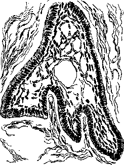
Figure 1. Adamantinoma (one of the cells of the tumor).
however, mostly in cystic forms, the capsule is absent, and the tumor lies in bone cavities. The mass of the tumor macroscopically appears as a spongy tissue, dirty-gray or reddish in color. Sometimes in A. a properly formed tooth or so-called odontoma was found. On cross-section, a loop-like structure is visible. A. solidum gives a characteristic microscopic picture: the basis of the tumor is formed by fibrous connective tissue rich in spindle-shaped cells, and in it are located large, irregularly shaped cells and as if branching strands of epithelial cells; the arrangement and appearance of the latter are very typical (see Figure 1); along the very periphery of the cell or strand lie high cylindrical cells, from them inward - flat and more polygonal cells, closer to the center of the cell transforming into a network of process-bearing star-shaped cells, between which there is liquid. This picture is very similar to the structure of the developing enamel organ of the tooth. The morphological difference from the normal tooth germ lies mainly in that in the parenchyma of A. there is no regular formative process that characterizes the process of tooth development. Cystic A. develops from solid due to
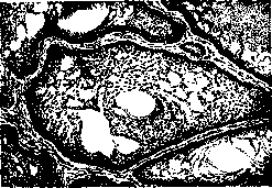
Figure 2. Epithelial vegetations and sheets (observed by I. Lukomsky).
liquefaction of the central parts of the epithelial cells, with the larger cysts retaining only the peripheral layer of cylindrical cells from the epithelium. - There are three theories of the origin of A.: some think that A. develops from excessive or incorrectly laid down in
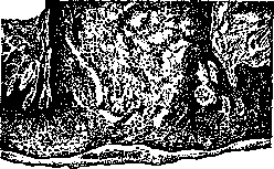
Figure 3. From the epithelium of the oral mucosa
a wide strand departs, forming the tumor (observed by I. Lukomsky). embryonic life of the enamel organ germ or tooth follicle; others consider them to originate from those epithelial islands (debris epitheliaux paradentaires Mallassez), which are found in the root sheath of the tooth; finally, others believe it possible for them to develop from the gum epithelium - sinking processes of epithelium can, as in embryonic life, differentiate into a likeness of the enamel organ. The last two theories must be considered the most probable. - Recognition of A. should be based on the symptoms given by the macroscopic description. The condition of the dentition sometimes helps to distinguish A. from other jaw tumors. In fibromas of the jaw or in periosteal sarcomas, the dentition appears displaced, tilted due to pressure from the tumor. In A., on the contrary, the dentition is usually not displaced and the teeth are not loosened, since the tumor develops in the direction of least resistance, as if growing around the roots of the teeth in the affected area. Treatment of A. should be surgical. Since spontaneous fracture of the jaw may occur due to thinning of its walls, A. must be removed as early as possible; due to the tendency of adamantinoma to recur, its removal should be performed as radically as possible.
Related articles
Mentioned in
Cite this page
“Adamantinoma.” Soviet Medical Encyclopedia. English translation of Bolshaya Meditsinskaya Entsiklopediya, 1st ed. (Moscow, 1928–1936), ed. N. A. Semashko. https://sovietmedicalencyclopedia.pages.dev/article/adamantinoma/