Odontoma
Historical document, translated for reference. It reflects medical knowledge of the 1920s–30s and is not medical advice.
Summary
Odontoma is a tumor consisting of a conglomerate of various soft and hard dental tissues, classified as hamartoblastomas due to developmental abnormalities in tooth germs. These tumors can be simple or complex, with histological structure resembling normal tooth tissue but with disrupted topographical relationships.
Encyclopedia article (1928–1936)
ODONTOMA (odontoma), a tumor representing a conglomerate of various soft and hard dental tissues. O. are predominantly localized in the area of the molar teeth, most commonly on the lower jaw corresponding to the position of the wisdom tooth, and are located either in the area of the alveolar process or centrally within the jaw bone. O. belong to the type of tumors known as hamartoblastomas, as they arise from disturbances in the development of tooth germs, both those that are laid down in the course of normal ontogenesis and those that are excessive. Some O. develop under the condition that paradental tissues retain the ability to form tooth germs in that period when the function of odontogenesis is generally already completed. This type of O. is closely related to those pathological formations in which the formation and development of new teeth continue far beyond the period of normal odontogenesis, manifesting as repeated (after surgical removal) formation of collections of a large number (up to 150-200 or more) of small teeth in the jaw bones. If upon examination of O. it turns out that the neoplasm represents only one deformed tooth or tooth germ, then one speaks of a simple O.; if, however, several teeth or germs are found in the composition of the tumor, then O. is called complex. Simple O. are divided into complete and incomplete (partial). C o m p l e t e O. have the appearance of an amorphous mass, in which it is impossible to establish the contours of an entire tooth or part of it; they arise as a result of a disturbance in the development of the tooth germ as a whole. Incomplete O. in some cases are located corresponding to the crown of the tooth, and the root(s) retain generally the correct configuration—crown O. (odontoma coronarium); if the neoplasm occupies the area of the root(s) with a normally formed crown, then such a tumor is defined as root O. (odontoma radiculare). Incomplete O. arise as a result of a disturbance in the development of only a part of the tooth germ—its crown or root department. Almost all O. described in the literature belong to hard O., i.e., to mature forms, which are in their main mass built from calcified, hard, dental tissues—enamel, dentin, and cement. Theoretically, the possibility of the existence of so-called s o f t O. is permissible, which should represent early stages of tumor development, preceding the formation of hard dental tissues. However, due to the fact that these forms do not manifest clinically in any way, in practice they are not encountered, and the few descriptions of soft O. (Partsch) cast doubt on the actual belonging of these to this type of tumor. The mass of O. as a rule consists mainly of dentin, and in it there are sometimes areas of connective tissue having the structure of dental pulp. In some cases, when enamel and cement are completely pushed into the background, one can speak of a dentinoma. There are also tumors consisting for the most part of enamel—adamantinomas; neoplasms consisting of tissue similar to the cement of tooth roots—cementomas (see) can hardly be considered as one of the types of O., although some authors do include them in this group. According to histological structure, the tissues of O. do not differ from the tissues of a normal tooth, and their development in general completely corresponds to the process of formation of dental tissues in the course of normal odontogenesis. Only the topographical relationships between individual tissues are disturbed, so that in sections or ground sections of O. areas of enamel are often found among masses of dentin, or an alternation of these tissues is observed. Such topographical disturbances are more sharply expressed in complex O., in which sometimes tooth-like formations with a distorted arrangement of enamel and dentin are encountered: a central cavity, imitating the pulp cavity, is surrounded by a layer of enamel, and the latter is covered by a mass of dentin (Figure 5). More often, however, complex odontomas represent a conglomerate of partially fused together by cement or bone tissue, often separate, surrounded by connective tissue, small teeth, having the structure, and often the configuration of normal teeth. The mentioned disturbances in topography of hard dental tissues in mature odontomas are explained by the distortion of topographical relationships between the epithelial (enamel organs) and connective tissue (dental papillae) parts of tooth-like formations in the early period of development of O., when they are in the stage of the tooth germ; in the course of normal odontogenesis, the dental papilla invaginates into the enamel organ (see vol. XI, art. 119-120, Figs. 7 and 8), and in the formed tooth enamel covers dentin (see Teeth, histology and embryology); in the development of individual areas of complex O., reverse relationships may occur: enamel organs turn out to be invaginated into areas of connective tissue corresponding to dental papillae (Fig. 4); with further differentiation of such areas, enamel is found enclosed within dentin, and the central cavity, at first glance imitating the pulp, corresponds to the position of the enamel organ (Fig. 5). In simple O., the arrangement of tissues in general differs little from normal. In those O. in which, along with hard dental tissues, there are rudimentary tooth formations, areas may be found where epithelial cells—enamel organs—are grouped in one place; such areas show great similarity in structure to adamantinoma (see) (compare Fig. 6 with Fig. 1 in volume I, art. 140), which should be kept in mind when diagnosing by biopsy; the difference becomes noticeable in later stages of development when the deposition of hard dental tissues begins in the odontoma. " From the surrounding bone substance of the jaw, O. is usually separated by a layer of fibrous connective tissue, which in the literature is defined by the term "capsule" (Figure 3). However, this designation is justified only for mature O. with completed development; study of the early stages of tumor development showed that this connective tissue is not simply a passive capsule, but is essentially a matrix of the neoplasm containing epithelial and connective tissue elements, from which in the process of differentiation numerous tooth-like formation germs arise (Ryvkind) (Figure 3). Just like a normal tooth germ, the mass of O., upon reaching a certain stage of development, begins to "move" toward the oral cavity (Figure 1). The process of eruption of the tumor, associated with violation of the integrity of bone and soft tissues, may be accompanied by the formation of pockets and inflammatory processes i^i™t1ЧЯ IBh i .)
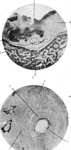
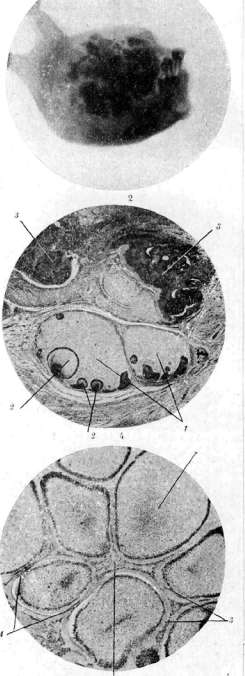
6. Fig. 1. Half of the lower jaw. o. a. - radiograph of an odontoma in a young child: 1-odontoma that has erupted into the oral cavity; 2-granulation tissue bulging into the defect of bone corresponding to the fistula opening in the area of the jaw angle; 3-deformation (outward bulging) of the jaw body. Fig. 2. Radiograph of the specimen from Fig. 1. Fig. 3. Microscopic preparation: 1-hard masses of the tumor; 2-"capsule"; 3-embryonic tooth formations; 4-hard tooth tissues in the "capsule"; 5-bone substance of the jaw. Fig. 4. Area 3 in Fig. 3 at strong magnification: 1-areas of connective tissue corresponding to dental papillae; 2-enamel organs protruding into the tissue of dental papillae; the relationships are the reverse of those occurring in normal tooth development (cf. Figs. 7 and 8, Vol. XI, pp. 119-120); 3-deposits of hard tooth tissues. Fig. 5. Area 4 of the same tumor, representing as it were a mature stage of the area in Fig. 1: 1-location of the left organ; 2-enamel ring; 3-dentin. Fig. 6. Area 5 of the same tumor; great similarity in structure to adamantinoma (cf. Fig. 1, Vol. I, p. 40): 1-enamel organs; 2-layers of connective tissue; 3-deposits of dentin; 4-beginning of enamel deposition (black lines). Odontomas, with symptoms (similar to those that occur in the difficult eruption of wisdom teeth), with consecutive osteomyelitis of the jaw and formation of fistulas (Figs. 1 and 2). These secondary phenomena are observed so frequently that they can serve, along with other symptoms, as a diagnostic sign (Wolf). Odontomas occur very rarely, predominantly in young age; they usually proceed without pain, and small ones generally without symptoms. The latter are most often discovered accidentally during extraction of teeth or roots or during operative intervention undertaken on the diagnosis of difficult eruption of wisdom teeth or in connection with an impacted tooth. In these cases, the presence of O. is a factor that complicates the operation. Large complex O. located centrally in the jaw manifest as a painless swelling of the jaw bone: due to the gradual increase in size of the tumor, there is resorption of bone substance from within the jaw and simultaneously periosteal apposition of new bone tissue, which leads to deformation, as it were, a bulging of the jaw (Fig. 1). The duration and benign course (decades), the eruption of the tumor into the oral cavity, the phenomena of chronic osteomyelitis with fistulous tracts facilitate diagnosis. The presence of a hard O. is confirmed by radiograph, on which the "capsule" is sometimes clearly outlined (Wolf). Treatment - operative: complete removal of the tumor together with the "capsule"; otherwise, the embryonic tissues contained in it (see above) may become the center of new tumor growth. In case of sharp thinning of the jaw bone due to tumor growth or as a result of complicating purulent osteomyelitis - resection or disarticulation of the corresponding half of the jaw. O. also occur in animals, most often in horses, then in cattle.
Related articles
Mentioned in
Cite this page
“Odontoma.” Soviet Medical Encyclopedia. English translation of Bolshaya Meditsinskaya Entsiklopediya, 1st ed. (Moscow, 1928–1936), ed. N. A. Semashko. https://sovietmedicalencyclopedia.pages.dev/article/odontoma/