Teeth
Historical document, translated for reference. It reflects medical knowledge of the 1920s–30s and is not medical advice.
Summary
This article provides a comprehensive overview of the evolutionary development, structure, and classification of teeth across vertebrates, with a focus on mammalian dentition. It details the transition from simple, homodont, polyphyodont systems in lower vertebrates to the specialized, heterodont, and diphyodont systems found in mammals, including the paleontological theories of tooth evolution.
Encyclopedia article (1928–1936)
TEETH. The teeth of vertebrates, in their structure and development, are completely similar to the placoid scales covering the entire skin of shark-like fish. Since the entire oral cavity, and partly the cavity of the pharynx, is lined with ectodermal epithelium, typical placoid scales can also cover the mucous membrane of these cavities in shark-like fish. On the jaws, and in the majority of lower vertebrates, including amphibians, as well as on various covering bones of the skull base and partly on the visceral apparatus, these scales developed more significantly and gave rise to teeth. The main mass of a tooth (like that of a placoid scale) consists of a very hard tissue—dentin, which is permeated by a multitude of tubules originating from the internal cavity of the tooth, which is filled with connective tissue rich in nerves and vessels—the dental pulp. The outer surface of the tooth can be covered with a layer of very hard enamel, consisting of calcified fibers fused together, the enamel prisms. Finally, over the enamel, there is a thin dental cuticle. The base of the tooth is anchored in most vertebrates to the underlying bone by means of connective tissue. On the jaws, teeth are usually attached along the edge of the jaw (acrodont teeth); in some lizards, however, they are attached by their outer edge to the inner edge of the jaw (pleurodont teeth). In crocodiles and some fossil reptiles, as well as in mammals, teeth are anchored (implantation, gomphosis) in special sockets of the jaws (thecodont teeth). Usually, in lower vertebrates, teeth function for a relatively short time and are replaced by new ones as they wear down. Such a change of teeth can occur throughout life (polyphyodontism). In mammals, the majority of teeth are replaced only once at a young age (diphyodontism), and sometimes there is no replacement at all (monophyodontism). However, in some mammals, traces of other replacements have been found, namely, the rudiments of a pre-milk generation of teeth, lying in the dental lamina outside the rudiments of the milk generation, and the rudiments of a post-definitive generation, lying on the inner side of the rudiments of the permanent teeth. Thus, the origin of the mono- and diphyodont dental system of mammals from the polyphyodont system of lower vertebrates has been proven. In mammals, only the incisors, canines, and anterior premolars are replaced once. The posterior, or "true molars," have no replacement. It is usually considered that they do not have milk predecessors. However, their later development is more likely explained by a lack of space in the short jaw of a young animal, and at the present time, many researchers are inclined to think that the "true molars" represent only the later-erupting and more powerful posterior members of the same series as the milk premolars. In some mammals, the replacement is reduced further, and in marsupials, only the last premolar is replaced. The reduction of the dental system also usually begins with the complete loss of tooth replacement (edentates, cetaceans).

The simplest form of a tooth is conical, and in lower vertebrates, teeth are small and numerous. In predatory shark-like fish, however, various cutting forms of teeth are encountered (in the form of ridges, tridents, etc.), and in rays that feed on mollusks, the teeth have the appearance of crushing plates. In the vast majority of vertebrates, all teeth are identical (homodont system), and only in very few fish and reptiles (especially in fossil theriodonts) is there a differentiation into teeth of different shapes (heterodont system). Conversely, in mammals, the latter is the rule: the anterior teeth are chisel-shaped, cutting ("incisors"), numbering up to 3 pairs in each jaw; then follows 1 pair of conical "canines," behind them are more complex tubercular or folded premolars (in primitive forms, 4 pairs each), and usually even more complex "true molars" (up to 3 pairs). Incisors are developed especially significantly in rodents; canines protrude strongly and are a weapon of attack in predators (hence the name dentes canini); the chewing molars are equipped with cutting ridges in predators; in omnivorous and herbivorous mammals, the chewing molars are tubercular or folded, reaching significant power in herbivorous ungulates and especially in elephants. The differentiation of teeth in mammals is accompanied not only by a change in shape according to their function in various parts of the jaws but also by an increase in their size and more secure anchoring in individual sockets. Larger chewing teeth are anchored especially firmly by means of a more complex root (hence the name "molar" teeth). The increase in the size of individual teeth is naturally connected with a reduction in their number. Thus, even in primitive mammals, the number of teeth is already very small compared to other vertebrates, usually reaching 3 pairs of incisors, 1 pair of canines, 4 pairs of premolars, and 3 pairs of true molars in each jaw. Thus, the full dental formula of placental mammals has the following appearance: 3 1 4 3 / 3 1 4 3. The number of teeth in more specialized mammals usually decreases, and sometimes a toothless gap is formed—a diastema—between the incisors and the molars (rodents, etc.). In monkeys, the dental system also turns out to be already reduced, expressed in broad-nosed monkeys of the New World by the formula 2 1 3 3 / 2 1 3 3
and in narrow-nosed monkeys of the Old World—by the human formula 2 1 2 3 / 2 1 2 3; at the same time, however, the canines are developed much more strongly than in humans, and this applies especially to males. In some cases, the special development of individual teeth turns out to be a secondary sexual characteristic: such are, for example, the canines in boars and especially in the male babirusa, in which huge canines pierce the upper lip and curve upward and backward over the skull; such are also the upper canines of walruses, forming huge tusks, the pair of upper incisors forming the tusks of elephants, and finally, the highly original tusk of the male narwhal (a cetacean), directed straight forward, reaching a length of 2 meters and representing the only tooth developing in it—the left canine of the upper jaw. The growth of mammalian teeth is completed by the formation of a root (sometimes with several branches) with a relatively thin canal inside. In rodents (incisors) and partly in herbivores (molars), teeth that wear down heavily possess continuous growth, and a root does not form in them. The complex shape of the molars of mammals developed, as has been proven paleontologically, from a simple conical form through a process of gradual differentiation of its crown. According to the most well-founded theory of paleontologists Cope and Osborn (Cope, Osborn), the initial conical form of the tooth (haplodont form; Fig. 1) was initially complicated by the formation of additional vertices in the form of projections in front of and behind the main vertex (protodont form; Fig. 2). With an increase in the size of the additional vertices to the size of the main one, this form transitioned into a three-toothed (triconodont; Fig. 3) form, and by shifting the vertices, which arranged themselves at the corners of a triangle, into a three-tubercular (tritubercular form; Fig. 4). These latter forms of teeth are also present in some modern insectivores. Then, an additional tubercle develops on the so-called "heel," and the tooth acquires a four-tubercular form (Fig. 5), which lies at the basis of various types of teeth in modern mammals. If the vertices are connected to each other by sharp cutting edges, then a cutting (secodont) form of teeth develops in insectivores and predatory animals. In omnivorous animals, a similar blunt-tubercular (bunodont) form of teeth is encountered (Fig. 6). Through a change in the shape of the tubercles, with each of the 4 tubercles bending at an angle (with the vertex inward), the lunate teeth of ruminants developed (Fig. 8). By pairing the tubercles, transverse ridges are obtained, which characterize the folded teeth of odd-toed ungulates (Fig. 7). An increase in the number of transverse ridges (which can be traced paleontologically) leads to the complex folded teeth of elephants (the same in rodents). In higher herbivores (horse, ruminants), the molars acquire continuous growth, and accordingly, the crown becomes high, and the tubercles and ridges are stretched into long prisms, between which cement is deposited. With the gradual wearing down of the surface, the hard enamel walls of the tubercles and ridges always protrude most on the latter, inside which dentin islands are exposed.
I. Schmalhausen. II. Anatomo-topographical data. Teeth (dentes) are located at the entrance to the oral cavity, on the border between the vestibule of the mouth (vestibulum oris) and the oral cavity proper (cavum oris), where they are arranged in the form of two arches, placed one against the other, and are anchored in the sockets (alveolae) of the upper and lower jaws (Figs. 9 and 10). Their function consists mainly in the act of chewing. The task of the latter consists in the grinding
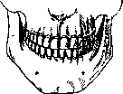
Figure 9. Jaw bite from the front (according to Tandler).
of food and giving it with the help of saliva a consistency suitable for swallowing. In this process, one group of teeth (incisors and canines) serves for cutting and tearing food, while the other (molars) serves for chewing. Teeth are hard formations, outwardly resembling bone but surpassing it in strength. Teeth in humans first appear between six and thirty months after birth; these are so-called temporary, variable, or milk teeth (dentes caduci, decidui, s. lactei), 10 in each dental arch. At the end of the 6th year of life, another series of teeth begins to grow, which replaces the milk teeth. These are so-called permanent teeth (dent. permanentes). There are 16 of these teeth in each dental arch. Thus, humans belong to the so-called diphyodonts. The average length of teeth is 2-2½ cm (canines - 3 cm). Teeth are planted in the jaws in a generally vertical position and so closely to each other that in humans, unlike many animals, there are almost no gaps between teeth (diasteme). On each tooth, the crown (corona dentis), facing the oral cavity, and the root (radix dentis) with the apex, sitting in the alveolar socket, are distinguished, and between the crown and root a slightly narrowed place is noted, called the neck of the tooth (collum dentis) and covered by the gum (fig. 11).
Due to the difference in the shape of the crown, teeth are divided into series: 1) incisors (dent. incisivi, s. incisores), 2) canines, or angular teeth, or cuspidate teeth, due to the sharply protruding in the middle of the cutting edge of one cusp (dentes canini, s. d. angulares, s. cuspidati), and 3) molars, which in turn are subdivided into premolars, or bicuspidate teeth, or small buccal teeth (dentes praemolares, s. bicuspidati, s. buccales minores) and molars, or multicuspidate teeth, or large buccal teeth (dent. molares, s. multicuspidati, s. buccales majores). Incisors and canines belong to the so-called front teeth, while molars belong to the back teeth. By roots, teeth are divided into single-rooted (front teeth) and multi-rooted (back teeth).

There are 16 permanent teeth in each dental arch; of these, 4 incisors, 2 canines, 4 premolars, and 6 molars. In the milk jaw - 10: of these, 4 incisors, 2 canines, and 4 molars. There is a so-called dental formula, which is written in such a way that the numbers of the series of the upper dental arch are written with the initial letters of the names of the series above the line, and the numbers of the series of the lower dental arch are written under the line; therefore for permanent teeth the formula is as follows: 2 1 2 2 4 6 2 1 2 2 4 6 = 32. Since the teeth in both halves of the jaws are placed symmetrically, the formula is written only for half of the upper and lower jaws. Thus, omitting the initial letters of the names for the upper jaw, 2 1 2 2 4 6 and for the lower jaw 2 1 2 2 4 6 are written. This formula is often used in comparative anatomy. When comparing it with the formulas of other mammals, it turns out that the human dental arch is far from complete. The most complete dental formula is found in an extinct mammal that lived in Patagonia and is known under the name Homalodontotherium; its formula for half of the jaw is expressed as follows: S1 4 3 3 3 3 3 3 3 4 3 3 3 3 3 3 3 4 = 44 or 44 in both dental arches. On this basis, the appearance of supernumerary teeth in humans is by many considered an atavism.
In addition to the above comparative anatomical formula, there is also a so-called practical formula, where teeth are denoted by a number corresponding to the place occupied by the tooth, counting from the midline, and for the right half of the dental arch the numbers are placed from the midline backward, and for the left - from the back to the midline; further, for the upper dental arch the numbers are placed above the line, and for the lower - under the line. Thus, the practical formula for the milk jaw will be, going from left to right: 5 4 3 2 1 1 2 3 4 5 and for the permanent jaw: 8 7 6 5 4 3 2 1 1 2 3 4 5 6 7 8. The practical formula for half of the jaw is more often used; then the right side is taken. Therefore, for the milk jaw it will be: 1 2 3 4 5 and for the permanent 8 7 6 5 4 3 2 1.
Among dentists, the method of denoting individual teeth with a corresponding number, separated by an angle open towards the side from which the tooth is taken, is widespread; for the right side of the upper row, one line of the angle is placed under the number, and the other behind the number; for the left side of the same row - one line at the bottom, and the other in front of the number. For the lower row of teeth on the right side, one line of the angle is placed above the number, and the other behind; for the left side - one line above the number, and the other in front. Thus, the upper right canine is denoted |3, and the upper left 3|, the lower right |3, the lower left 3|. When describing crowns of teeth, on front teeth the cutting edge and four surfaces are distinguished: labial (facies labialis), lingual (f. lingualis), and two contact surfaces: medial, or proximal (f. medialis, s. proximalis), and lateral, or distal (f. lateralis, s. distalis). On back teeth - the chewing surface (f. masticatoria) and four lateral: buccal (f. buccalis), lingual (f. lingualis), or palatine (f. palatina), and two contact surfaces: anterior, or proximal, and posterior, or distal (f. anterior et posterior).
Permanent teeth. Series of incisors (fig. 12) - there are eight of them, four in each dental arch; of these, two central and two lateral. The shape of the crown is chisel-shaped, with the sharp edge facing the opposite jaw. On this sharp cutting edge of unworn teeth, three small protrusions are noted. The labial surface is convex. The widest part of the labial surface is at the cutting edge. The lingual surface is concave, and on the upper incisors the so-called marginal ridges are noted, which, going from the medial and lateral sides at the neck, converge and form a tubercle (tuberculum dentis). In the lower incisors there are no such ridges. The widest part of the tooth surface is also at the cutting edge. The contact surfaces of one tooth with another represent triangular platforms, with the apex facing the cutting edge, and the arched base facing the neck. The angles formed by the cutting edge and the contact sides are different: the medial angle is acute, and the lateral is rounded. If both angles are rounded, the lateral is always more rounded. The roots of these teeth are cone-shaped, rounded, and somewhat compressed from the sides; this compression is especially expressed in the lower incisors. The root is not set perpendicularly to the cutting edge, but is deviated laterally. As for the size of these teeth, the largest are the upper central incisors,
Fig. 12. a-upper right central incisor from the front; b-the same from the side; c-from behind; d-upper right lateral incisor from behind; e-lower right central incisor from the front; f-the same from the side; g-from behind. (According to Tandler.)
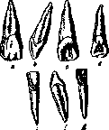
Fig. 13. a-upper right canine from the front; b-the same from the side; c-from behind; d-lower right canine from the front; e-the same from the side; f-from behind. (According to Tandler.)
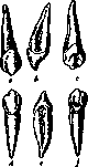
and the smallest are the lower central ones. To determine from which half of the dental arch a tooth is taken, Mühlreiter has proposed three signs: 1) the sign of curvature of the labial surface (its medial half is more convex than the lateral), 2) the sign of angles (the lateral angle of the cutting edge is more rounded than the medial), and 3) the sign of the root (the root deviates from the perpendicular restored to the cutting edge to the side from which the tooth is taken). Canines (d. canini; fig. 13) - there are 4 of them, 2 in each dental arch. The upper ones are usually more developed than the lower ones; their crown is somewhat shorter, and the root is longer; in the lower ones - on the contrary; the cutting edge in both is broken, and therefore in canines there are two cutting edges standing at an angle: the medial edge and the lateral, the first being shorter and lying more horizontally than the second. The angle where the edges meet is the middle of the three protrusions that were noted on the cutting edge of the previous series - greatly developed into a cusp, which is why canines are called single-cuspidate teeth. The labial surface is strongly convex in the transverse direction. This convexity is strengthened by a ridge going from the apex of the cusp to the neck.
1
The lingual surface is concave, and on it there is also a ridge running along the middle from the tip of the tubercle to the neck. On the sides of this surface there are marginal ridges that, when they meet at the neck, form a tuberculum dentis similar to that noted in the incisors, but more developed than in the latter. The contact surfaces are similar to those of the previous series, and as for the root, it is well-developed, especially in the upper teeth, and has an oval shape. The root is usually single, rarely bifid in the lower canine. To determine on which side a given canine is taken, the same Müller's criteria are used as for the incisors. The premolars (d.praemolares, s. bicuspidati; fig. 14 A)-there are 8, 4 in each jaw; placed behind the canines, 2 on each side. They are called first and second premolars. In these teeth, the cutting edge has been transformed into a chewing surface due to the strong development of the tub. dentis of the previous series, as a result of which two tubercles are noted here: buccal and lingual, separated by a longitudinal groove (chewing groove) running along the chewing surface closer to the lingual side, so that the area of the lingual tubercle on the chewing surface is smaller than the area of the buccal one. The groove does not reach the edges of the chewing surface, so at the ends of the groove there remain ridges connecting both tubercles. In the lower premolars, the lingual tubercle is less developed, as a result of which the chewing surface is inclined toward the oral cavity. In the upper ones, this tubercle is higher, and the chewing surface is almost horizontal, but in any case, in these teeth, the development of the lingual tubercle never reaches the development of the buccal one. The shape of the crown is transversely oval, with its long axis running from the buccal side to the lingual. The root of the upper first premolar is more often double, in the others-with some exceptions relating to the upper second premolar-single. In the upper teeth, the root is flattened from the sides, while in the lower ones it is more rounded. The molars (d. molares, s. multicuspidati, s. buccales majores; fig. 14 B). There are 12 in humans-3 on each side in each dental arch. These are teeth that have no predecessors, i.e., milk teeth, and complement the permanent jaw. They are the most massive and have the largest chewing surface; located behind the premolars and differ from each other in the place they occupy, counting from the premolars backward. The last, or third molar, is also called because of its late eruption the wisdom tooth (d. sapientiae, s. d. serotinus). The shape of the crown of these teeth is generally cuboidal, gradually decreasing from the first to the last molar. The chewing surface of the first molars is quadrangular, closer to rhomboid, the second is trapezoidal, and the third is irregularly triangular. On the chewing surface, 4-5 tubercles are noted, separated from each other by chewing grooves, the appearance of which in the upper teeth is H-shaped, and in the lower ones cross-shaped. Thus, two buccal tubercles and two lingual ones are formed, with the buccal tubercles being higher in the upper jaw, and the lingual ones in the lower. Quite often, in the first upper molars, in addition to the four tubercles mentioned, an additional tubercle appears, tubercul.anomale Carabelli, located on the lingual surface in front of the lingual tubercle. The apex of this tubercle usually does not reach the level of the chewing surface. The chewing surface of the second upper molar, whose crown is usually smaller, often deviates from the type described above. As for the wisdom tooth, both its chewing surface and the entire tooth are subject to so many variations and anomalies that it is impossible to classify it under any laws. It can be said that the inherent lack of any definite size and shape is its distinguishing characteristic. This tooth can be well-developed, and on its chewing surface there can be from 3 to 8 tubercles, but it can also be represented in the form of a pin, where there is no question of a chewing surface. The chewing surface of the first lower molar differs in that it has five tubercles (95%): 3 buccal and 2 lingual. This occurs due to the forked splitting of the buccal half of the transverse groove. The same splitting (however rarely) can also affect the lingual half of the transverse groove (cases with 6 tubercles). The chewing surface of the second molar is smaller in size and, with few exceptions (10-15%), has four tubercles. As for the lower wisdom tooth, it, like its antagonist, shows a tendency to variations, although to a lesser degree. In any case, this tooth should also be classified as degenerating. The number of tubercles can be very different (from 1 to 7), but the shape and size of the crown are more constant than in its antagonist. The buccal and lingual surfaces of all molars are moderately convex in the direction from front to back, with the buccal surface being less convex than the lingual, and its convexity in the anterior half of the surface is greater than in the posterior, which allows the application of Müller's curvature criterion to these teeth. When passing into the anterior contact surface, the buccal surface forms an acute angle, but when passing into the posterior one, it does not form such an angle, but presents a rounding. The lingual surface is smaller in size, more rounded, and when passing into the contact surfaces, it does not form angles at their border.-The molars belong to the multi-rooted teeth; in the upper jaw there are 3 roots: 2 buccal, anterior and posterior, and one palatal. The buccal roots run vertically, are flattened from front to back, the anterior one is wider and longer than the posterior one, both are bent backward, and grooves are often observed on their surfaces. The palatal root is deviated toward the oral cavity; it is the most massive, round, and on its palatal side a groove is observed. In the first and second upper molars, sometimes fusion of one root with another is observed. As for the wisdom tooth, here too all kinds of variations are possible in relation to the roots; there can be one root and several (5-7). The direction of these roots is also different. In the lower molars there are 2 roots-anterior and posterior. Both are flattened from front to back and bent to the lateral side; the anterior one is more massive and longer than the posterior one. Rarely, fusion of the roots can be observed in the first and second lower molars
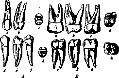
Figure 14A. a-upper premolar tooth from the front; b-the same from the side; c-its chewing surface; d-lower premolar tooth from the front; e-the same from the side; f-from behind; g-its chewing surface.
Figure 14B. a-upper molar tooth from the front; b-the same from the side; c-from behind; d-its chewing surface; e-lower molar tooth from the front; f-the same from the side; g-from behind; h-its chewing surface. (According to Tandler.)
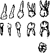
tshsh
Figure 15. Milk teeth (according to Altukhov). The upper wisdom tooth usually has three roots, but sometimes there is a fusion of the front root into two prongs. The roots of the lower wisdom tooth are subject to the same variations as its antagonist; therefore, there can be either one root with traces of fusion or several. Milk teeth (figure 15), being predecessors of permanent teeth, generally have the form of the latter with few exceptions. Their distinctive features are: smaller size, bluish-white color (instead of the yellowish-white of permanent teeth), enamel bulge at the root edge of the crown and formation of a thickening, due to which the boundary between the crown and neck is sharply marked, greater roundness of the roots of incisors and canines and divergence of the roots of molars, greater thinness of the walls of teeth and relatively large pulp cavity and root canal and finally less strength of the constituent parts of teeth. Since incisors and canines in form correspond to their permanent successors already described, only the posterior ones, i.e. molars, deserve a separate description. The upper ones have three roots, while the lower ones have two. The first upper molar differs by a crown elongated from front to back, due to which the chewing surface has the appearance of a quadrangle, on the surface of which a deep longitudinal groove runs closer to the palatal side, dividing this surface into two cutting edges or tubercles. The buccal tubercle is divided by transverse depressions into three tubercles; the buccal and palatal tubercles are connected to each other by ridges in front and behind. The buccal surface is lower than the lingual, and on it a prominence is noted on the front part of the buccal tubercle, which Zuckerkandl named tuberculum molare. The palatal surface is considerably rounded, the contact surfaces are also rounded, with the posterior being more convex than the anterior. The second upper molar in appearance is completely like the first permanent molar of the upper jaw, with only the difference that besides its smaller size on its chewing surface there is an enamel fold connecting the posterior buccal tubercle with the anterior palatal. The tuberculum Carabelli inherent in the first molar is also noted here, even more often than in the permanent one. The first lower molar has a crown in the form of an elongated quadrangle; the chewing surface is narrow and divided by a longitudinal groove into buccal and lingual halves, on which two cutting edges are noted, separated by deep notches into individual tubercles; on the buccal edge there are 2-3, and on the lingual 2. The second lower molar is in the form of its crown and the appearance of its surfaces similar to the first lower molar of permanent teeth in reduced dimensions; the crown is oblong-quadrangular, equipped with 4, sometimes 5 tubercles, of which 2-3 are buccal and 2 lingual.
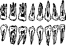
Figure 16. Dental cavities (according to Altukhov).
As for the roots of milk molars, both in number and in location they fully correspond to the permanent molars, i.e. in upper teeth there are three roots: of them 2 buccal and one palatal; in lower teeth 2 roots: anterior and posterior. Their peculiarity is the strong divergence to accommodate between them the germs of permanent teeth. In both milk and permanent teeth, as has already been indicated, there is a space known as the dental chamber, or cavity of the tooth (cavum dentis; figure 16). This cavity continues by means of special tubes into the roots (canalis radicis dent.) and opens at their apices through openings (for. apicis dentis). As can be seen from the figure, the cavity of the tooth in general repeats its external form, and in it they note the upper wall, or cover of the cavity, then the floor and four walls, the name of which corresponds to the name of the buccal, lingual and two contact surfaces. The cover generally corresponds to the cutting edge of anterior or the chewing surface of posterior teeth, and according to the number of tubercles there are convexities in the cover called horns, and the higher the tubercle on the surface of the tooth, the larger the horn. Regarding the floor, it should be said that in single-rooted teeth it essentially does not exist, since the cavity of the tooth without a special boundary passes into the root canal. The same can be in teeth with two canals, if the latter divide at the apex of the root; in those teeth where the division of canals is high, the floor is saddle-shaped; such a saddle-shaped floor from the lingual to the buccal surface is also present in large molars of the upper jaw and from front to back in large molars of the lower jaw. The other four walls are convex corresponding to the convexity of the external surfaces of the tooth. As for the size of the cavity of the tooth, in young people it is larger, and in older people smaller due to the continuous deposition of dentin on the walls of the cavity. The cavity is located in the tooth or in the lower part of the crown at the neck or at the beginning of the root, so that the cover of the cavity lies approximately in the middle of the crown or somewhat lower. The root canals repeat their form. In single-rooted teeth there is one canal each, exception is made by small molars, lower incisors and canines, where there can be two canals. In teeth with a larger number of roots there is a corresponding number of canals, exception can be made by upper large molars, where in the anterior buccal root instead of one there can be two canals, and lower large molars, where in the anterior root there can also be two canals. As shown by corrosion casts, the root canal can give blind processes into the dentin, into which of course the pulp goes; in two- and three-rooted teeth two neighboring
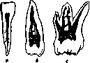
Figure 17. a-incisor; b-small molar; c-large molar tooth (according to Vorobiev).
canals can be connected by anastomoses or can merge and form a cavity (fig. 17). Finally two opposite walls of the canal can in a certain part fuse and form one or more partitions. At the apical opening sometimes a delta-shaped branching of the root canal is encountered. Teeth are fixed by their roots by a special joint called impaction (gora-phosis) in the so-called alveolar, or dental, processes of the upper and lower jaws, in special cone-shaped depressions called alveoli or cells (alveolae), which are an exact cast of the roots sitting in them (fig. 18). For single-rooted teeth these depressions are single, for multirooted they are divided by partitions according to the number of roots of a given tooth. Thus, for large molars of the upper jaw there are two partitions, of which one goes from front to back and divides the depression into buccal and palatal halves, and the other divides the buccal half into two parts, anterior and posterior. For large molars of the lower jaw there is one partition, placed in the bucco-lingual direction and dividing it into anterior and posterior parts. The buccal and lingual walls of the alveoli consist of bony plates and are thin, especially the buccal one, forming on the facial surface of the alveolar processes protrusions (juga alveolaria); the partitions, both between neighboring alveoli and inside complex alveoli, consist of spongy substance covered on the side of the alveoli with a bony plate studded with small openings, of which some go horizontally in the partition, and others vertically to the edge of the alveolus. These openings are intended for vessels and nerves going to the root sheath and to the gums. At the bottom of the alveolus there is an opening (sometimes two), intended for the passage of vessels and nerves going through the apical opening of the root into the cavity of the tooth (apical openings can also be two). Blood (see separate table, fig. 2) to the dental apparatus goes from the internal maxillary artery (art. maxillaris interna), which is a branch of the external carotid. This artery gives for the lower teeth a branch (arteria alveo
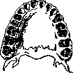
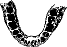
Fig. 18. Dental sockets (according to Kolanovsky). The inferior dental artery (art. alveolaris inferior) originates from that portion which curves around the articular process of the mandible. This branch, having separated from the inferior surface of the trunk of the internal maxillary artery, descends between the ramus of the mandible and the medial pterygoid muscle, reaches the mandibular canal going in the bone below the floor of the sockets, and passes through this canal; and upon reaching the mental foramen, it divides into two branches, one of which emerges through this opening to the outside, while the other continues its course in the canal beneath the incisors. Passing along the canal, art. alveolaris inferior gives off at the floor of each socket two groups of branches, namely: proper dental twigs, rami dentales, passing in the form of one, more rarely two, through the opening in the floor of the socket into the root canal and into the dental pulp; and interalveolar arteries, rami interalveolares, which are much thicker than the dental ones and which, passing through the septa between the sockets, give off twigs to the bone itself and to the periodontium, and also through the vertical openings mentioned above, they proceed to the alveolar border of the jaw in order to supply the gums (rami gingivales). Zukkerkandl mentions twigs which pass from the lateral walls of the socket to the periphery of the root, perforate the cement and dentin, and reach the pulp. For the upper dental arch, blood is carried by art. alveolaris superior-for the posterior teeth and by art. infraorbitalis-for the anterior ones. The first of these trunks arises from that part of the internal maxillary artery which lies on the lateral pterygoid muscle and, descending, enters the openings on the temporal surface of the maxilla (by one or more twigs), penetrates to the posterior teeth and, similar to the inferior maxillary, gives off proper dental twigs and branches for the bone, gums, root sheath, and for the mucous membrane of the maxillary sinus. The end of the internal maxillary artery and art. infraorbitalis, passing through the corresponding canal, gives off twigs for the anterior teeth through the dental openings located on the floor of the canal. The distribution of these branches is the same as for the posterior teeth.
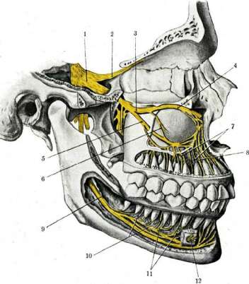
Veins carrying blood away from the dental apparatus begin in the pulp by means of venae dentales, emerging through the apical foramen, and venae interalveolares, going through the openings in the interalveolar septa and carrying blood from the root sheath, as well as venae gingivales, proceeding from the gums. All these branches proceed from the lower row of teeth through special openings in the mandibular canal and form the inferior maxillary vein, which empties into the facial vein; from the upper row of teeth they proceed into the so-called deep branch (ramus profundus), which beneath the zygomatic bone unites with the facial vein. Lymph vessels begin in the coronal pulp, according to some (Schweitzer) in the form of small canals (vessels), and according to others (Izachik) in the form of clefts, which flow into larger trunks lying in the root pulp, between the blood vessels and nerves, it should be mentioned that both the pulp lymph vessels and those of the periodontium, periosteum, and gums are devoid of valves (the efferent lymph pathways of the gums are already provided with valves), which has important practical significance. In multirooted teeth, each root receives lymph from a definite part of the coronal pulp lying over this root. Then from the root canal the lymph vessels proceed either through the apical foramen or through lateral openings in the walls of the root into the lymph vessels of the periodontium (pericementum). From the periodontium for lymphatic vessels there are two paths: the first-along the blood vessel canals going from the sockets into canalis alveol. infer. for the lower teeth and into can. alv. sup. et can. infraorb. for the upper teeth; the second path-along canals leading from the sockets to the periosteum and gums, and thence to the corresponding lymphatic glands (e.g., to the mental glands lying in the space between the anterior bellies of m. digastr. and os hyoid.). To their lymphatic area (according to Partsch) belong the 4 lower frontal teeth. The submaxillary (glandulae lymphaticae submaxillares) in number three are located between the lower border of the mandible and the submaxillary salivary gland. The anterior of these glands lies at the anterior border of the salivary gland, the middle-in front of the internal maxillary artery passing here, and the posterior-immediately behind this artery, at the posterior end of the salivary gland. To the lymphatic area of these three glands belong (according to Partsch) both rows of teeth with the exception of the lower incisors. The described groups of glands give off their lymph partly to the parotid lymph glands, and these latter-to the superficial and deep cervical glands, and partly directly to the latter. The groups of cervical lymphatic glands connect with the supraclavicular lymph glands, the vessels of which empty into the jugular trunk, which on the right empties into ductus lymphaticus dexter, and on the left-into ductus thoracicus. Innervation of the teeth (see separate table, fig. 1) is from n. trigeminus, namely: for the upper jaw from the second branch, and for the lower-from the third. The second branch of nervus maxillaris gives off n. alveolaris superior posterior, which goes together with the artery of the same name into the canal on the temporal surface of the maxilla; then the terminal branch of n. maxillaris, s. n. infraorbitalis, gives off 2 more dental branches: n. alveolaris superior medius et n. alveolaris superior anterior. The ramifications of these branches in the anastomose with each other and form the dental plexus, and the lower part of these anastomoses appears in the form of an arch, from which through special canals pass branches for the spongy substance of the alveolar process and interalveolar septa, going then to the gums, and branches going through the openings on the floor of the sockets for the dental pulp. The third branch of the trigeminal nerve, n. mandibularis, s. n. alveolaris inferior, proceeds from the foramen ovale downward to the opening of canalis alveolaris, enters it, and there at the level of the floor of the socket gives off the same branches as the superior dental nerve. At the mental foramen the nerve divides like the accompanying artery into two branches, one of which continues its course in the canal to supply the incisors, and the other through the mental foramen emerges to the outside. c. Stoppvitsvii.
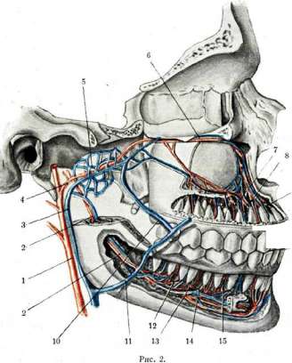
Fig. 1. Nerves of the teeth. 1 - ganglion semilunare Gasseri; 2 - n. ophthalmicus; 3 - n. maxillaris; 4 - n. infraorbitalis; 5 - n. alveolaris superior; 6 - n. alveolaris superior medius; 7 - rami dentales superiores; 8 - rami gingivales superiores; 9 - n. alveolaris inferior; 10 - rami gingivales inferiores; 11 - rami dentales inferiores; 12 - n. mentalis. Fig. 2. Vessels of the teeth. 1 - a. carotis ext.; 2 - a. alveolaris inferior; 3 - v. facialis post.; 4 - a. maxillaris int.; 5 - plexus pterygoideus; 6 - v. infraorbitalis; 7 - a. dentalis superior; 8 - a. gingivalis superior; 9 - v. dentalis superior; 10 - v. anastomotica facialis; 11 - v. facialis ant.; 12 - a. dentalis inferior; 13 - a. gingivalis inferior; 14 - v. dentalis inferior; 15 - v. gingivalis inferior. Histology and embryology. The composition of the teeth includes the following tissues: dentin (see), which forms the main mass of the tooth and determines its shape, enamel, which covers the crown, and cementum, which covers the root of the tooth; these are hard tissues; in addition, the tooth cavity is occupied by delicate fibrous connective tissue - the dental pulp, and on the surface of the cementum there is a dense connective tissue sheath - the periosteum of the tooth (peri-cementum, or periodontum, root sheath). Enamel is a modification of epithelial tissue and, according to its microscopic structure, consists of elongated formations - enamel prisms - modified epithelial cells. On a ground section of the tooth, dark bands can be distinguished in the enamel, running obliquely through the thickness of the enamel to the border with the dentin - Retzius bands, often pigmented, and another system of lines that gives the ground section of the enamel a resemblance to moiré fabric: these lines, visible in reflected light, run through the thickness of the enamel to the border with the dentin - Schreger's lines [see separate table (pp. 119-120), fig. 3]; these systems of lines are an expression of the winding course of the enamel prisms, and are partly due to pigment. In chemical composition, enamel is the tissue richest in inorganic salts in the entire organism, and contains only 2-3% of organic compounds, as a result of which an adult tooth, upon decalcification, usually never retains its enamel covering. The salts include carbonates and predominantly lime phosphates, to which small amounts of silicates are also added. In its density, enamel approaches apatite. On ground sections of teeth, the composition of enamel from five- or six-sided prisms can be clearly seen, the course of which, while generally directed from the outer surface to the border with the dentin, appears very tangled, which is why it is never possible to trace an individual prism throughout its entire length; usually this course is conceived as follows: an individual prism, starting from the border with the dentin, first passes in a direction perpendicular to this border, then turns obliquely and closer to the surface of the tooth again takes a direction perpendicular to the surface; at the same time, such a curved enamel prism cannot be laid in one plane, but appears twisted; the enamel prisms are arranged in groups, bundles, and in adjacent bundles the direction of their course is opposite. With such an arrangement of prisms, their crossings are obtained, which in general form a system of curved, mutually supporting fibers, which in mechanical terms significantly gains in strength and distributes pressure from one point over a large surface of the underlying parts (fig. 19). Each enamel prism is also twisted along its axis; partly due to this, and partly due to the uneven deposition of salts, under the microscope the enamel prisms appear transversely striated Fig. 19. [see separate table (pp. 119-120), fig. 4]. Between the prisms there is a small amount of intermediate cementing substance, somewhat larger at the base of the prisms, at the border with the dentin, and negligible - closer to the surface; the cementing substance is also calcified, however somewhat less than the prisms; this difference is clearly evident (as well as the lines) when the surface of the ground section is carefully acted upon by mineral acids. The surface of the enamel is covered by a cuticular formation, the enamel cuticle, or Nasmyth's membrane. The border between the enamel and the underlying dentin does not run evenly, but in the form of a wavy line; in places where the dentin extends relatively deeply into the enamel layer, the dental tubules pass for some distance into the enamel, into its cementing substance [see separate table (pp. 119-120), fig. 1]. In young teeth, the enamel prisms contain significantly less inorganic compounds, and in this case, on decalcified preparations, it is possible by splitting to isolate individual prisms entirely and study their shape. Cementum, both in structure and composition, closely resembles ordinary bone, with the only difference that, with rare exceptions, it completely lacks Haversian canals and the bone lamellae forming it are weakly expressed; their arrangement, however, can be determined by the arrangement of bone cells; only in some cases on old teeth, closer to the apex of the root, in thick deposits of cementum, Haversian canals can also be seen, very few in number. In addition, a distinctive feature of the structure of cementum is the abundance of Sharpey's fibers, passing through the thickness of the cementum into the periodontum (peri-cementum - root sheath) and then extending through a layer of loose connective tissue into the periosteum of the tooth socket [see separate table (pp. 119-120), fig. 2]; they thus connect the root of the tooth with the bony wall of the socket. Cementum, covering the root of the tooth, in the region of the neck borders with the enamel and here slightly overlaps it. Pulp. The cavity of the tooth is occupied by loose connective tissue, which for a long time retains an embryonic character, connecting through the root canal with the loose connective tissue lying under the tooth. The pulp is relatively poor in thin bundles of collagen fibers and contains a relatively large number of small cellular elements, anastomosing with each other by their processes. At the border between the pulp and the dentin there is a layer of large pear-shaped cells, also connected by processes both among themselves and with the underlying cells - the so-called odontoblasts (a name that has remained for these cells due to their participation in the development of tooth tissues). From these cells, thin long processes extend outward into the dentinal tubules and along them reach the border with the enamel, where they partly extend into its layer (see separate table, fig. 5). In the loose tissue of the dental pulp, blood vessels passing through the root canal form a dense network of capillaries; the veins formed from them exit the cavity of the tooth by the same path into the underlying loose connective tissue lying under the apex of the root. Together with the vessels, nerve twigs enter through the root canal, forming a plexus in the pulp, from which the terminal twigs are directed to the layer of odontoblasts, to which they are traced. The presence of nerve twigs in the thickness of the dentin, as well as the manner of termination of nerves in the tissues of the tooth, have not yet been sufficiently studied, due to the great difficulty of the research technique. Most authors tend to believe that the sensitivity of the dentin is due to the irritation of Tom's fibers - processes of odontoblasts. Periosteum of the tooth, peri-cementum, or periodontum. The surface of the tooth root, as stated above, is covered by a layer of dense connective tissue, in the upper part of the tooth socket directly passing into the periosteum of the tooth alveoli; closer to the apex of the root, a loose layer delimiting the root sheath from the periosteum of the tooth socket becomes increasingly distinct, and in the region of the apex of the root and under the tooth, a layer of loose connective tissue is already distinctly developed, forming a soft padding under the root of the tooth, containing the vessels and nerves passing into it. In the region of the neck of the tooth, a special system of dense collagen fibers can be noted, extending from the edge of the tooth socket to the neck of the tooth, encircling the neck like a tourniquet and then extending again to the edge of the tooth alveolus; this system of fibers forms around the neck of the tooth a circular ligament - ligamentum suspensorium dentis, firmly attaching the tooth to the edges of the tooth socket and, at the same time, due to its slight elasticity, allowing some mobility of the tooth in the vertical direction - a circumstance of no small importance in terms of maintaining proper blood and lymph circulation in the tissues of the tooth (massage when chewing solid food). Development of the tooth. In terms of its development history, the tooth is a complete analogue of a dermal papilla or papilla of the tongue (especially closely resembling the filiform papillae of carnivores). The difference consists only in that the connective tissue base here partially ossifies, and the epithelium, instead of keratinizing, is impregnated with mineral salts. The composition of the tooth germ in higher vertebrates includes an epithelial outgrowth of the mucous membrane of the oral cavity (modified skin) and a connective tissue papilla, growing from the depth to meet the epithelial germ (fig. 20). In humans, the first signs of the tooth germ can be found at the end of the second month of intrauterine life (according to other authors - at the beginning of the third). At this time, the lip is not yet separated from the jaw.
At the edge of the jaw arch, the multilayered flat epithelium of the oral cavity forms a thickening in the form of a ridge extending from the midline of the body to the ends of the jaw—the dental ridge. Soon this thickening sinks inward, and in place of the ridge a semilunar groove is formed—the dental groove, which in the thickness of the jaw tissue corresponds to a plate of flat epithelium—the dental plate, thicker in the middle and gradually thinning toward the back. The flat epithelium of the dental plate in its middle part gives off two outgrowths: the anterior, deeper one, in its middle part begins to keratinize and, splitting into two epithelial layers, separates the lip from the jaw gub—a lip plate, while the posterior one—the actual dental plate—gives pear-shaped outgrowths, the primordia of enamel organs (see separate table, fig. 6). Further from the midline, the lip and dental plates stand A
B
v
Г_____
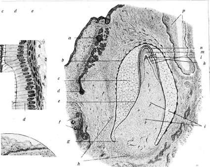
Fig. 1. k jaw; b- *g VU4J dental lamina; B-second stage: c-epithelial buds; d-connective tissue papillae; C-third stage: e-dental groove; f-enamel organs; D-fourth stage: k-free edge of the dental lamina; l-necks of the enamel organs. They are located at a small distance from each other, and between them a narrow strip of oral mucosa is formed. Here, the labial lamina, as it grows, separates the inner surface of the cheek from the jaw tissue. The pear-shaped outgrowths of the dental lamina, as they grow into the tissue, soon encounter a collection of round mesenchymal cells growing towards the epithelial outgrowth; thus, the dental papilla is formed - the rudiment of dental pulp and dentin (see separate table, fig. 7). As many of these papillae form under the epithelial rudiment as there will be cusps on the tooth. Development of dentin. The cells of the dental papilla do not remain the same as they were at first: at the border with the cells of the inner epithelium of the enamel organ (see below), a layer of larger pear-shaped cells, arranged like an epithelium, is isolated on the surface of the papilla; subsequently these cells form dentin, and for this reason they are called odontoblasts (see separate table, fig. 8 and 9). From their pear-shaped body, thin processes extend to the periphery, directed towards the epithelial rudiment; from the basal end, processes extend connecting adjacent odontoblasts with each other, and thin thread-like processes also extend into the dental papilla, connecting the odontoblasts with the deeper-lying cells. The first deposits of dentin appear in the form of a uniform thin layer (membrana praeformativa, praedentin), in which then thin collagen bundles differentiate (however, not all researchers agree with this, and some believe that odontoblasts deposit fibrous substance from the very beginning). When studying young dentin on tangential sections or on teased preparations, one can see that the thin bundles of collagen fibers form a dense mesh lying in a plane perpendicular to the direction of the odontoblast processes (Tomes' fibers), and therefore parallel to the surface of the dentin. In the layer of young dentin, the deposition of lime salts begins, which with the ground substance first form small, then large spherocrystals, initially soft, capable of merging with each other, and then hardening. The deposition of lime and the formation of spherocrystals proceed unevenly, and the spheres deposited earlier have time to harden before new deposits appear. As a result, the latter may not merge with the earlier ones, and between them remain small gaps of ground substance, devoid of lime or very poor in it; on a skeletonized tooth, such areas are empty, filled with air, and therefore black, and are called interglobular spaces (see Dentin); at the border with the cement, the slowly proceeding process gives small crystals in the form of grains, as a result of which the gaps between the salt deposits turn out to be very small; thus Tomes' granular layer is formed. Development of enamel. After the first deposits of dentin appear at the apex of the dental papilla, the formation of enamel prisms from the epithelial rudiment begins. In the so-called enamel organ from the very beginning, a layer of basal cells can be distinguished, differing in their height and regular arrangement. Corresponding to the apex of the dental papilla, these cells form a spherical cluster-the enamel nodule, which subsequently passes into a cell cord extending from the apex of the papilla towards the narrow neck connecting the enamel organ with the dental lamina-the enamel cord. It is assigned the role of a cord that holds the epithelium in the area of the apex of the dental papilla and gives the epithelial rudiment the appearance of a bell. The cells adjacent to the dental papilla of the enamel organ-the inner epithelium of the enamel organ-are arranged in a regular row and are significantly elongated in length; due to their activity, enamel prisms are formed, and for this reason they are called ameloblasts [see separate table (for the article Excretions), figures 9-10]. In the depth at the base of the dental papilla, the inner epithelium of the enamel organ passes without a sharp boundary into the outer epithelium of the enamel organ, the cells of which are strongly flattened; the epithelial cells lying in the middle, closer to the inner epithelium, are applied in several layers densely to the latter, and the rest undergo a very peculiar change, making them resemble rather reticular connective tissue than an epithelial formation-the cell bodies remain very small; from them numerous processes extend, connecting the cells with each other, and fluid accumulates in the interval; as a result, a very delicate layer is obtained between the outer epithelium and the layer of ameloblasts, obviously having a protective significance for the unclear layer of ameloblasts from the pressure of the surrounding parts and the growing mass of tooth-pulp of the enamel organ. The place of transition of the inner epithelium of the enamel organ into the outer epithelium gradually grows inward and outlines the contours of the future tooth; however, below the neck of the tooth, ameloblasts do not form enamel and subsequently disappear, sometimes remaining in the form of small cell groups (debris epitheliaux paradentaires), which are a source of the formation of cysts and other epithelial proliferations. The cells of the inner epithelium of the enamel organ, also starting from the apex of the dental papilla, elongate and at the border with the formed dentin deposit in their bodies drops of substance impregnated with lime salts and blackening from osmic acid; more and more such drops appear; the area of their deposition gradually moves from the base to the apex of the ameloblast; a small amount of these drops can also be seen in the ground substance between the ameloblasts. The inner ends of the enamel cells appear thinner and elongated, and a clear boundary can be seen between the young enamel prisms, in which mineral salts are deposited (in connection with the mentioned drops), and the unchanged body of the ameloblast; it seems that the forming enamel prisms push the layer of epithelial cells outward. The thin central ends of the ameloblasts, which turn into enamel prisms, are called Tomes' processes. The process of salt deposition in the enamel prisms proceeds gradually to the distal end of the ameloblast, and its remnants form on the surface of the prism a cuticular membrane, merging with similar neighboring cells into one common covering of the enamel skin (Nesmith's membrane). From the apex of the papilla, this process gradually spreads to its base until the entire enamel layer of the crown is formed. During the development of enamel prisms, the surface of the enamel increases significantly, as a result of which the outer ends of the prisms turn out to be thicker than the inner ones; moreover, apparently not all enamel prisms reach the border with dentin, and some of them end in the middle layer of enamel. The gaps between the prisms, occupied by ground substance, in the deep parts of the enamel layer turn out to be wider. The outer epithelium of the enamel organ, consisting of flattened cells, at the end of tooth development undergoes atrophy, first partially, in islands, so that a perforated epithelial plate is obtained, through which connective tissue easily penetrates to the young tooth. Subsequently, all this epithelium, as well as the inner epithelium of the enamel organ, completely disappears; however, in some places its remnants may remain-the future glands of Serre. The process of deposition of dentin, and subsequently of enamel, as stated, begins at the apex of the dental papilla, therefore the young tooth at the beginning of development appears in the form of islands (tooth germs), of which there are as many as there will be cusps in the corresponding tooth. Gradually these cusps grow in depth and then merge (in multi-cusped teeth), forming a common crown, tooth. With an increase in the thickness of the dentin layer, odontoblasts Figure 1. Border of enamel and dentin at high magnification: a-dentin; b-dentinal canals extending into enamel; c-enamel. Figure 2. Grinding through the root of the tooth; a-cement; b-dentin; c-light lines-boundaries between cement plates; d-Tomes' granular layer; e-cement cells; f-Sharpey's fibers. Figure 3. Longitudinal grinding of a tooth (incisor): a-enamel; b-dentin; c-cement; d-pulp cavity; e-lines of Retzius; f-Schreger's lines; g-interglobular spaces in the dentin of the crown; h-Tomes' granular layer. Figure 4. Grinding of the tooth crown: a-enamel; b-dentin; c-lines of Retzius; d-crossing of enamel prisms; e-longitudinally cut enamel prisms with transverse striation; f-wavy boundary between dentin and enamel. Figure 5. Pulp, section of a decalcified tooth: a-dentin; b-pulp; c-layer of odontoblasts; d-blood vessels in longitudinal and cross section; e-bundles of pulp fibers; f-pulp cells. Figure 6. Early stage of tooth rudiment: a-oral epithelium; b-dental lamina; c-bell-shaped outgrowth of epithelium-rudiment of enamel organ; d-collection of connective tissue cells-rudiment of dental papilla; e-bone. Figure 7. Later stage of developing tooth: a-neck of the enamel organ;
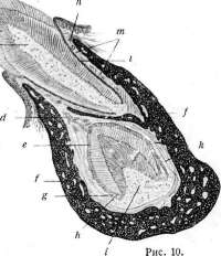
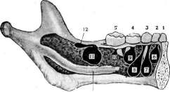
b, s, d-enamel organ (d-internal epithelium; s-external epithelium, b-pulp of the enamel organ); e-dental papilla. Figure 8. An even later tooth bud: b-external epithelium of the enamel organ; e-internal epithelium of the enamel organ; s-pulp of the enamel organ; d-transition of the external epithelium of the enamel organ into the internal; k-layer of ameloblasts; p-thin cap of young enamel; t-cap of dentin (larger than the enamel cap; its light inner part-unmineralized dentin); l-layer of odontoblasts; t-dental papilla-future dental pulp; a-remains of the dental lamella and in it concentric accumulations of cells-pearls; d-germ of permanent tooth; /-bone of the jaw; p-flat epithelium of the lip and gum; Ji-dental sac. Figure 9. Part of the previous preparation, indicated by the rectangle, at high magnification: d-ameloblasts, at the top having deposited a thin layer of enamel (s), below the layer of ameloblasts-lagged behind due to shrinkage of the preparation; e-intermediate layer; /-pulp of the enamel organ; b-dentin; a-odontoblasts; d-pulp (dental papilla). Figure 10. Relative position of deciduous and developing permanent tooth: o-enamel of deciduous tooth; b^-dentin of deciduous tooth; J-cement of deciduous tooth; s-pulp of deciduous tooth; p-gum; m-root sheath of deciduous tooth; /-bone of alveolar process; d-neck of permanent tooth bud, remnant; e-enamel organ of permanent tooth; d-external epithelium of enamel organ of permanent tooth; h-internal epithelium of enamel organ of permanent tooth; i-dental papilla of permanent tooth; k-dental sac of permanent tooth. Figure 11. Left half of the lower jaw of a three-year-old child, opened from the inside (germs of permanent teeth-black): 1 and 2-deciduous incisors; 3-deciduous canine; 4 and 5-deciduous molars; 6 and 7-germs of permanent incisors; S-germ of permanent canine; 9 and 10-germs of permanent molars; 11-germ of first molar; 12-germ of second molar; 13-canal of lower jaw. (From Rauber.) To the illustrations of the article Teeth.
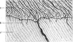
Figure 1.

Figure 2.
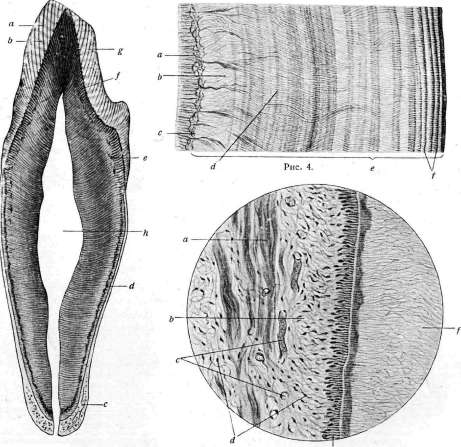
Figure 3.
Figure 5. To the article Teeth. Figure 9. Figure 6. 6-J

I.)
To the article Teeth. gradually pushed deeper, their number decreases; the size of the dental papilla also decreases; remnants of the dental papilla tissues form the pulp 3., for a long time retaining its character of embryonic connective tissue in the developed 3. At the time when the crown has already formed, there is still no root, and the pulp communicates widely with the underlying connective tissue; blood vessels of the dental papilla freely communicate with the deep vessels of the jaw. The continuing growth of the enamel organ epithelium gradually outlines the contours of the root, and the superficial cells of the papilla (odontoblasts) form the dentin of the root; initially this layer of dentin is very thin, and the dental pulp communicates with the underlying tissue by a broad bundle; then the dentin layer thickens, the length of the root increases, and in the final form the pulp communicates with the deeper tissue only through the narrow root canal. Cement. The connective tissue surrounding the 3. germ forms a dense mass around it, enveloping the tooth germ in the form of a sac-the dental sac. This dense connective tissue, on the one hand, protects the 3. from pressure of surrounding parts, and on the other hand, serves as material for the development of the third hard component of the 3.-cement. With atrophy of the external epithelium of the enamel organ, the inner layer of the dental sac grows through it to the 3. and in the area of the root it ossifies, giving cement; bundles of Sharpey's fibers, captured by the ossification process and remaining unmineralized, turn into Sharpey's fibers. The outer layer of the dental sac forms the periosteum of the tooth (root sheath, periosteum). In the area of the neck, this layer closely adheres to the wall of the dental alveolus and merges with its periosteum, further between them there remains a thin layer of loose tissue, more abundant under the apex of the root. As the 3. forms, it begins to move toward the outer surface of the gum; The dental lamella in humans initially gives 10 protrusions in each jaw corresponding to the number of deciduous teeth; after the laying of the second premolar, the posterior end of the dental lamella grows backward and forms the germ of the first molar, on the continuation of the dental lamella another germ is formed for the second molar (see separate table, figure 11), and then the posterior end of the lamella turns into the germ of the third molar - wisdom tooth; occasionally the dental lamella grows further, and then a germ of another additional 3. may form. After all tooth germs are laid, the dental lamella atrophies, first turns into a perforated layer of epithelial tissue, and then completely disappears; remnants of its epithelium can also give groups of epithelial cells in the gum and serve as the starting point for the formation of epithelial proliferations of one kind or another. Germs of permanent 3., replacing deciduous 3., depart with a long stalk from the neck of the enamel organ of deciduous 3. and descend medially from it deep into the jaw, where they are placed in a small niche of the dental cell under the forming deciduous 3. (see separate table, fig. 10). This germ of permanent 3. lies, especially in incisors, almost horizontally. There are 26 germs of 3. in each jaw in humans; sometimes this number increases, and the number of 3. reaches the complete dental formula of other animals (see above); in other cases, some tooth germs may not develop, resulting in a reduction in the number of teeth. In the upper jaw, which develops from three germs, a violation of the normal number of teeth can be caused by the failure of these germs to fuse or by the splitting and loss of tooth germs.
V. Fomin. IV. Eruption of teeth and their statics. For the eruption of both deciduous and permanent teeth, there are certain definite periods, during which teeth erupt in groups with certain intervals. Eruption generally begins first in the lower jaw and then in the upper. Table of eruption times for deciduous teeth. Central incisors........... from 6 to 8 months. Lateral »............ » 7 to 9 » First molars.......... » 12 to 14 » Canines................ » 15 to 20 » Second molars........... » 20 to 30 » There may be deviations from this table in one direction or another: thus, earlier eruption is observed as hereditary, due to the more superficial location of the germinal lamina. Cases have been observed of a child being born with erupted teeth. Later eruption may depend on the deep location of the germinal lamina, but mainly occurs in pathological processes affecting mineral metabolism and associated with endocrine organs (rickets, myxedema, mongolism). After the last, i.e., second molar, has erupted, there is a certain pause in the development of deciduous teeth, during which the roots and crowns of deciduous teeth are finally formed. This period lasts until 6 years, at the end of which the first permanent molar appears as a harbinger of the beginning of the shedding of deciduous teeth and their replacement by permanent ones. This process proceeds in the same order as the eruption of deciduous teeth, preceded by a series of changes in the roots of deciduous teeth (see below). The time of appearance of permanent teeth is shown in Dietlein's table (see next article). Cases of earlier eruption than indicated in the table are rarely observed. More common are cases of delayed eruption. Appeared and finally formed permanent teeth undergo further deposition of dentin from the side of the pulp cavity, which leads to a decrease in the size of this cavity, constriction of the pulp, its atrophy, and atrophy of the vessels and nerves located in the tooth cavity, and consequently to a gradual decrease and then cessation of nutrition of the teeth. Simultaneous obliteration of the periodontal vessels deprives this tissue of nutrition. Such wholesale deprivation of nutrition leads to loosening and loss of teeth, and after that to resorption of the alveolus, which is also deprived of nutrition due to obliteration of the vessels. However, cases are not uncommon when permanent teeth do not fall out and remain until deep old age. Lost or extracted permanent teeth are not replaced by new ones, and those cases described in the literature as proof of a third dentition should be considered (as Scheff points out) either as a delay in the development of permanent teeth, due to which they erupt very late, or (according to Kollmann's opinion) as the development of extra teeth (hyperdentition), and such teeth appear only after the loss of permanent teeth. Cases of the appearance of new teeth were also noted by Eustachius, Fallopius, and others, but a separate case is that of Hufeland, where in a 116-year-old old man 8 teeth appeared, which fell out very soon, and after them again appeared several short-lived teeth, which were in turn replaced. Such phenomena of hyperdentition cannot be interpreted as a third dentition, since it has never been observed in the study of the development process of teeth that the dental lamina, contrary to the theoretical assumption of Rose, gave primordia for three successive series. On the arches formed by teeth, two surfaces are distinguished: the anterior labial-buccal, convex, and the posterior-lingual, concave; two edges: of them one is free, occlusal, the other, facing the alveoli, is root. The height of the crowns in these rows, starting from the incisors, gradually decreases (the canines are an exception), while their volume from front to back increases up to the first molar, and then decreases towards the wisdom tooth. The upper jaw arch forms half of an ellipse, the short axis of which is the line connecting the posterior surfaces of the wisdom teeth, and half of the long axis is the line going from the middle of the previous one and passing in the sagittal direction through the space between the central incisors. The location of teeth in the alveolar processes of the upper jaw is inclined forward and outward. Therefore, the occlusal edge forms an arc of a larger radius than the root edge. In addition, the teeth are somewhat inclined in the distal direction. The lower jaw arch forms a parabola. The location of teeth in the alveolar processes of the lower jaw is opposite to that in the upper. Here the anterior teeth stand vertically, and the posterior teeth are somewhat inclined inward, due to which the occlusal edge forms an arc of smaller dimensions here than the root edge. Also here, as on the upper jaw, the teeth are somewhat inclined in the distal direction. The closure of both jaw arches is called articulation. In so-called correct articulation, the following relationship of teeth of both jaw arches occurs: due to the different inclination of the teeth of the upper and lower jaws and the different sizes of the arches in the transverse direction, the teeth of the upper jaw cover the lower teeth so that the lower incisors with their cutting edges touch the palatal surfaces of the upper incisors, and the upper molars with their buccal tubercles protrude towards the cheek, while the lower ones with their lingual tubercles - towards the oral cavity; the lingual tubercles of the upper molars lie in the longitudinal groove of the lower jaw arch, and the buccal of the lower molars - in the longitudinal groove of the upper. Due to the unequal sizes of the cutting edge of the central incisors of the upper and lower jaws, the upper central incisor touches not only the lower central one, but also the medial half of the lateral one, and the upper lateral incisor not only with the distal half of the homonymous one, but also with the medial canine. This relationship continues to the wisdom teeth, where due to the larger size of the lower wisdom teeth, the position equalizes and the distal surfaces of both these teeth already lie in the same plane. Due to such a relationship of teeth of both jaw arches, each tooth of the upper and lower jaw arch articulates with two opposite teeth, i.e., has two antagonists, of which the homonymous one is called the main antagonist, and the heteronymous one is the secondary. For the teeth of the upper jaw, the main antagonists will be in front, and the secondary ones distally, and for the teeth of the lower jaw, the main ones are behind, and the secondary ones are medial. Due to the same unequal sizes of the cutting edge of the central incisors of both jaws, the spaces between the teeth of the upper and lower jaw arches will not coincide, and each space between the teeth of one jaw will come in the middle of the cutting edge or occlusal surface of the other tooth. The described relationships of teeth of both jaw arches are called correct occlusion, but not all people possess it. Quite common is so-called straight occlusion, when with the teeth closed, the upper row does not cover the lower, as is inherent in normal occlusion, but the cutting edges of both rows coincide. This occlusion depends either on the more vertical direction of the upper teeth or the forward inclination of the lower ones. With straight occlusion, the anterior teeth are preserved longer, as they loosen less, because when closing, the impact is along the axis of the teeth, not from the side, as in correct occlusion, where the lower incisors strike the lingual surface of the upper ones. The disadvantage of straight occlusion is the relatively rapid wearing of the anterior teeth. Straight occlusion is so common that it is more correct to attribute it to normal.
S. Stotsky. V. Pathology of teeth. Malformations, anomalies of development and dystrophies. The human dental system during the period of formation and growth is under the influence of factors of heredity, embryonic life of the organism and the various diverse influences of endogenous and exogenous character, which it experiences from the moment of birth of a person until the time of final development of the chewing apparatus. The multiplicity, diversity and complexity of these influences give rise to a whole series of deviations from the norm. I. As a true malformation, doubling of the upper or lower jaw or absence of one of them is noted. Doubling of the jaw is expressed in the presence next to the normal dental row of another row. Such malformations are explained either by a developmental defect of one individual (polygnathia) or by the fact that the additional jaw is a remnant of an undeveloped second embryo (epignathia). II. Anomalies relate to the number, order and time of eruption, position, forms, sizes of individual teeth, as well as closure or bite. Anomalies of the number of teeth: 1) supradentia-congenital excess of teeth of this or that group, 2) adentia-congenital deficiency of this or other teeth.-Anomalies of eruption of teeth: 1) early eruption of 3., 2) delayed eruption of 3., 3) simultaneous eruption of homonymous 3. of the right and left sides.-Anomalies of position of 3.: 1) Position of supraanomalia of 3., when the cutting edge (in frontal 3.) or chewing surface (in premolars or molars) protrudes beyond the chewing plane of the dental row. 2) Position of infraanomalia of 3., when the cutting edge or chewing surface does not reach the common chewing plane of this dental row. 3) Position of labial and buccal anomalia; 3. is protruded from the dental arch toward the lip or cheek. 4) Position of lingual and palatal anomalia; 3. protrudes from the dental arch on the lower jaw toward the tongue, on the upper jaw toward the palate. 5) Position torto-anomalia-the entire 3. is rotated to one or another number of degrees around its long axis. 6) Diastema and trema. Under the first is meant a free space between two neighboring 3. existing in the presence of a full dental row, which sometimes can reach a significant size (over 6 mm). When there are spaces between three or more neighboring 3., they speak of trema. 7) Transposition-congenital displacement of 3. from its usual place to the place of a 3. of another group, for example, a premolar to the place of a canine or molar, etc., or position of 3., reverse to normal: crown upward, and root downward, and such a 3. can erupt in the nasal, maxillary cavity and other places. Sometimes such 3. do not erupt at all, remaining in the thickness of the jaw bone, and their presence is clarified
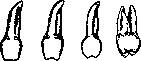
Figure 21. a - tooth of Fournier; b - screw-shaped form of tooth of Hutchinson; c - conical form of tooth of Hutchinson; d - tooth of Pflilger.
can only be determined by an X-ray. The cause in this case should be considered as incorrectly positioned (heterotopia) tooth germs in the early period of embryonic life. - Cases of other position anomalies can arise due to early extraction of deciduous teeth, when neighboring permanent teeth occupy the place of the corresponding normal ones. Anomalies of the shape of individual teeth: 1) teeth with a notch on the cutting edge. This dystrophy of shape can occur on each of the eight incisors. It is characterized by the presence of a semilunar notch on the cutting edge of otherwise normal in shape tooth. The notch is caused by hypoplasia of its enamel. 2) Fournier's tooth, - characterized by a screwdriver-like shape, i.e. the cross-section of the tooth near the gum is wider than at the cutting edge; the latter is normal. The name Fournier's tooth applies only to the central upper incisor (figure 21a). 3) Hutchinson's tooth (fig. 21b) - defined by a screwdriver-like shape of the central upper incisor, the presence of a semilunar notch on its cutting edge and sometimes a reduction in the size of the tooth (see Hutchinson's triad). A similar shape of the central lower incisor and all lateral incisors is not given the significance of Hutchinson's tooth. 4) Barrel-shaped (or semi-barrel-shaped) tooth. - has arched labial surfaces, causing the greatest cross-section at the middle of the crown height. In the semi-barrel-shaped form, the width of the tooth in the neck area and at the middle of the crown height is the same, and then a sharp narrowing begins towards the cutting edge. If the central upper incisor has this shape, it is considered a variety of Hutchinson's tooth, and the same significance is attached to it (figure 21c). 5) Spindle-shaped tooth, resembling a wedge. The shape of this tooth is essentially the most pronounced form of Fournier's tooth, and therefore, when it occurs on the central upper incisor, it should be given the significance of Fournier's tooth. 6) Saw-toothed teeth: teeth with an uneven, worn-saw-like cutting edge, the formation of which is explained by the weakness of the enamel structure. On each of the incisors in normal condition at their eruption, there are 3 large tubercles, which should not be counted as saw-toothed teeth. 7) Teeth twisted along the long axis. 8) Pfluger's teeth: the first molars, characterized by a narrower palato-buccal diameter of the crown in the area of the chewing surface compared to this same diameter of the crown near the neck of the tooth (fig. 21d). 9) 4-cusped first lower molar. Normally this tooth has five cusps: three buccal and two lingual. 10) 6-cusped first lower molar. 11) Upper first molar with a pronounced tuberculum anomale Carabelli. Anomalies of tooth size. They speak of 1) macrodentia of a tooth, when the size of the crown of one or another tooth exceeds the absolute normal size of the crown for a tooth of this kind, 2) microdentia - the size of this tooth does not reach the absolute normal size of teeth of this group. If the proportionality of the crown is violated due to a sharp decrease in its height with normal or exceeding normal width, they speak of 3) hypoalthism of the tooth. - Anomalies also include: a) reduction or increase in the number of roots of multi-rooted teeth, division of roots of canines and incisors, change in shape and direction of roots (rotation of the root around the axis, corkscrew-shaped root), b) fusion of two neighboring teeth to each other by means of cementum in the area of only the roots or by means of dentin along the entire length of the tooth. The dental cavity in such cases can be common or double. These anomalies are explained by the too close position of neighboring tooth germs. From fused teeth, they distinguish c) double teeth, formed from one dental sac. Anomalies of bite can have the following variants. 1. Prognathia. Under this is meant such an anomaly of position of the entire upper dental arch, in which it protrudes sharply forward. The lower front teeth bite deeply behind the upper ones, sometimes directly into the palate and due to the lack of natural support they lengthen excessively, giving a convex line along the cutting edge. The normal relationship between the molar teeth is preserved. -2. Progenia. Protrusion of the front teeth of the lower jaw in relation to the front teeth of the upper jaw. The relationship between the molar teeth is not violated. Prognathism results from excessive development of the upper jaw bones compared to the lower one. Progenia is characterized by significant development of the lower jaw. The described prognathia and progenia refer to the so-called "true"; in other cases they can be "apparent", namely, when the upper jaw is properly developed and the upper teeth are in the correct position, while the lower jaw is small and its front teeth when closed occupy a place behind the upper ones; this condition is called lower opisthognathia (opisthognathia infer.). The opposite phenomenon - when the lower jaw is normal and the upper one is underdeveloped - is called upper opisthognathia (opisthognathia superior). - 3. Partial straight bite. The front teeth of the upper and lower jaws touch directly with their cutting edges, while the molar teeth have normal relationships. -4. Open bite (mordex apertus Carabelli). It is characterized by the presence of a free, sometimes larger, sometimes smaller gap between the front teeth of the upper and lower jaws in their closed position, while the posterior ones can articulate normally. -5. Partial cross bite. The front teeth of the right side articulate correctly, while on the left the lower ones protrude in front of the upper ones or there are reverse relationships of the front teeth; the molar teeth maintain normal relationships. - 6. Mixed bite. It includes all other irregularities of the relationships of front teeth with preserved normal relationships of molar teeth. It should be noted that in orthodontics another so-called practical classification of bite anomalies, developed by Angle, is also accepted. In his system, the first molar of the upper jaw ("key of articulation") is taken as the basis. According to the relationship of this molar with the same one of the lower jaw, Angle divides all anomalies into 3 groups, or classes: to class I belong all anomalies in which the mesio-distal relationship of the cusps of the first molars is normal (neutral bite), i.e. the mesiobuccal (buccal) cusp of the upper one falls upon closure into the groove between the buccal cusps of the lower one. All incorrect positions of individual teeth often occur in this class, and even an open bite belongs to this group with the correct relationship of the molars. Class II can be diagnosed when there is a distal bite, i.e. when the lower molar, and with it the entire lower dental arch, is more distal than normal. The upper incisors and canines tilt forward, protrude strongly; the lower incisors press against the palate or near the necks of the upper ones, are elongated, may be tilted inward; the second division of this class is also characterized by a distal bite, but the upper incisors are bent inward with their cutting edges. Both divisions of distal bite can be bilateral (1st subdivision) or unilateral (2nd subdivision). Thus, all forms of prognathia belong to this class. Class III is characterized by a medial bite, i.e. the mesiobuccal cusp of the upper molar falls upon closure into the space between the 6th and 7th teeth or even deeper. The lower front teeth protrude in front of the upper ones, the entire lower jaw is strongly developed. The anomaly is often associated with opisthognathia of the upper jaw. This violation of bite can also be bilateral (1st subdivision) and unilateral (2nd subdivision). According to the nomenclature of pathological anatomy, this deformation is called progenia. Class III is the most severe lesion of the bite, which can be corrected only in the initial stage; in the later period, help can be provided only surgically and not always completely. - Causes of bite anomaly are divided into congenital and acquired. The first include abnormally large and small sizes of the upper and lower jaws, lengthening and shortening of the alveolar processes of the jaw, irregularities from the angle of the lower jaw, abnormal frenulum of the lips. The second include premature loss of deciduous teeth, delayed resorption of roots of deciduous teeth and late eruption of permanent teeth; early loss of the first large molar teeth before the formation of the entire dental row; constant mouth breathing, habit of sucking fingers or biting the lower lip, and such general causes as rickets.
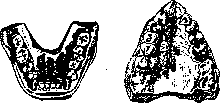
Figure 22. Rickets
Figure 23. V-shaped
lower jaw (atypical
upper jaw (atypical
opacity. of the jaw). thickness of the enamel. 2. Spotted enamel; on the enamel of individual 3. single or multiple milk-white shiny spots of various sizes and shapes are visible. 3. Erosions; on the enamel 3. there are cup-shaped depressions, sometimes larger, sometimes smaller in size, mostly darker in color than the rest of the enamel. 4. Grooved form; a depression in the enamel, caused by its insufficient development, has the appearance of a groove running around the crown of the 3. at one or another height; this depression, encircling the crowns of the 3., may be of varying width. 5. Stepped form; the latter is characterized by the presence on the enamel of the crowns of teeth of a series of grooved depressions, located one above the other like a ladder.-Dystrophies of tooth color. 1. Milky-white, chalky color of the 3., observed in hyperfunction of the thyroid gland. 2. Dark yellow color of the 3. with a reddish
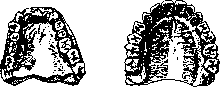
Figure 24. Rickets
Figure 25. Normal-
upper jaw
upper jaw. shape of the upper
jaw. tint on the chewing surface of molars, depends on hyperfunction of the adrenal glands (see below). 3. Pink, brown-pink, brown-purple color of the 3. is observed in congenital porphyrinuria.- Dystrophies of jaw structure. 1. Rachitic lower jaw: atypical- has the shape of a trapezoid, the short side of which is formed by the front 'teeth, the diverging lateral sides are formed by premolars and molars (fig. 22); in typical- in addition to the described form, there is an inclination of the molars toward the tongue. 2. Rachitic upper jaw is characterized by compression of it along the premolars, and sometimes also of the canines and the first molar (compare figs. 24 and 25). 3. V-shaped upper jaw B. m. E. t. XI. owes its origin to mouth breathing, for which reason it also bears another name, namely "jaw of the mouth breathing type". Its signs are: a) convergence of the alveolar processes at an angle in the area of the central incisors and b) gothic palate (figure 23). 4. Acromegaly of the lower jaw is recognized by a) disproportion in its size compared to the size of the upper jaw and the entire skull, b) rare arrangement of 3. In acromegaly, the growth of the horizontal part of the jaw occurs after the replacement of the 3. has already taken place; therefore, free spaces are formed between the latter. 5. Dwarf jaws differ from normal ones by a reduction in all dimensions. 6. General dystrophic jaw-a collective concept; various deviations from the normal shape of the upper and lower jaws that do not lend themselves to classification are placed under it. Anomalies and dystrophies of the dental system are of great importance in human life. When sharply expressed, they disrupt the physiol. function of the chewing apparatus and create favorable conditions for the development of caries of the 3. and so-called alveolar pyorrhea. Various types of anomalies are important diagnostically. Clinical medicine teaches us that Hutchinson's tooth, Fournier's tooth, the knife-like upper central incisor are very reliable signs of congenital syphilis. Of somewhat less value for this disease are Pflüger's teeth, the 4-cusped first lower molar, diastema (when its size exceeds 4 mm and it is not caused purely by local causes), congenital absence of the upper lateral incisors. The changes in the 3. in congenital syphilis are so diverse and constant that their very multiplicity is considered a very probable sign of this ailment. No less frequent cause of dystrophies is rickets. Hypoplasias of the enamel are very often observed in rachitic children precisely on those 3., the ossification of which coincides with the onset of the rachitic process. In general, it should be noted that the dental system quite often contains a number of valuable signs of past or existing diseases.
n. Agapov. Heredity of anomalies of 3. Until recently, teeth were studied very little from the point of view of heredity. This is explained by the fact that 3. are an extremely unfavorable object for compiling family-hereditary tables; as is known, genealogical data in any field of anthropogenetics is very difficult to collect. In odontology, however, with rare exceptions, this data cannot be obtained at all, since a person is usually very little informed about the number, shape, size, and other features of his chewing apparatus; as for his relatives, in this respect he is in the vast majority of cases completely uninformed. The possibility of systematic work on the heredity of 3. therefore appeared only since the introduction into anthropogenetics of the method of studying twins.
TEETH
The essence of the twin method consists in that with respect to any trait, both monozygotic (homologous) twins and dizygotic twins are examined. When evaluating from a genetic point of view the material obtained in this way, first the para-variability (dependence on external factors) is established by determining how often non-matching (discordant) pairs appear in monozygotic twins compared to completely matching (concordant) pairs. Then, in order to judge the hereditary determination of this phenomenon, it is necessary to compare the ratio of concordant to discordant pairs in monozygotic twins and in similar dizygotic twins. The hereditary dependence is the greater, first, in that concordant pairs in monozygotic twins exceed discordant pairs (or are exclusively present), and second, in that the ratio of concordant to discordant in monozygotic twins is greater than that in dizygotic twins (due to segregation in dizygotic twins). In recent years, a whole series of physiological and pathological phenomena in the masticatory apparatus have been intensively studied by many researchers using the twin method, and to the present day a rather significant amount of material has already accumulated on this question. According to Korkhaus's data, out of 35 pairs of monozygotic twins, in 17 pairs the first tooth appeared in both twins on the same day, in 10 pairs there was a difference of 1-2 days, in 6 pairs a difference of 1-2 weeks, and only in 2 pairs a difference of 2 months. Out of 30 pairs of dizygotic twins, only in 4 pairs did the first tooth appear simultaneously, in 7 pairs a difference of 1-2 days, in 8 pairs a difference of 1-2 weeks, in 2 pairs a difference of 1 month, in 7 pairs a difference of 2 months, and in 2 pairs a difference of more than 2 months. The predominance of coincidence (concordance) in eruption in monozygotic twins and frequent non-coincidence (discordance) in dizygotic twins speaks for a high degree of hereditary determination. This position is clearly applicable also to dentitio praecox, the hereditary nature of which is recognized by many authors; as for dentitio tarda, which is often observed in rickets, it must be admitted that the latter undoubtedly plays a major role in the late appearance of milk teeth; but since rickets itself is to some extent hereditarily determined and besides there are cases of late eruption without rickets, it must be assumed that in dentitio tarda idiopathic factors also play a certain role. The time of replacement of milk teeth by permanent ones, according to observations by Korkhaus, Kosters, Praeger, and Gelman, is regulated by idiopathic factors. As for the color, shape, and size of teeth, according to the latest data by Kadner, the size and shape of teeth are highly hereditarily determined. With regard to tuberculi Carabelli (an additional fifth cusp on the first molar), some authors held the view that it was caused by congenital syphilis. But this theory is refuted by twin studies. These data (see table 1) - almost complete coincidence in monozygotic twins with significant differences in them (due to segregation) in dizygotic twins - well illustrate the hereditary determination of this anomaly. Enamel hypoplasia is usually associated with rickets. There are twin studies on this anomaly by Siemens, Riepenhausen, and Kosters. The results of these authors' research confirm the view of the non-hereditary nature of this anomaly (almost half of the cases are discordant in monozygotic twins). Nevertheless, a certain hereditary predisposition exists (there are significantly more discordant cases in dizygotic twins, both relatively and absolutely). On the other hand, Pfluger's case is interesting, in which marked enamel hypoplasia is inherited in four generations according to a dominant type. Therefore, one must admit the existence of hypoplasia of different etiology. Along with hereditary forms, there are apparently all transitions to purely paratypic forms. - Deficiency of teeth occurs in two types: 1) symmetrical, when the corresponding tooth is absent in the right and left halves of one jaw, and 2) asymmetrical, when the tooth is absent on one side. Twin studies speak for the hereditary determination of the symmetrical anomaly (complete concordance in monozygotic twins and 4/5 concordance in dizygotic twins). Genealogical data speak in the same direction. Thus, the symmetrical absence of upper lateral incisors (alternating with abnormally small size) was observed many times in families (Praeger); the same was true for molar teeth (Konrad). The author personally also had the opportunity to observe the symmetrical absence of teeth 112 in a brother and sister. Quite different is found in the case of asymmetrical absence of teeth, which, according to the data of many authors, is not subject to any hereditary influence (excess of discordant cases in monozygotic twins, the same ratio of concordant to discordant in monozygotic and dizygotic twins). - Rotation around the long axis of the tooth can also be symmetrical and asymmetrical. Table 2. Rotation | Twins + + + 0 Symmetrical Asymmetrical {д: 54 23 40 28 29 74-82 135 According to the given data (see table 2), symmetrical rotation of the tooth around its long axis in monozygotic twins is discordant in more than half of the cases and thus depends to a large extent on paratypic factors. But hereditary predisposition is still clearly evident from the fact that discordance is significantly more frequent in dizygotic twins (37/2 times more discordant than concordant). In the case of asymmetrical rotations, hereditary determination is significantly less (in monozygotic twins there are 2 times more discordant than concordant). But if external factors play the main role here, it is still possible to detect the influence of hereditary rudiments (significantly more discordant in dizygotic than in monozygotic twins). The symmetrical anomaly of lateral incisors apparently shows a high degree of hereditary dependence (complete concordance in monozygotic twins and 4 times more discordant than concordant in dizygotic twins). Dislocation (eruption of any tooth outside the dental arch) can be symmetrical and asymmetrical. Table 3. Dislocation Twins + + + 0 Symmetrical | \ д 1 ( o Asymmetrical | < д 10 4 18 4 9 18 41 77 In symmetrical dislocation in monozygotic twins (see table 3) there is almost equilibrium between concordant and discordant, in dizygotic twins discordant exceed concordant by 4 1/2 times. Thus, one can assume a medium degree of hereditary determination here. In asymmetrical dislocation, the influence of hereditary factors recedes even further into the background (a huge excess of discordant in monozygotic and dizygotic twins), and paratypic factors play the predominant role in its occurrence. - Symmetrical diastema (an elongated distance between any pair of teeth except the central incisors), according to available material, is strongly hereditarily determined (absolute concordance in monozygotic twins with complete discordance in dizygotic twins). The asymmetrical anomaly, on the contrary, is much less associated with hereditary rudiments (the same number of concordant and discordant in monozygotic twins), but apparently still does not do without the influence of genotypic factors (significantly greater discordance in dizygotic twins). - Judging by the data in the literature, one must think that tremata (an elongated distance between the central incisors of the upper or lower jaw) is determined not only by properties of the genotype (as was previously thought), but to a large extent also by paratypic conditions. Differences between tremata of the upper jaw and that of the lower jaw apparently do not exist. - Symmetrical deviations of the long axis of the tooth (medially, distally, buccally, palatally), according to available data, are quite clearly genotypically determined (excess of concordant in monozygotic twins with a sharp excess of discordant in dizygotic twins). - Asymmetrical deviations, however, are of non-hereditary origin (no concordant in monozygotic twins). - Kantorowicz, on the basis of studying the family tree of the Habsburgs (9 generations), comes to the correct conclusion that progeneia is dominant. He holds the same opinion regarding progathia. The summarized data on the study of twin pathology do not coincide with the widespread opinion that the type of bite is determined exclusively by heredity. Table 4. 1 Authors Twins + + + ( + ) + 0 Siemens .... { S: 28 19 13 5 6 5 Praeger .... o. Weitz . Gelman .... - - Sum { S: 44 19 22 5 16 5 As for caries, Siemens, on the basis of his data (see table 4), comes to the conclusion that 'the similarity of monozygotic twins in this respect is hardly greater than in dizygotic twins,' and therefore 'it is not necessary to attach any importance to hereditary rudiments for the occurrence of dental caries.'
On the contrary, Veits and especially Preger vigorously defend the role of the genotype in the origin of caries. Veits especially emphasizes the circumstance that caries in monozygotic twins affects the same teeth. The material presented in Table 4, although small in number, confirms the view of Siemens. Summarizing the data of all authors, one must admit that since the percentage of coincidence (complete or incomplete) in dizygotic twins is even higher than in monozygotic twins, the main cause of this phenomenon should be sought in the influence of exogenous factors. The role of the genotype here is not noticeable. In relation to alveolar pyorrhea, twin studies are lacking. The available small genealogical material allows us to consider at least some cases to be hereditarily conditioned.
S. Gelman. The role of the neuro-glandular system in the pathology of diseases of the teeth and oral cavity is especially evident in diseases of the endocrine glands: Basedow's and Addison's disease, etc. Changes in the secretion of the thyroid gland, besides the general phenomena of delayed growth and the associated deformation of the bones of the skull, also cause mild morbidity of the teeth, lengthening of the period of replacement of milk teeth by permanent ones, and the appearance of rudimentary teeth. Erdmann, and then Kraus, experimentally studied the influence of hyperthyroidism on the development of the jaws and teeth, and in guinea pigs and rabbits deprived of the thyroid gland, they obtained delayed growth and deformation of the teeth. Hyperthyroidism, obtained by feeding thyroid gland preparations, caused an increase in regenerative ability in fractures of the teeth. Along with the so-called "trophic" disorders in other skin appendages in Basedow's disease, atrophy of the gums and alveolar cells of the jaws is occasionally observed, followed by the loss of teeth. Myxedema is accompanied by characteristic dough-like thickenings and swelling of the tongue and gums with the deposition of a mucin-like interstitial substance, and there is a strong tendency to develop caries, atrophy of bone cells with subsequent loosening and loss of teeth. Due to changes in bones in the sense of acromegalic growth, including the jaws, large spaces are formed between the teeth (Hopaek). In congenital and infantile myxedema, teeth appear very late, have a tendency to early caries and anomalies of position. Shortening of the base of the skull promotes the development of prognathism; teeth protrude from the rows, some of them are not at the same level; the incisors are cone-shaped and small. It is often possible to determine a deficiency or, conversely, an excess of teeth, arranged in a second row (preservation of milk teeth). In the field of odontology, the anomaly of mineral metabolism (more precisely - calcareous) is of great interest, which is associated with impaired function of the parathyroid glands and, more recently, the nervous system. The greatest significance in the disorder of calcareous metabolism of the teeth is played by rickets and tetany. In the analysis of these diseases, the topographical difference is clearly evident: if in rickets there are pathological processes in the dentin, quite analogous to the disorder of calcification and growth of bones, then in tetany changes are observed exclusively in the enamel substance. This localization fully corresponds to the long-established fact (Erdheim, Fleischmann, Busch and others) that in tetany the ectodermal elements (nails, hair, lens) suffer, while the enamel represents a derivative of the ectoderm. In childhood tetany, defects of the enamel, as well as transverse parallel cracks (Frdheim, Fleischmann) are characteristic; apparently those teeth are affected whose plates were not yet laid at the time of the disease. By removing the epithelial bodies in rabbits, corresponding changes in the enamel can be obtained. Usually 6-10 weeks after parathyroidectomy, white spots (enamel defects) appear. The affected teeth become very brittle; after transplantation of parathyroid glands, normal development of the teeth is restored. The tissues and vessels of the teeth have visceral innervation, just like any other organs; therefore, it is natural to assume that they will react in one way or another to processes in which lesions of the vegetative apparatus are observed. It is necessary to note the important significance for biology and pathological histology of odontological biopsy material, which is extremely grateful for the study of some still unresolved questions, for example the relationship between epithelium and connective tissue, histogenesis of inflammatory proliferate and giant cell granulomas, metaplasia of tissues, etc. The connection established by Hunter (Hunter) between focal infection, in particular periapical granulomas, and diseases of the joints, kidneys, blood vessels, psychoses, polyneuritis is an extremely important factor in medicine. One cannot but recognize that the doctrine of oral sepsis, with the most restrained and critical attitude, has created a close connection between odontology and other medical disciplines. Representatives of the latter have become more interested in the condition of the teeth in their patients and to attach greater importance to dental diseases. In connection with this, after the removal of diseased teeth, many cases of healing or significant improvement of diseases of various visceral organs, neuritis and even psychoses have been established. V. Mogilshchsky. Mechanical injuries to the teeth. These include dislocations, fractures, maiming, abrasion, professional erosions and injuries. Fractures of the teeth (fractura dentis). A distinction is made between fractures caused by direct (fall, push, blow, gunshot wounds) and indirect effects (blow to the chin). Fractures often occur during the chewing act - when biting on a hard object, especially on premolars and molars, if these teeth have large and improperly placed fillings. Depending on the direction of the fracture line, transverse, oblique and longitudinal (through the crown and root) fractures are distinguished. With strong impacts, comminuted fractures can also occur, and usually the alveolar socket is also damaged and the gum is wounded; dislocations of adjacent teeth and fractures of the alveolar process can be added to this. Sometimes only enamel fractures are observed. Fractures of the upper incisors, especially the central ones, are most common. In isolated root fractures, especially if the fracture site is near the root apex, the tooth itself may remain immobile. Constant complaints of pain when using the teeth should arouse suspicion of a root fracture; in such cases, an X-ray is necessary. Treatment. In fractures that have not reached the pulp, the defects are corrected by grinding, porcelain and gold inlays, or ordinary filling. In fractures that have reached the pulp, the latter is necessarily removed (under local anesthesia), and the defect is corrected either by the method just mentioned or by the insertion of a pin tooth or crown. If the fracture extends deep into the socket, extraction of the tooth has to be resorted to. In rare cases, healing of the fracture can occur due to the formation of a dentin (from the side of the odontoblasts of the pulp) or bone (from the side of the root sheath) callus. The prerequisite for the formation of a dentin callus is a living and viable pulp. Tooth mutilation is quite widespread among wild peoples, especially among Australians, Asian Malays and Negroes. The following groups of mutilations are encountered (Schroder): 1) simple sharpening of the teeth, 2) sawing out of teeth and gaps, 3) knocking out a tooth, 4) horizontal sawing or amputation of the entire dental crown, 5) all kinds of coloring of the teeth (sometimes in combination with sawings) and 6) decorating the teeth with metal inlays and stones (which also occurs among civilized peoples). Abrasion of the teeth (abrasio dentium) can occur as a physiological phenomenon, for example, abrasion of the cusps of permanent incisors and a certain degree of abrasion of the cutting and chewing surfaces of the teeth in older people. The latter is especially sharply manifested in those with the so-called straight bite. In other cases, defects occur due to diligent use of a toothbrush in connection with the use of poor-quality, strongly abrasive tooth powders; this mainly concerns the vestibular (buccal-labial) surfaces. First the enamel, and then the dentin, is subjected to abrasion. Such surfaces appear polished, shiny. Of special attention are the so-called wedge-shaped defects observed on the necks on the vestibular (labial) surface of the front teeth, more rarely - other teeth; this is a triangular-shaped loss of enamel and dentin substance with a smooth, polished, uncolored surface. The defect develops very slowly and is rarely affected by carious processes. Its etiology should be attributed to the toothbrush and powder; attempts by some authors to connect this with other factors, for example bacterial ones, are unfounded. In all the described cases, the pulp reacts by depositing reparative dentin, so no independent exposure of it is observed. In far advanced cases of abrasion, sensitivity to mechanical and chemical stimuli appears. If the defects are not deep, they are cauterized with zinc chloride or lunar caustic (keep in mind the staining of teeth with lunar caustic); large ones are filled. With rapidly progressing abrasion, raising the bite with artificial crowns is considered. On professional erosions and injuries to the teeth, see below - professional diseases of the teeth.
g.
Kovarsky. Other pathological processes of the teeth. Pathological processes can affect the entire tooth or its individual tissues. The former include various anomalies of the teeth (see above) and such processes as the development of irregular and reparative dentin, alveolar pyorrhea, dislocations and fractures of the teeth, and tumors originating from the teeth (adamantinoma, cysts, etc.). Pathological processes of individual tooth tissues include diseases of the hard tissues (enamel, dentin, cementum) and soft tissues of the teeth (pulp and peri-cementum). - Vascular disorders of the pulp. 1. Anemia of the pulp is observed in general anemia, as well as as a local phenomenon due to compression of the arteries at the entrance to the tooth canal. The latter may result from narrowing of the canal lumen due to cementum hypertrophy and the development of reparative or irregular dentin, as well as various types of tumors within the apex. The pathological-anatomical picture is characterized by the pulp's poverty of blood, degeneration and atrophy of cells. 2. Hyperemia of the pulp can be in the form of active, arterial hyperemia and passive, venous. The latter occurs in general congestive phenomena in the body, but can be observed as local congestion when a vein is compressed at its exit from the canal. Pathological anatomy: the pulp is hyperemic, red, the blood vessels are dilated and filled with blood. 3. Hemorrhage into the pulp; in fresh cases the tooth appears colored pink, and later in dark purple and other shades. The pulp and dentinal tubules are impregnated with blood, which, undergoing further breakdown, leaves traces of pigmentation (grains of hemosiderin).- All the listed disorders occur, besides the noted causes, more often as a result of thermal, mechanical, infectious and other irritations. They manifest as poorly expressed painful sensations in the tooth, sensitivity to temperature fluctuations and percussion of the tooth. Therapy consists of eliminating the causes and providing rest for the teeth, applying counter-irritants (T-ra Jodi), prescribing pyramidon in some cases, etc., and filling carious cavities. - Inflammation of the pulp - see Pulpitis. Regressive processes of the pulp. These include degeneration of the pulp, atrophy, necrosis and pathological deposits. 1. Degeneration of the pulp (fatty, hyaline, etc.) is observed when its nutrition is weakened due to anemia, in some forms of chronic pulp inflammation. Under the microscope, a picture corresponding to the type of degeneration is found. 2. Atrophy of the pulp - it can be primary and consecutive. The first is observed in milk teeth during their resorption and in the teeth of elderly people as a result of the pulp's diminishing function (senile atrophy); this atrophy occurs either due to a general decline in the vital activity of tissues or due to impaired nutrition of the pulp as a result of compression or changes in the walls of blood vessels. Consecutive atrophy of the pulp is observed either as a result of certain pulp inflammations or as a result of impaired blood circulation in the pulp. Pathological anatomy: the pulp is pale, dry, inelastic. Under the microscope, a decrease in the number and size of cellular elements, proliferation of connective tissue, and sometimes its transformation into a reticular tissue are noted. Clinical manifestations in degeneration and atrophy of the pulp remain either unnoticed by patients or are reduced to localized or diffuse neuralgic pains of varying intensity in the area of the branches of the trigeminal nerve. 3. externally it may be intact. In the absence of precise localization of pain by the patient himself, the diagnosis is facilitated by testing the tooth's sensitivity to induction current. In fresh cases, the reaction of the affected tooth to current will be more lively than that of other groups of teeth; in cases of advanced pulp changes, the tooth does not react to induction current. An X-ray may reveal a picture of obliteration of the pulp chamber. Treatment consists of trepanation of the tooth followed by its depulpation and appropriate filling. 3. Deposition of lime in the pulp (concrementosis pulpae) is caused by the same factors that occur in other tissues of the body, i.e., it occurs with reduced vital activity of the tissue, in cases of chronic pulp inflammation, in areas of sclerosis and in pulp atrophy. Impregnation with lime can be diffuse or in the form of separate lumps and grains. Lime is deposited among the cellular elements of the pulp as well as in the walls of blood vessels; cases of deposition in the myelin sheath (G. Fischer) have been noted. Concrementosis of the pulp can proceed for years without manifesting itself, but it is often discovered as a result of patients' complaints of pulpal or neuralgic pains in the area of a specific tooth or an entire nerve branch. The tooth is often externally undamaged. The diagnostic method and therapy are the same as in pulp atrophy. Necrosis of the pulp - see Pulpitis. - Regressive processes of the pulp; these include denticle, irregular and reparative dentin. - Diseases of the peri-cementum (periodontitis). Here, as in the pulp, there are circulatory disorders, as well as inflammatory processes. Other diseases of the peri-cementum have no independent significance. Tumors associated with the teeth - see Adamantinoma, Dental cysts. Granulomas of the teeth - see Granulomas.
D. Evdokimov. Professional Diseases of Teeth. - Professional diseases of the dental apparatus can be divided into three groups: 1) diseases associated with professional injuries, 2) diseases caused by the effect of various types of dust, 3) pathological processes in teeth, associated with the action of chemical substances on them. To the first group belong various injuries to teeth from single trauma - blow, fall, etc. These injuries, being purely accidental, are not characteristic of any particular profession. With frequent repetition, even if each time weak, injuries (as well as with constant pressure on teeth), certain changes in teeth may occur, characteristic of this profession. Here belong facets on teeth of weavers who during work hold a metal hook in their mouth; small abrasions from biting off threads in workers of the textile industry, tailors, shoemakers (hold nails between teeth). Large abrasions of central incisors are found in glass-blowers who hold the tube between these teeth. More profound changes in the entire dental apparatus are found in persons whose profession is connected with heavy physical labor, because during work they tightly clench their jaws, in consequence of which there occurs overstrain of the dental apparatus. The above-mentioned circumstances lead to rapid wearing down of teeth and to pathological changes in the periodontium (paradentoses). The most characteristic representatives of this group are boiler-makers-pneumatics, hammerers, loaders, fighters, etc. The most studied professional hazards, caused by the effect of various kinds of dust, deserve the greatest attention. Here one can single out a whole series of professions where dust particles settle on the surface of teeth, without however causing destruction of the tissues of teeth. In workers of metal factories a dark coloring of teeth is found, especially of the front ones. This so-called "metallic plaque" which with prolonged stay of the worker at the plant reaches great intensity, covering the entire surface of teeth (mainly front ones). The same dark coloring may be taken on by teeth of workers who deal with emery. In professional-pathological respect much greater attention is deserved by professions in which workers are subjected to the effect of organic dust. Here we have to do not only with mechanical settling of dust particles but also with chemical effect on the tissues of teeth, sometimes leading to profound destructions. Of such professions the first place in harmfulness is occupied by confectioners, then come bakers, millers. It is assumed that from sugar dust settling on the surface of teeth, under the influence of bacteria, acids are formed which act in a dissolving way on the inorganic constituents of teeth. According to other authors, organic dust serves only as a good nutrient medium for acid-forming bacteria. When by one way or another the inorganic constituents of teeth are destroyed, subsequently various kinds of putrefactive bacteria penetrate into the pulp, producing profound destructions of teeth. First of all in "sugar" caries the front teeth are affected; premolars and molars are better protected from dust by the mucous membrane of the cheeks, and therefore are affected much more rarely - as a rule only with prolonged work at the plant. The localization of caries - mainly the neck of teeth (cervical caries), because here dust is deposited first and is more difficult to wash away by saliva and during mouth rinsing. In the first place the lower front teeth are affected. Confectioners and other workers dealing with sugar dust have a huge percentage of carious teeth. Bakers, especially those baking only bread, are affected to a much lesser degree. The introduction of sugar in large quantities as food (workers of beet-sugar factories) has no special significance for the occurrence of caries: sugar dissolves quickly during the act of chewing and is easily washed away from teeth by saliva. Dust on cotton mills, in rope productions, etc., settling at the gingival margin, first causes hyperemia of the papillae, then stomatitis and later may serve as a cause of the occurrence of inflammatory diseases of the periodontium of a marginal character. To the third group belong pathological changes of the dental apparatus in persons engaged in various chemical productions or generally dealing with various kinds of industrial poisons. The most profound lesions of the tissues of teeth are observed in workers engaged in acid productions; here also on the basis of acid burns stomatitis may arise, creating a favorable soil for disease of the periodontium. Phosphorus productions (white phosphorus) do not have a destructive effect on the tissues of teeth, but in the presence of carious teeth phosphorus penetrates through the canal into the tissues of the periodontium and causes there necrotic processes extending to the jaw bone ("phosphorus necrosis"). A whole series of industrial poisons do not act directly on teeth, but, being excreted with saliva, change the chemical conditions existing in the oral cavity in the normal state, and thereby cause pathological processes in teeth (haloids, carbon disulfide, mercury).-PROPHYLAXIS. Professional-hygienic and general hygienic measures undoubtedly play a great role in the prevention of professional diseases of teeth with all the above-mentioned harmfulness. Careful care of the mouth, extraction of carious roots, filling of teeth affected with caries, are elementary necessary measures contributing to the protection of teeth from the effect of production hazards, especially in harmful dusty and associated with the effect of professional poisons productions.
V. Uvarov. VI. Operative Surgery of Teeth. The most frequently used in dentistry operations are the following: extraction of teeth, trepanation of roots and teeth, artificial displacement (reduction) of teeth, resection of the root apex, replantation of teeth, operation for dental cyst and operation of root section. All these operations are performed almost always under local anesthesia. The technique of it is simple. Here deserves description the mandibular anesthesia in extractions. Immediately behind the last molar and somewhat outward from it is a triangular bony area - trigonum retromolare (fig. 9). The outer side of this triangle is formed by the anterior sharp edge of the coronoid process of the lower jaw, descending here downward and under the name linea obliqua externa lying on the lateral surface of the horizontal branch of the lower jaw. The inner side of the triangle is formed by a prominence described in anatomical manuals under the name crista buccinatoria or cr. temporalis. The base of the triangle serves as the posterior edge of the alveolus of the last molar. The outer edge of this triangle is easily palpated in every person under the thin, very movable mucous membrane and serves as a recognition point for the place of needle puncture. This place at the beginning of injection is marked by the index finger of the left hand (fig. 10), the surface of the nail being turned inward (toward the oral cavity), and the pulp outward (toward the cheek). The injection is made either with an ordinary Rekord syringe or with a Fisher syringe (fig. 1-p. 145-146); the length of the needle is 42 mm. The syringe is placed in the space between the chewing surfaces of the premolars of the opposite side (fig. 9), and the needle is guided across the entire mouth in a horizontal direction to the resting on trigonum nail of the index finger of the left hand. Then the needle is inserted immediately at the edge of the nail, 1 cm above the chewing surface of the last molar, inevitably pressing into the middle of the above-mentioned triangular area, after which the needle is advanced to the inner edge of the area-crista buccinat., while not yet pressing on the piston. Only after making sure that the needle has slipped from this inner edge (fig. 9, position II), about 7 cm3 of solution is injected, since in this place the lingual nerve passes immediately under the mucous membrane. Then the syringe is penetrated to a depth of another 1-2 cm, all the time trying not to lose the sensation of bone and caring that the needle is in a plane parallel to the chewing surface of the lower molars. Under these conditions the needle inevitably enters the area of the inferior alveolar nerve in that place where the latter penetrates into the lingula-covered mandibular canal (fig. 9, position III). Here the remainder of the contents of the syringe is emptied (112-2 cm3) and one waits for the onset of anesthesia, which usually comes after 5-25-30 minutes. For anesthesia of the nerve on the left side the operator stands to the right and behind the patient, and the place of puncture is marked by the left thumb, which at the same time pulls the cheek.
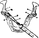
Figure 9. 1-lingula mandib.; 2-lin. obi. ext.; 3-proc. coron.; 4-crista buccin.; 5-trigonum retromolare; I, II and III- positions of the needle. c-^/l
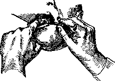
Extraction 3. Indications for the application of this operation are classified as follows: 1) indications for the removal of deciduous teeth, 2) indications for the removal of permanent 3.: a) healthy (non-carious) and b) diseased. Deciduous teeth are removed a) during the eruption of the corresponding permanent teeth, b) during the resorption of the roots of deciduous 3., when the 3. becomes loose and causes pain, even if the corresponding permanent teeth have not yet erupted, c) in the presence of acute diffuse inflammatory processes caused by a diseased tooth, d) in cases of being born with teeth to avoid injury to the mother's nipples, e) in adult individuals (in the not infrequent delay of the shedding of deciduous 3. for several years), if an X-ray reveals the presence of a formed, ready-to-erupt permanent 3. - Non-carious permanent 3. are extracted: 1) in certain anomalies of tooth position, 2) if they are supernumerary and interfere, 3) severely loosened teeth in alveolar pyorrhea, in acute purulent osteomyelitis, 4) in difficult eruption of the lower 3. wisdom teeth, accompanied by repeated acute inflammatory processes with trismus, 5) in the preparation of prostheses (strongly protruding and isolated 3. that interfere with the proper fabrication of the prosthesis are removed), 6) in the presence of neoplasms directly adjacent to the 3. or arising from their periosteum-, and 7) in cases of retained 3., if the latter cause the occurrence of neuralgias and other painful phenomena. An indication for the extraction of carious permanent 3. are the following cases. 1. Acute septic osteomyelitis of the jaw of odontogenic origin (a vital indication for the removal of the offending 3.). 2. Acute osteomyelitis of the jaw or alveolar process without signs of sepsis-an absolute indication for the removal of the offending 3. Extraction is postponed only in those cases when a deep (intra- or extraoral) incision of soft tissues provides an outlet for pus and controls the process, as well as when there are indications that the process is already on the decline. 3. Acute inflammation of the maxillary sinus of dental origin-an absolute indication for the removal of the offending 3. In chronic maxillary sinusitis, the 3. can be preserved if resection of the affected apex is possible. 4. Chronic osteomyelitis of the jaw and alveolar process-in all those cases when the affected 3. for some reason cannot be preserved by methods of conservative dental surgery, i.e. with the help of resection of the root apex, replantation, etc. 5. Tuberculosis and actinomycosis of the jaws-also an absolute indication for the removal of gangrenous roots and teeth of the corresponding side.-Relative indications. 6. Pulpitis and peri-cementitis (periodontitis) of wisdom teeth. 7. Acute osteomyelitis of the socket, i.e. periodontitis (in new terminology-parodontitis)-in cases where conservative treatment is ineffective. 8. Chronic periodontitis, if conservative treatment is ineffective, and operative treatment (resection of the root apex) is not feasible or for some reason contraindicated. There are no absolute contraindications to the extraction of 3. (in acute osteomyelitis of the jaw, in so-called 'flu' extraction, on the contrary, is indicated).- Relative contraindications to extraction are the following conditions: pregnancy, breastfeeding, menstrual period, hemophilia and other blood diseases (chlorosis, leukemia), scurvy, as well as hysteria and epilepsy. Before performing the extraction, a plan of the operation is made, preparation of instruments, hands, and the operative field is carried out according to the general rules of surgery, and finally local anesthesia of the corresponding area. When anesthesia has occurred, the extraction itself is begun, and under this operation is meant the forcible removal of the 3. (or only the root) from its socket after preliminary rupture of the connective tissue bundles of the root periosteum, connecting it with the bony alveolus. To overcome the resistance of the socket, it is necessary to firmly grasp the 3., which is achieved by grasping the 3. with forceps, the beaks of which correspond to the anatomical features of the 3. or at least of a group of 3. (pp. 85-86, figs. 12 and 13); for removing roots, in addition to forceps, various kinds of levers are used.
Figure i.
according to the general rules of surgery, preparation of instruments, hands, and the operative field is carried out, and finally local anesthesia of the corresponding area. When anesthesia has occurred, the extraction itself is begun, and under this operation is meant the forcible removal of the 3. (or only the root) from its socket after preliminary rupture of the connective tissue bundles of the root periosteum, connecting it with the bony alveolus. To overcome the resistance of the socket, it is necessary to firmly grasp the 3., which is achieved by grasping the 3. with forceps, the beaks of which correspond to the anatomical features of the 3. or at least of a group of 3. (pp. 85-86, figs. 12 and 13); for removing roots, in addition to forceps, various kinds of levers are used.
The patient should be seated so that the oral cavity is well illuminated, and the head
Figure 12.
is fixed by a headrest. If there is a dental chair, the operator's left hand fixes the patient's head to the headrest (when extracting upper teeth) or fixes the lower jaw with the left hand (when extracting lower teeth). The free fingers of the left hand push aside the patient's lips and cheeks or grasp the alveolar process from both sides. When extracting on the upper jaw, the patient's head is tilted back, on the lower jaw-it is placed vertically with the calculation that when removing upper 3., the patient's head is at the level of the operator's shoulder, and when removing on the lower jaw, the patient's mouth is not higher than the elbow of the operator's lowered hand. The operator stands for all extractions on the upper jaw to the right of the patient, somewhat in front. For extracting lower teeth on the right side-to the right and in front (with horizontal forceps) or to the right and behind (with vertical forceps); for extracting lower left teeth-also in front or to the left (when extracting with vertical forceps). There are two main methods of grasping forceps. According to the old method (Schef) the forceps rest on the index and middle fingers, the thumb is applied to the upper part of the lock, the 4th and 5th fingers are inserted
Figure 13.
Figure 14.
between the beaks so that their dorsal surface touches the upper inner edge of the beak (figs. 11 and 12). When applying the forceps, the 4th and 5th fingers are removed from between the beaks, and the forceps are grasped with the whole palm (fig. 13). According to the Parks method, forceps for both lower and upper 3. are grasped in the same way: one beak (left) is grasped with the thumb
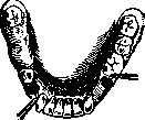
Figure 15.
Figure 16. and with the index fingers (inserted from the inner side of the beak), with the end of the beak pressing into the palm of the hand. The opening of the other beak is done with the middle finger, with the little finger and 4th finger grasping the outer side of the beak, and the nail of the middle finger sliding along the inner side of the same beak (fig. 14). The extraction operation itself 3. breaks down into the following moments: 1) application of forceps, 2) advancement, 3) closure of forceps, 4) dislocation of the tooth and 5) its removal from the socket-traction. Application is always done in such a way that one cheek is applied from the labial (buccal) side of the 3., the other from the lingual (palatal). The cheeks of the forceps must necessarily be applied along the axis of the root. Advancement of the cheeks is the most important moment and is continued until the operator feels that the edges of the cheeks have rested against the edge of the alveolus. At the moment of closing the forceps, the entire system, i.e. the 3., forceps and operator's hands, becomes a single one-armed lever of the second kind, and the moment of dislocation of the tooth-luxation occurs. For this, either rotational movements along the axis of the root (for conical roots, e.g. upper incisors) or side-to-side swinging movements toward the least resistance, i.e. outward (only when extracting lower molars-inward) are performed. When it is possible to tear the tissues connecting the cell with the root, overcoming the resistance of the cell, the tooth is freely removed from the socket-traction is performed. For extracting roots, the cheeks of the forceps have to be pushed even deeper, partially entering the space between the gum and the root; the cheeks of root forceps are made thinner, and the ends of the cheeks meet (pp. 85-86, figs. 12c and 13d).-The general rules for performing extraction with root forceps are the same. For extracting tightly fused roots of multi-rooted 3., it is sometimes necessary to first separate the fusion with a suitable bur or special separating forceps (fig. 7, pp. 145-146); this can also be done with a chisel. If during root extraction the walls break and repeated attempts to push the cheeks deeper fail, extraction is completed with the help of resection forceps or with the help of levers. Root extraction with resection of the alveolus is performed as follows: with specially constructed forceps (fig. 8, pp. 145-146) with strong cheeks, with sharp edges, they penetrate between the gum and alveolus (after preliminary vertical or flap incision of the gum) to a sufficient depth, grasping the root through the wall of the socket at a level where it can withstand the pressure of the cheeks without being crushed, and the root either pops out according to the law of action of inclined planes or the cheeks of the forceps actually cut off (resect) part of the alveolus they have grasped, after which they firmly grasp the root itself. All kinds of elevators, by the method of action, represent a lever of the 1st kind (double-armed), the fulcrum of which is either an adjacent 3., or a strong root, or even the wall of an adjacent socket. (The most commonly used forms of elevators are shown in figs. 4 and 5, pp. 145-146).-With respect to the position of the patient, operator and fixation of the jaw-the rules are the same as for extraction with forceps. The method of action is clear from figs. 15 and 16. For extracting lower wisdom teeth, the Lecluse lever (fig. 2, pp. 145-146) is successfully used, and sometimes the Parson's turning chisel, which is a combination of such a lever with a chisel (fig. 3, pp. 145-146).-Extraction of upper roots is often successfully performed with the help of screws (fig. 6, pp. 145-146). At the end of extraction, broken fragments of the wall and granulations are removed from the socket, it is iodized and bleeding is stopped; the latter is achieved by applying a cotton or gauze swab of appropriate size to the wound, with the patient being asked to bite on it for 1-2 min. Persistent bleeding is stopped by tight packing of the socket with iodoform gauze. If there are sharp edges protruding in the socket, they are nipped off with Luers bone forceps.
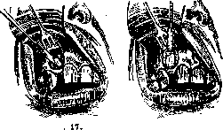
Fig
Figure Trephining of roots. If extraction cannot be performed by any of the above methods, the operation of trephining is resorted to, which consists in that after exposing the alveolus from the outside, it is opened, the thus exposed root is exposed even more until it becomes accessible for grasping
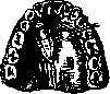
Figure 19
it with forceps or dislocating it with a suitable lever or the same chisel. The objects for the application of a chisel and hammer are also fragments of broken or simply deeply destroyed roots covered with gum granulations, as well as poorly erupted wisdom teeth and all kinds of retained {delayed} teeth. Preparations for these operations and anesthesia are the same as for ordinary extraction. The technique of trephining roots is clear from figs. 17 and 18. Trephining of retained teeth (especially from the palate side) is somewhat more complicated, as they are usually hidden by a fairly thick layer of bone. (Technique of the operation-see fig. 19). Particularly difficult is the trephining of lower wisdom teeth when they are in a horizontal (transverse) position (fig. 20). If a retained tooth is near the edge of the jaw, it is trephined by an extraoral (through the skin) incision. Forced correction is used mainly for aligning teeth. With the help of this operation, two kinds of anomalies are corrected: first, teeth that have erupted but are in the wrong position, and second, those teeth that have fully developed but have not erupted and remained retained in the jaw. The operation is performed only in youth or childhood, if there is enough space for the teeth being positioned. The technique is as follows: the tooth is grasped with suitable forceps and carefully pushed into the correct position, with the apex of the root as much as possible not being displaced (so as not to cause rupture of the vascular-nerve bundle of the pulp); in this position, the tooth is fixed to adjacent teeth with wire ligatures for 10 days. Retained teeth are put in their place with the help of reposition only if the retention was incomplete. In the opposite case, it is necessary to first expose such a tooth by chiseling away the bone and only after that implant it into an artificially created socket. In view of the possibility of complications associated with pulp necrosis, reposition is used very rarely with the current development of orthodontics. Resection of the root apex (apicotomy), replantation, as well as trans-, im-plantation and other similar operations are used for treating chronic granulating inflammations of the periosteum of the root, as well as for dental cysts (inflammatory and retention).-The operation of root sectioning, proposed by Schuster, is

Fig.
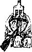
Figure 21.
used in cases where it is necessary to save a tooth with a broken-off nerve extractor or needle during treatment in the depth of the canal. The technique is simple: after exposing the alveolus in the usual way, a window is chiseled along the root in it, after which in the exposed root a thin rose-shaped bur drills a slit down to the canal (fig. 21), and the foreign body is removed from it with an excavator. The slit, after preliminary introduction of a temporary post into the canal (to keep it patent), is filled with amalgam, and the wound is sutured tightly. g. Kovarokiy. VII. The role of teeth in forensic medicine. Anthropometric studies (Wilga and others) showed that the development of jaw bones and teeth is subject to significant racial and individual variations and that these dimensions are in an undoubted connection with some anthropological features. Being the most persistent organ in the human body in relation to the action of decay and fire, teeth have great importance in forensic-medical respect. In the literature there are cases when, based on the study of teeth, the identity of a deceased person could be established even in cases of complete destruction of the body by flame or decomposition. In identifying the identity of a corpse, as well as the identity of a living person, dental fillings and prostheses, individual and racial features of teeth, their anomalies and professional changes have great importance.
Related articles
Cite this page
“Teeth.” Soviet Medical Encyclopedia. English translation of Bolshaya Meditsinskaya Entsiklopediya, 1st ed. (Moscow, 1928–1936), ed. N. A. Semashko. https://sovietmedicalencyclopedia.pages.dev/article/teeth/