Pulpitis
Historical document, translated for reference. It reflects medical knowledge of the 1920s–30s and is not medical advice.
Summary
Pulpitis is inflammation of the dental pulp, most commonly resulting from dental caries and associated irritations and infections. The article describes the causes, including bacterial invasion through dentinal tubules, and the clinical manifestations characterized by spontaneous pain that intensifies with thermal stimuli.
Encyclopedia article (1928–1936)
PULPITIS (pulpitis), inflammation of the dental pulp, most commonly is a consequence of dental caries and the associated irritations and infections affecting the pulp through the carious cavity. As is known, in the presence of caries, the dentinal tubules are inhabited by bacteria; these latter, as well as generally bacteria living in the oral cavity, mainly Streptococcus mitior, or Viridans, penetrate through the dentinal tubules into the pulp, forming there toxins which cause inflammation. Thus, the main causative agents of P. are streptococci, they are found in all types of pulp inflammation; at the same time, other types of bacteria are also found in the inflamed pulp—staphylococci, Bacillus fusiformis and other rods, still not sufficiently identified1. There are also indications of hematogenous P., but they are extremely rare and hardly have any practical significance. Among irritants, thermal factors must be mentioned first, which have enormous importance, then trauma and chemical factors. As soon as the carious cavity deepens sufficiently into the dentin (caries media), the pulp tissue begins to react to thermal irritations; cold and heat cause significant pain. With the deepening of the carious cavity and with the approach of its floor to the pulp, these irritations significantly intensify and, combined with infection of the pulp through the dentinal tubules, cause inflammation of it—P. Trauma to the pulp—fracture of the tooth crown near the pulp horn, injury to the pulp during preparation of carious cavities—also causes P. Among chemical factors, arsenic acid must be mentioned first, used for devitalization of the pulp, as well as a number of acids used for disinfection of carious cavities (arsenic acid before devitalization of the pulp causes its inflammation). All the mentioned irritations can act through the dentinal tubules, being transmitted to the pulp; medicinal substances are absorbed by the pulp through the dentinal tubules: the pulp generally has the ability to resorb substances that enter the dentin. Pathological anatomy. Depending on this or that type of pulp inflammation—acute, chronic, partial, general, etc.—under the microscope is observed greater or less engorgement of capillaries and small vessels, exudation of blood plasma through the walls of damaged capillaries and emigration of leukocytes, then proliferation of tissue and multiplication of cells (proliferation). Alterative and exudative phenomena accompany acute pulpitis, while proliferative phenomena predominate in chronic P. In general, it can be said that P. proceeds in acute and chronic forms, and acute P. can proceed with a limited inflammatory focus—partial P., or the entire pulp tissue is involved—general P. General acute P. can pass into chronic, which can also have various forms. It is necessary to note that this or that condition of the pulp is directly dependent on the depth of the carious cavity, on the nature of the infection, etc. In all cases, the odontoblasts undergo changes first, and their degenerative changes often appear before the penetration of bacteria into the pulp, which depends on the participation of odontoblasts in the formation of reparative dentin (see Dentin).
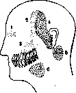
Clinical P. The beginning of pulp inflammation is characterized first by the appearance of spontaneous pain, which, having persisted in the form of an attack for a short time, again repeats after a certain interval; from cold and heat the pain significantly intensifies, and thermal irritations cause a prolonged attack, which distinguishes pulpitis from simple caries: in the latter, pain radiating along the course of the trigeminal nerve from cold and hot quickly passes as soon as the cause is removed, while in P., on the contrary, any irritation, and especially thermal, causes a prolonged attack; even moving from a warm room to the street and vice versa causes pain. The pains have the character of tearing, beating pains and intensify toward evening, reaching maximum intensity when lying down; this is due to the fact that when lying down, blood, flowing to the head, further engorges the pulp and the pressure in it increases. With further development of the process, as larger and larger areas of the pulp become involved, pains from the upper pulpitis teeth; 4-6 zones in pulpitis of the lower teeth. Irradiation is extremely pronounced in pulp inflammation of the molars. (According to Miscn'y.)
The pain attacks become increasingly prolonged, while the intervals between them become shorter. By the time the entire pulp is affected, the pain lasts almost continuously day and night, and is significantly intensified by even the slightest irritation; to this is added the loss of pain localization; the patient can no longer determine which tooth is aching; irradiation of pain along the tract of the trigeminal nerve begins (Fig. 1). If the affected tooth is in the upper jaw, the pain is transmitted to the temporal region of the corresponding side; if the affected tooth is located in the lower jaw, the pain is transmitted to the parotid region. Often the patient indicates that the tooth on the opposite jaw is aching, so unclear is the localization of pain. This incidentally is one of the main symptoms of general pulpitis. If the exudate has a purulent nature, the pains intensify even more, take on a pulsating and throbbing character, and the patient feels how each beat of the pulse coincides with the throbbing or pulsating painful sensation. Sometimes with purulent pulpitis, a febrile condition is also observed. Classification and diagnosis of pulpitis. When considering pulpitis, various stages in the development and course of the inflammatory process in the pulp tissue clearly emerge. Corresponding to one or another patho-anatomical picture, various clinical symptoms are revealed, both subjective and objective. For a long time, there has been an effort to find a possibility for the clinical determination of one or another form of pulpitis. At present, there are a number of classifications of pulpitis, some of which are based mainly on patho-anatomical data, while others also on clinical symptomatology. However, it must be said that to this day there is still no precise differential diagnosis that would fully correspond to both patho-anatomical and clinical data. Meanwhile, for the practicing physician, the clinical classification is of particular importance. All pulpitis, according to their course, are divided into acute and chronic. Acute is divided into: 1) pulpitis acuta partialis, 2) pulpitis acuta totalis, 3) pulpitis acuta purulenta. These three forms occur with a closed pulp chamber. Chronic pulpitis is divided into: 1) pulpitis chronica simplex, 2) pulpitis chronica hypertrophica, s. granulomatosa, 3) pulpitis chronica gangraenosa. These three forms occur with an open pulp chamber. Each of these forms has its differential symptoms, with acute pulpitis being predominantly of subjective nature and chronic pulpitis of objective nature. Thus, with partial acute pulpitis we have: 1) short pain attacks, 2) prolonged intervals, 3) clear localization of pain in a specific tooth. With general acute pulpitis, on the contrary, the following are observed: 1) prolonged pain attacks, 2) short intervals, 3) loss of pain localization (irradiation of pain along the tract of the second or third branch of the trigeminal nerve, depending on whether the upper or lower tooth is affected by pulpitis). On examination, in the first case, with partial pulpitis, probing the floor of the carious cavity causes a painful sensation at a specific point, in the direction of the inflamed area of the pulp; in the second case, with general pulpitis, probing causes pain throughout the floor of the carious cavity. It goes without saying that the main differential symptoms are the duration of the attack and the irradiation of pain along the tract of the trigeminal nerve. Acute purulent pulpitis gives continuous pulsating pain and the floor of the carious cavity is easily pierced on probing with the appearance of a drop of pus, after which the pain completely ceases. As for chronic pulpitis, even in its first form there is a characteristic objective symptom: the pulp chamber is open at one point and the probe freely enters the chamber, injuring the pulp, which gives slight bleeding. The second form—granulomatous pulpitis—is easily identified by the presence of pulp proliferations in the carious cavity—a pulp polyp. The third form—gangrenous pulpitis—is identified by the presence of pulp necrosis in the pulp chamber with simultaneous sensitivity of the root pulp to probing. It should be emphasized that the greatest difficulty and interest lies in establishing partial and general pulpitis, which occur with a hermetically sealed pulp chamber and therefore do not allow examination of the pulp itself. The classification presented is simple and applicable to everyday practice. If there can sometimes be any deviations in the course of certain forms of pulpitis, they are rather rare, so in general this classification can serve as a working scheme for the differential diagnosis of pulpitis. The latest classification by Siegmund and Weber in this part generally comes down to the same forms, with acute simple pulpitis being divided by them into pulpitis acuta circumscripta, which corresponds to pulpitis partialis, and pulpitis acuta diffusa, which corresponds to pulpitis totalis of the above classification. To all classifications, it is also necessary to add such forms as pulpitis traumatica, pulpitis idiopathica, pulpitis concrementosa, of which the first is easily diagnosed on the basis of anamnesis. As for the idiopathic form of pulpitis, it arises in intact teeth depending on causes that more or less disrupt the nutrition of the pulp tissue—from physical, bacterial, etc. causes. Pulpitis concrementosa is a consequence of the formation of solid deposits in the pulp tissue, which by their pressure on the blood vessels and nerves of the pulp cause its inflammation.

The treatment of pulpitis consists in devitalizing the pulp, amputating it up to the entrance to the root canals, or extirpating it, i.e., removing all the pulp tissue from the root canal to the apical foramen, and subsequently filling the tooth. To this day, there are still no methods for treating the inflamed pulp itself to achieve restitutio ad integrum or at least restitutio incompleta. For a long time in odontology, the view has firmly taken root that the inflamed pulp necessarily dies, therefore the pulp that has once manifested acute independent pain must be devitalized. We treat the pulpitis tooth, not the inflamed pulp. However, not everyone reconciled themselves with this, and a number of authors at one time proposed the so-called conservative method of treating pulpitis, or covering the pulp without preliminary devitalization. As early as the 18th century, Pfaff proposed covering the exposed pulp with a gold cap, under which supposedly it could be cured. Witzel proposed an asbestos cap for covering. Then many other proposals were made in this direction, but all of them gave no positive results. Miller proposed preserving the pulp in the case when there is no diffuse inflammation in any part of it. This form can be defined as pulp irritation, or pulp hyperemia, and this stage is transitional from deep caries of dentin to pulpitis. Apparently these cases gave Miller and other authors satisfactory results when treating them without preliminary devitalization of the pulp. This can often be observed in the clinic even with rarely occurring short spontaneous pains in carious teeth; it is sufficient to thoroughly excavate the softened dentin, disinfect the cavity, and cover its floor with a paste of zinc oxide with eugenol, and in 2-3 days the tooth can be successfully filled. Apparently, cleaning and sterilization of the cavity are sufficient in such cases to eliminate the beginning, but not yet fully developed, inflammation of the pulp. In those cases where the pulp is already inflamed, even if only in its most limited part, these measures lead to nothing: severe throbbing and tearing pains occur and the case ends with purulent inflammation of the pulp. Such an outcome of pulpitis apparently depends on the unfavorable anatomical position of the pulp, on the insufficient outflow through the excretory vessels of the pulp; the latter dies from the products of its own metamorphosis during inflammation. Devitalization of the pulp is carried out by means of arsenic acid, from which a paste of the following composition is prepared: Acid arsenicosum, Morphii muriatum, Cocainum and Kreosoti fagi q. s. to obtain the paste. There are other combinations as well, but in all mixtures the main active ingredient is Acidum arsenicosum. Even a small amount of this paste, the size of a pea, is sufficient. This paste is called arsenic paste.
Figure 2. Application of paste: 1 - sealing the pulp chamber head, sufficient to cause devitalization of the pulp (figure 2). Arsenic acid is a cellular and plasma poison. It acts 1) on the nerves of the pulp, 2) on the vessels and 3) on the cellular elements of the pulp. After contact of the poison with the pulp tissue, a slough from burning first forms. This necrotic slough, as some authors indicate, leads to paralysis of the capillary network in the cauterized area (Rebel). Already after 2-3 hours, the pulp vessels become engorged with blood and then thrombosed. As for the nerve elements of the pulp, in them there is a breakdown of their myelin sheath; the stroma of the pulp also undergoes significant changes, the nuclei of the cells, including odontoblasts, become pyknotic and lose the ability to be stained. In the end, necrosis of the pulp occurs. It is difficult to say with certainty to what the predominant value belongs in arsenic necrosis of pulp tissue - to the damage of vessels or nerves, but it can be assumed that the damage to the entire vascular-nervous apparatus of the pulp is the main cause of the effect of this poison, which has been used for this purpose in odontology for a hundred years. Devitalization of the pulp is usually accompanied by significant pain for 2-3 hours, which depends on bringing inflammation under the influence of arsenic paste to its climax. As soon as the vessels and nerves of the pulp are damaged, the pain stops either gradually or suddenly. For a long time instead of arsenic acid, cobalt-arsenic (metallic arsenic) was proposed, which acts much more gently and is less neurotropic and plasmotropic than the first. In general, however, it must be said that cobalt has no special advantages and in the practice of treating pulpitis teeth, arsenic acid is most often used. A very important question is the effect of arsenic on periapical tissues. Throughout a whole century, campaigns against this substance have arisen, and it has been established (especially experimentally on animals) that arsenic, placed in a carious cavity, causes inflammation of the periodontium, and sometimes even destructive processes in the jawbone itself. In the clinic, complications of pulpitis with periodontitis after devitalization of the pulp with arsenic are also sometimes observed. However, this depends in the vast majority of cases on not observing contraindications to the use of this substance: for some types of pulpitis (purulent, granulomatous and gangrenous) the use of arsenic acid is not indicated at all. The technique of application is also important: one should strive to use arsenic for the purpose of devitalizing the pulp in minimal doses, to seal the cavity well and to leave the paste in the tooth no longer than 24-48 hours. In general, it must be said that with correct indications and application, no serious damage to the periodontal tissue occurs. - Since devitalization of the inflamed pulp with arsenic has as its main purpose, besides stopping the pain, to make further surgical treatment of the pulpitis tooth painless, the idea arose of using local anesthesia for this purpose. Endosteal anesthesia is performed with a 4% solution of novocain-suprarenin, with the injection being made deeply into the interdental space to the interdental septum. After 10 minutes, pulp anesthesia occurs, which makes it possible to painlessly prepare the carious cavity, the pulp chamber, and even remove the pulp tissue from the root canal. However, this method has not yet received such wide application as arsenic, and is rarely used in mass dental treatment. Experimentally, some advantage of arsenic devitalization over local anesthesia has also been established. Amputation of the pulp. In any case, as soon as it becomes possible to operate painlessly on the pulpitis tooth, the carious cavity is cleaned of softened dentin, finally shaped, and then, under conditions of aseptic work, the container of the diseased pulp - the pulp chamber - is opened. First, the dentinal covering of the pulp - the floor of the carious cavity - is removed with a fissure bur, and then all parts of the dentin overhanging the pulp chamber are smoothed and ground down, and they proceed to the amputation of the pulp. By amputation of the pulp is meant the removal of the coronal part of the pulp up to its entrance into the root canals. This operation is performed with a sterile rose-shaped or round bur, and one must cut only the pulp, without touching the walls of the chamber itself. - Subsequently, the treatment of pulpitis teeth can be carried out by two methods: amputation and extirpation. The extirpation method consists in the fact that after amputation, all the pulp tissue is removed from the root canal to the apex itself, and the canal is then filled with some material. Removal or extraction, extirpation of the pulp is performed with special toothed needles - nerve extractors (figure 3) or,
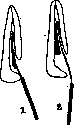
Fig. 3. Removal of pulp: 1 - the entire pulp has been wound around; 2 - only the apical part has been grasped.
or rather, with pulp extractors. This method is the oldest and even now by some authors and especially by the German and American schools is considered classical. However, it is so difficult that to perform it lege artis, especially in multi-rooted teeth, does not seem entirely possible. This depends mainly on the anatomical features of the root canals, which are often narrow, curved, have corroded areas and anastomoses, which do not allow for the removal of all the pulp right up to the apex. Therefore, it has long been believed that extirpation is a deep amputation: some larger or smaller part of the pulp still remains in the root canal closer to the apical part. The subsequent moment, the filling of the root canal, also does not present a fully resolved and easy problem: there is not yet any method that would guarantee the complete sealing of the root canal with reliable closure of the apical opening. This is why the second method for treating pulpitis teeth arose. The amputation method consists in that the pulp tissue, after amputation of its coronal part, is left in the root canals, and this root stump of the pulp is covered with antiseptic substances, which are believed to promote healing of the pulp amputation wound. WitzeГ (A. WitzeГ), who proposed this method in the early seventies, believed that the pulp remains alive in the root canal. Subsequently, however, he together with other authors came to the conclusion that the root pulp after amputation necrotizes, and with the help of antiseptic substances it is possible to cause its mummification. Formalin is recognized as the best substance for mummifying the pulp, which is included in almost all mummification pastes. Recently, the view of the fate of the pulp stump has changed radically. There are indications that after amputation, resorption of the pulp stump occurs under the influence of granulations growing into the root canal through the apical opening; This granulation tissue gradually transforms into cementoid tissue, which closes the apex and the entire canal. At present, a number of authors, based on experimental data, insist that the result of the biological processes occurring in the pulp stump is its metaplasia into osteoid tissue. It is on this last process, on the metaplasia of the pulp, that all the attention of experimental dentistry is now focused, and in this they see the resolution of the difficult problem of therapy for pulpitis teeth. That metaplasia of the pulp into osteoid or cementoid tissue is possible, in this no one now doubts, however, it cannot yet be said with certainty under what conditions this occurs and whether metaplasia of pulp tissue occurs in all cases. In this process, age and the tooth's participation in functional activity, and the resulting condition of the pulp undoubtedly have no less importance. The stage in which the pulp inflammation is located and even the nature of the infection that caused P. are also not indifferent here. Similarly, we have no reliable means of stimulating this biological process in the root pulp stump. In any case, at present the amputation method of treating P. is considered not only equivalent to the extirpation method, but it should even be preferred in mass work. Statistics showed that the extirpation method gives 62% successes and 38% failures, while the amputation method gives 85% successes and 15% failures; the percentage of complications with the extirpation method is more than twice as high. As long as there is a connection between the root part of the pulp and the periodontium, one can also be calm about the possibility of infections from the tooth. However, it must be taken into account that the extirpation method cannot be completely abandoned. There are cases of pulp inflammation, especially purulent and gangrenous forms, where it is necessary to achieve the removal of as much as possible of all pulp tissue from the root canal with subsequent filling of the root canal. It is also often necessary to retreat teeth, i.e., to unseal teeth treated by the amputation method, which give one or another complications. In such cases, naturally, all pulp has to be removed from the root canals, which is most often in some stage of inflammation and can no longer remain there without harm to the surrounding tooth tissues. After extirpation or amputation of the pulp, the tooth is filled in the usual way. Such inflammations of the pulp stump in the root canal are quite common and are characterized mainly by pain from cold in pulpitis teeth filled by the amputation method. The outcome of pulp inflammation is usually its death - gangrene of the pulp, which is characterized by cessation of pain and the presence of a foul odor from the carious cavity. The tooth is externally changed in color - it is usually darker than healthy teeth. Upon examination, it is found that no irritants - cold, heat, probing - cause any sensitivity, since the pulp is dead.
Related articles
Mentioned in
Cite this page
“Pulpitis.” Soviet Medical Encyclopedia. English translation of Bolshaya Meditsinskaya Entsiklopediya, 1st ed. (Moscow, 1928–1936), ed. N. A. Semashko. https://sovietmedicalencyclopedia.pages.dev/article/pulpitis/