Tongue
Historical document, translated for reference. It reflects medical knowledge of the 1920s–30s and is not medical advice.
Summary
A detailed anatomical and biological description of the tongue in vertebrates and humans, covering its development, structure, musculature, and function as described in the 1930s.
Encyclopedia article (1928–1936)
TONGUE, a mobile muscular organ of the oral cavity of vertebrates, helping them to grasp and swallow food. Already in fish, on the floor of the oral cavity, there is a fold of the mucous membrane supported by an unpaired outgrowth of the visceral skeleton and called the T., however, the T. of fish lacks its own musculature, and it moves only during the movements of the entire visceral apparatus. Only in terrestrial vertebrates can one speak of the T. as a separate organ. Here, its own musculature develops, which is a product of the isolation and differentiation of the hyoid musculature. Corresponding to its origin, the musculature of the T. is innervated by the hypoglossal nerve. In amphibians, the anterior part of the T. abounds in glands secreting sticky mucus, which helps in catching insects. In some cases, the T. can be thrown far forward. In higher terrestrial vertebrates, the T. is an even more complex formation. It develops in them from a posterior unpaired primordium, lying in the region of the II and III branchial arches and giving rise to the root of the T., and an anterior paired primordium, lying on the sides of the thyroid gland primordium and giving rise to the body of the T. proper. The shape and degree of mobility of the T. of reptiles and birds are highly varied. In lizards and snakes, it is usually long, deeply bifurcated at the end, and is distinguished by extreme mobility and sensitivity. In this case, at the base of the T., there is an annular fold forming its sheath. The hyoid apparatus forms the skeleton of the T., with its anterior unpaired element (basihyale) extending into a long process that penetrates deeply into the body of the T. (os entoglossum). In birds, the tongue is usually thin and not very mobile, distinguished by the strong development of the horny layer with numerous horny papillae. In mammals, the intrinsic musculature of the T. reaches its greatest development. Cornification is sometimes quite significant. The T. of mammals is also extremely rich in glands and is provided with papillae of various significance, partly sensory (with taste buds), partly cornified, and performs very diverse functions. Its role is especially significant during the act of swallowing, as well as as an organ of taste.
I. Schmalhausen. The tongue (lingua) in humans is a muscular organ of the oral cavity, representing a formation of a flat-oblong shape, connected by its lower surface to the floor of the oral cavity. Its anterior, rounded end—the tip of the T. (apex linguae)—passes without sharp boundaries into the body of the T. (corpus linguae). The body of the tongue is bounded posteriorly by the so-called terminal sulcus (sulcus terminalis) (Fig. 1); the right and left halves of this sulcus run obliquely backward to the midline and converge at the blind foramen of the T.
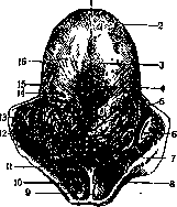
Figure 1. Dorsum of the tongue: 1—apex linguae; 2—margo lat. linguae; 3—dorsum linguae; 4—sulcus medianus linguae; 5—sulcus terminalis linguae; 6—foramen caecum linguae; 7—radix linguae; 8—plica glosso-epiglottica lat.; 9—plica glosso-epiglottica mediana; 10—epiglottis; 11—vallecula glosso-epiglottica; 12—folliculi linguales (tonsilla lingualis); 13—tonsilla palatina; 14—papillae vallatae; 15—papillae foliatae; 16—papillae fungiformes. (foramen caecum linguae) (Morgagni). The mentioned blind pit of the T. represents a remnant of the depression formed in the embryonic period during the development of the thyroid gland. It is precisely in connection with this developmental process that in some cases, one can observe remnants of the lumen of the thyroglossal duct (ductus thyreoglossus) at this site, the terminal part of which is called the lingual excretory duct (ductus lingualis). It is generally accepted to distinguish 2 parts of the T.—its anterior part, located exclusively in the oral cavity (pars buccalis), and the posterior part, territorially located mainly in the pharyngeal cavity (pars pharyngea). The boundary between these two parts is considered to be the mentioned angular figure formed by the vallate papillae (papillae vallatae). While the anterior part of the T. is mainly a taste organ and is provided with a large number of nerve endings, the posterior part is abundantly provided with lymphoid tissue (tonsilla lingualis).

The part of the T. behind the terminal sulcus is called the root of the T. (radix linguae). The upper surface of the T.—the back of the T., or dorsum of the tongue (dorsum linguae)—is provided in the middle with a well-defined, especially anteriorly, longitudinal groove (sulcus med. linguae). The body of the tongue has blunt lateral edges on the sides (margo lateralis linguae). The lower surface of this organ (facies inf. linguae) is completely free along the lateral edges and in the greater part of the anterior section.
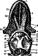
Figure 2. Lower surface of the tongue and floor of the oral cavity: 1—apex linguae; 2—glandula lingualis ant. (Nuhni); 3—m. longitudinalis inf.; 4—m. genio-glossus; 5—glandula sublingualis; 6—ductus submaxillaris; 7—frenulum labii inf.; 8—gingiva; 9—caruncula sublingualis; 10—plica sublingualis; 11—frenulum linguae; 12—plica fimbriata. While the upper surface of the T. is covered with a peculiar structure and has no submucosal layer, the lower surface is lined with a smooth mucous membrane facing the sublingual region (regio sublingualis). The mucous membrane of the lower surface of the tongue is provided with two narrow, anteriorly converging, low fringed folds (plicae fimbriatae) (Fig. 2). From the lower surface of the tongue, in the middle, a median fold (plica mediana) runs to the floor of the mouth, forming the frenulum of the T. (frenulum linguae). On both sides of the frenulum on the floor of the oral cavity is the so-called sublingual caruncle (caruncula sublingualis), in which the opening of the excretory duct of the submandibular gland is located. In the space between the frenulum of the T. and the glossopalatine arch, the so-called paired sublingual fold (plica sublingualis) is formed, which extends in a lateral direction from the sublingual caruncle. It is precisely on this fold that the small ducts of the sublingual gland (ductus sublinguales minores) open.
The pharyngeal part of the T. (pars pharyngea) extends posteriorly to the epiglottis, and its upper surface, unlike the anterior, horizontal surface, faces posteriorly, i.e., has an almost vertical direction. When the mouth is closed and breathing is calm, this surface is in contact with the uvula and the soft palate. Accumulations of lymphoid tissue under the mucous membrane of the posterior part of the tongue are so sharply defined that they can be visible to the naked eye in the form of special submucosal tubercles (folliculi linguales) with clearly visible excretory ducts. Posterior to the root of the T., 3 mucous folds extend in the direction of the epiglottis and the soft palate: 1) the unpaired plica glosso-epiglottica med. (the so-called frenulum epiglottidis) and 2) two lateral ones—plicae glosso-epiglotticae laterales. The T., in its main basis, consists of a whole series of mutually intertwining and running in different directions muscle groups, some of which both begin and end within the T. itself (Fig. 3) (e.g., longitudinal and transverse muscles of the T.—m. transversus linguae and m. longitudinalis linguae) (Fig. 4). Some, however, only begin from bone attachments and diverge in different directions. The muscles of the T. that begin from bone formations are the following: the styloglossus muscle (m. stylo-glossus). It begins from the styloid process and, running obliquely downward, forward, and inward, to the outer surface of the hyoglossus muscle, is divided into 2 bundles, one of which runs along the outer edge of the T., and the other is directed downward and inward. This paired muscle pulls the T. and its root upward and backward.
Figure 3. Musculature of the tongue and fauces: 1—apex linguae; 2—m. genio-glossus; 3—m. hyo-glossus; 4—m. stylo-glossus; 5—m. chondro-pharyngeus; 6—os hyoideum; 7—membrana hyo-thyreoidea; 8—cornu majus ossis hyoidei; 9—n. laryngeus sup.; 10—lamina sin.; 11—lig. crico-thyreoideum med.; 12—pars recta and pars obliqua m. crico-thyreoidei; 13—arcus cartilaginis cricoideae; 14—incisura thyreoidea inf.; 15—lamina dext.; 16—m. constrictor pharyngis inf.; 17—incisura thyreoidea (sup.); 18—lig. hyo-thyreoidea med.; 19—fat body; 20—m. stylo-glossus; 21—m. hyo-glossus; 22—m. longitudinalis inf.; 23—septum linguae. The hyoglossus muscle (m. hyo-glossus) acts in the same sense, which pulls the T. not only backward but also downward. Starting from the body and the greater horn of the hyoid bone, it extends upward and forward, gradually weaving into the bundles of the styloglossus muscle. An especially powerful muscle group is formed by the genioglossus muscle (m. genio-glossus). This paired muscle, located near the midline and parallel to it, is separated from the formation of the same name on the other side by a loose connective tissue septum of the tongue (septum linguae); the beginning of this muscle is the mental spine of the mandible (spina mentalis mandibulae). Diverging in a fan shape, the bundles of the m. genio-glossus
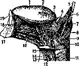
Figure 4. Musculature of the tongue and fauces: 1-dorsum linguae; 2-margo lat. linguae; 3-m. glosso-palatinus; 4-tonsilla palatina; 5-processus styloideus; 6-m. stylo-glossus; 7-m. stylo-pharyngeus; 8-m. glosso-pharyngeus; 9-m. constr. pharyngis med.; 10-m. hyo-glossus; 11-m. constr. pharyngis inf.; 12-n. laryngeus sup.; 13-m. thyreo-hyoideus; 14-lig. hyo-thyreoideum med.; 15-membrana hyo-thyreoidea; 16-m. genio-hyoideus; 17-mandibula; 18-m. longitudinalis inf.; 19-dens incisivus. The genioglossus muscles (Fig. 5 and 6) run posteriorly and upward, attaching partly to the body of the hyoid bone and partly to the epiglottis. A significant portion of the bundles of this musculature extends fan-shaped toward the back of the tongue and is defined throughout its entire length. The muscle pulls the tongue forward and downward. The tongue is united with the soft palate into a single system by the aforementioned fold (arcus palato-glossus), in the thickness of which the muscle of the same name is located. The inferior longitudinal muscle of the tongue (m. longitudinalis inf.), which shortens the tongue in the longitudinal direction, is a long and narrow formation located in the deep parts of the tongue and beginning under the mucous membrane of this
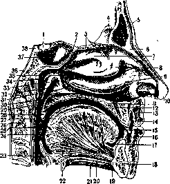
Figure 5. Sagittal section through the facial part of the head and the bodies of the 3 upper cervical vertebrae: 1-hypophysis; 2-recessus spheno-ethmoidalis; 3-lamina cribrosa; 4-crista galli; 5-sinus frontalis; 6-os nasale; 7-concha nasalis sup.; 8-concha nasalis media; 9-concha nasalis inf.; 10-apex nasi; 11-palatum durum; 12-canalis incisivus; 13-labium sup.; 14-vestibulum oris; 15-cavum oris proprium; 16-labium inf.; 17-tunica mucosa sublingualis; 18-mandibula; 19-spina mentalis; 20-m. genio-glossus; 21-m. genio-hyoideus; 22-os hyoideum; 23-epiglottis; 24-plica palato-pharyngea; 25-foramen caecum (Morgagnii); 26-epistropheus; 27-m. longitudinalis sup.; 28-uvula (palatina); 29-plica salpingo-pharyngea; 30-labium ant.; 31-palatum molle; 32-projection of m. levatoris; 33-torus tubarius; 34-recessus pharyngeus (Rosenmuelleri); 35-meatus naso-pharyngeus; 36-arch of the choanae; 37-septum sinuum sphenoidalium; 38-dorsum sellae.
organ. The main muscular mass of this muscle runs directly forward and ends in the mucous membrane of the lower surface of the tongue. The bundles of this muscle, which originate on the lesser horn of the hyoid bone, the so-called m. chondro-glossus, stretch upward and forward, between the bundles of the genioglossus musculature, and merge with the superior longitudinal muscle. The superior longitudinal muscle (m. longitudinalis sup.) is located directly under the mucous membrane, along the entire dorsum of the tongue; its fibers run in a longitudinal direction and bend the tongue in the same longitudinal direction, with a convexity upward. The transverse muscle of the tongue (m. transversus linguae) is located throughout its entire length and consists of frontally arranged bundles that interweave with the bundles of the genioglossus musculature. In a sagittal section, this muscle has a fan-like appearance with the direction of the fibers toward the mucous membrane, to the dorsum and the lateral edge of the tongue. It gives the tongue a convexity upward and in the transverse direction. The vertical muscle of the tongue (m. verticalis linguae) consists of weak muscle bundles located mainly near the lateral edges of the tongue. The fibers of this muscle penetrate the tongue from its upper to its lower surface. Blood supply to the tongue occurs mainly via the lingual artery (a. lingualis) (Fig. 7), which emerges from the external carotid artery at the level of the greater horn of the hyoid bone. Its branches are: 1) rami dorsales linguae, which supply blood to the root of the tongue; 2) a branch known as the a. sublingualis, which provides blood supply to the sublingual region; and finally 3) the deep lingual artery (a. profunda linguae), as the most powerful branch of the main trunk, which rushes to the dorsum of the tongue and its tip. It is interesting that both deep lingual arteries do not have well-defined anastomoses between them. Innervation of the tongue occurs via 3 types of nerve fibers,
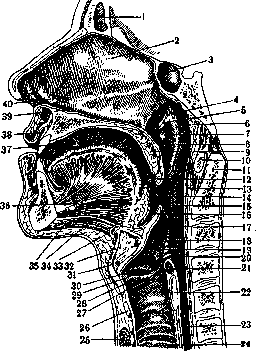
Figure 6. Paramedian section through the facial skull, as well as through the fauces and larynx: 1-sinus frontalis; 2-septum nasi; 3-sinus sphenoidalis; 4-recessus pharyngeus (Rosenmuelleri); 5-labium ant.; 6-tonsilla pharyngea; 7-projection of m. levatoris; 8-torus tubarius; 9-velum palatinum; 10-atlas; 11-dens epistrophei; 12-arcus glosso-palatinus; 13-foramen caecum; 14-tonsilla palatina; 15-arcus pharyngo-palatinus; 16-epiglottis; 17-plica ary-epiglottica; 18-vestibulum laryngis; 19-tuberculum cuneiforme; 20-tuberculum corniculatum; 21-m. arytaenoideus transversus; 22-cartilago cricoidea; 23-oesophagus; 24-trachea; 25-isthmus glandulae thyreoideae; 26-cartilago cricoidea; 27-processus vocalis; 28-plica vocalis; 29-ventriculus laryngis; 30-plica ventricularis; 31-cartilago thyreoidea; 32-lig. hyo-thyreoideum medium; 33-os hyoideum; 34-m. genio-hyoideus; 35-m. mylo-hyoideus; 36-m. genio-glossus; 37-cavum oris proprium; 38-vestibulum oris; 39-recessus naso-palatinus; 40-organon vomero-nasale (Jacobsoni).
The motor functions of this organ are performed by the hypoglossal nerve; the sensory properties of the tongue are provided by the lingual nerve (n. lingualis); finally, specific taste sensations are transmitted via the glossopharyngeal nerve and via the fibers of the lingual nerve (by the pathways of the chorda tympani). The lymphatic system of the tongue has 4 groups of vessels that drain into regional lymph nodes, the main ones of which are: the sublingual, deep cervical, and submental nodes. 1) The lymphatic vessels of the tip of the tongue in the form of 3-4 trunks run under the mucosa, along the midline, penetrate the frenulum, forming the truncus centralis, and drain into the lgl. submentales or posteriorly into the lgl. cervicales profundae, no
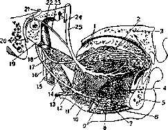
Figure 7. Topography of the arteries and veins of the tongue: 1-papillae vallatae; 2-m. longitudinalis sup.; 3-palatum durum; 4-m. genio-glossus; 5-mandibula; 6-m. mylo-hyoideus; 7-m. genio-hyoideus; 8-a. sublingualis; 9-a. prof. linguae; 10-corpus os. hyoidei; 11-m. hyo-glossus; 12-ram. dorsalis linguae; 13-a. lingualis; 14-m. stylo-pharyng.; 15-m. stylo-glossus; 16-n. vagus; 17-n. glosso-pharyngeus; 18-n. hypoglossus; 19-proc. styloideus; 20-n. facialis; 21-geniculum n. facialis; 22-a. carotis int.; 23-chorda tympani; 24-n. alveolaris inf.; 25-n. lingualis.
are, along the dorsum of the tongue, more sparse but also larger mushroom-shaped papillae (papillae fungiformes) (Fig. 8); they are especially numerous on the anterior half of the tongue; similar cone-shaped projections on the lateral edges of the organ are sometimes strongly flattened and then bear the special name - papillae lenticulares. The largest papillae in size are the vallate papillae (papillae vallatae), which usually number 7-11; they are arranged in the shape of a Roman numeral five in front of the terminal sulcus, almost close to and parallel to it, such that the most posterior vallate papillae are close to the foramen caecum of the tongue. Usually, these are small cylindrical elevations surrounded by a ring-shaped groove and a fold of the mucous membrane. Foliate papillae (papillae foliatae) are visible on the lateral edges of the tongue, in front of the base of the glossopalatine arch; these formations have the appearance of small, almost vertically arranged mucosal folds,
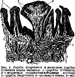
musculature of the tongue.
barely rising above the general level of the tongue; on each side there are on average from 5 to 8 of them. The physiological functions of the tongue are divided into two directions: firstly, it is an auxiliary digestive organ, mechanically facilitating the moistening and grinding of food. The act of chewing is significantly facilitated by the fact that the tongue places food boluses between the chewing surfaces of the teeth. By appropriate movements of the tongue, food that has been mechanically and chemically processed in the oral cavity is moved from it into the pharyngeal cavity; besides this, the tongue also serves to grasp the food itself from the outside; it also determines its qualitative and taste properties. The second physiological function of the tongue consists in its active creation of articulate human speech. However, clinical observations show that even with total or subtotal extirpation of this organ, speech can remain sufficiently intelligible. Pathology of the tongue. Developmental defects of the tongue primarily include its complete or almost complete underdevelopment; thus, one may speak of either so-called aglossia or aplasia of the tongue. Congenital defects also include longitudinal, more or less sharply expressed, splitting of the posterior belly of the digastric muscle. 2) The lymphatic vessels of the lateral edges and the lower surface of the tongue, after the merging of the small lymphatic capillaries of each side, form a paired duct—the marginal trunk. This lymphatic trunk drains into the submandibular lymph nodes, in the area of the submandibular triangle, and also into the superficial and deep upper cervical groups of nodes. 3) The lymphatic vessels of the base of the tongue drain into 4) the lymphatic vessels of the dorsum of this organ. The latter is provided with an extensive thin network of lymphatic capillaries, merging with each other into larger pathways, especially in the posterior direction; some of them entwine the papillae and here turn into even larger trunks, which are directed toward the epiglottis and tonsils and from there go into the depths. Both halves of the lymphatic system of the tongue anastomose very widely with each other, as a result of which even with unilateral pathological processes on this organ (for example, in malignant tumors), the lymph nodes of both sides of the neck or the nodes on the side opposite to the primary process turn out to be affected (so-called crossed lymph nodes in cancers of the tongue). The mucous membrane of the tongue (tunica mucosa linguae) is covered on the dorsum of this organ with a significant number of small elevations that differ in shape, called papillae of the tongue (papillae linguales). Among them, filiform papillae (papillae filiformes) are distinguished; these formations of a narrow-conical shape, sometimes with brush-like appendages on their tips, cover the entire dorsum of the tongue and extend to its lateral edges up to and including the tip. Among the multitude of mentioned papillae, similar in shape but rarer conical papillae (papillae conicae) are scattered. Among the above-mentioned papillae are scattered 733
its longitudinal splitting, the so-called bifid tongue; the number of lobes formed in such cases sometimes increases to 3-4 (Alfeldt). From comparative anatomy, however, it is known that a split tongue in some animals and birds turns out to be a completely physiological phenomenon; this property is observed especially sharply in some reptiles. In humans, splitting of the tongue is sometimes combined with clefts of the palate, lips, and jaws. Congenital developmental defects also include so-called adhesions of the tongue, or the so-called tied tongue, with which newborns are born. The limitation of the mobility of the tongue in such cases is explained by separate membranous adhesions connecting the lower surface of the tongue with the floor of the oral cavity; this defect is easily eliminated by simple separation of the mentioned adhesions. One should not confuse this defect with congenital shortening of the frenulum of the tongue, i.e., the so-called ankyloglossia, in which the frenulum is too short and fixes the tongue to the floor of the oral cavity not by scar formations, but by a shortened mucosal fold. Simple incision of this fold with careful hemostasis leaves no trace of this developmental defect. Congenital defects of the opposite order include the so-called too long tongue, or too long frenulum, which pathologically increases the mobility of the tongue, especially posteriorly, as a result of which there arises the danger of "swallowing one's own tongue" with symptoms of suffocation. Congenital excessive mobility of this organ with a too long frenulum has been described by a number of authors (Petit and others). In some cases, the displaceability of the tongue is so great and its pathological congenital mobility is so sharply expressed that a person can reach the posterior surface of the soft palate with the tip of the tongue and touch the vault of the pharynx. An abnormally large tongue, or so-called macroglossia, is sometimes observed as a congenital developmental defect, but can be considered as such only in the case of purely muscular hypertrophy of this organ. Under this name, however, other pathological processes that increase the size of the tongue (for example, lymphangiomas) often appear. Congenital hypertrophies of the tongue can affect only one half of it and be combined with congenital hypertrophies of one half of the face. Girod described a true, huge congenital macroglossia, in which the hanging of the tongue beyond the edges of the lips extended to 7 cm. Congenital defects should also include the excessive enlargement or gaping of the foramen cecum of the tongue. Congenital anomalies of the tongue that have no particular practical significance include the so-called folded tongue (lingua plicata, "scrotal tongue"), when deep, coarse folds and epithelium-lined fissures noticeably furrow the upper surface of this organ in all directions. With a sharply expressed anomaly of this type, the impression of a torn tongue (glossitis dissecans) is obtained. The only practical significance that this defect has is the diagnostic errors accompanying it, when, for example, the folding of the tongue in healthy people is mistaken for a syphilitic lesion of it. To the same harmless developmental defects belongs the map-like tongue, the so-called geographic tongue (see Glossitis, glossitis migrans). Unusually typical is the so-called black or hairy tongue (lingua nigra; nigrities linguae), in which a thickened, sometimes covered with hyper- LK
With keratoses or pigment spots, the mucosa of the dorsum of the tongue acquires a felt-like or hairy character as a result of pigmentation and thickening of the covering epithelium of the filiform papillae. Traumatic injuries of the tongue are diverse; among wounds of this organ, puncture wounds are quite common, usually inflicted by sharp objects, with varying depths of the wound tract. Incised wounds of the tongue, in addition to being inflicted by cutting instruments, are especially often observed in children as a result of pulling stiff grasses with sharp edges between half-opened lips. Cases have been described in pediatric practice of even complete amputation of parts of the tongue under such circumstances. Most frequently observed are bite wounds of the tongue, which are of varying degrees of severity, ranging from minor injuries and biting of the tongue during eating to very serious injuries of this organ, especially during epileptic seizures. Relatively rare are so-called obstetric injuries of this organ, observed during manual extraction of the fetus with the insertion of the obstetrician's finger into the child's oral cavity (Veicherovsky maneuver). All these injuries present a danger from the point of view of immediate and subsequent bleeding from the tongue. Late hematomas of the tongue, sometimes 3-4 days after the injury, can be sudden in nature, life-threatening, with symptoms of suffocation and the need to perform an emergency tracheotomy. Among the numerous complications accompanying injuries of this organ, the lodging of foreign bodies in the thickness of the lingual muscles is not uncommon. Some of the lodged splinters, needles, seeds, and other foreign bodies are expelled over a long period, some remain for a long time or forever and sometimes serve as a source of diagnostic errors when recognizing neoplasms of this organ. Traumatic injuries of lesser practical importance include ulcerations of the tongue in children when newborns or nursing infants are born with already erupted lower front teeth. The sharp edges of the incisors in such cases, lying close to the lower surface of the tongue, at the site of attachment of the lingual frenulum to the lower surface of this organ, cause typical but easily eliminated ulcerations. Similar tissue defects at the frenulum linguae (ulcus frenuli linguae) are observed in children with whooping cough from constant friction of the mucosa of the lower surface of the tongue against the edges of the teeth. Ulcerations of the pressure sore type on the tongue are observed in adults either during severe illnesses, from constant contact of the immobile, sometimes edematous tongue with the inner surfaces of the teeth, or with carious teeth systematically cutting the tissue with sharp edges. Similar defects on the outside or in the thickness of the organ are often seen from constant pressure on the tissues and with poor dentures; in such cases, it is also a matter of subsequent acute or chronic inflammatory limited induration, sometimes with the appearance of a truly carcinomatous ulceration as a result of prolonged damage to a section of the mucosa. Burns of the tongue by thermal and chemical agents can reach severe degrees. They are usually accompanied by acute pain, edema, and in sharply pronounced cases, the formation of blisters, necroses, ulcerations, and subsequently disfiguring scars. Relatively great rarities, which however have practical significance, are insect bites of the tongue (bees, wasps), usually accompanied by a violent reaction, sometimes taking on a threatening character (acute edema of the tongue), especially if the bite occurs in the posterior parts of this organ and is accompanied by symptoms of difficulty breathing (tracheotomy). When using stagnant water for drinking, cases of bites and attachment of leeches to the tongue have been described, which are easily removed by applying cotton wool soaked in ether to them; it is recommended to carefully ensure that the killed leech is not aspirated through the glottis (Briming). For acute inflammation of the tongue, see Glossitis. Chronic, non-specific inflammatory conditions of the tongue mucosa include so-called leukoplakia (see). Until recently, serious diagnostic significance, as a sign of late syphilis of the tongue, was attributed to its changes known by the term smooth atrophy of the tongue (atrophia laevis linguae). Clearly atrophic changes of this organ in such cases relate to its root and occur due to the flattening of the epithelial cover and the almost complete disappearance of lymphoid tissue. These changes are observed mainly in very old age. Studies by Seifert, Goldschmidt, and others have proven that smooth atrophy of the tongue is by no means always of syphilitic origin, especially since atrophy of the entire mucosa of this organ is encountered in a whole range of debilitating diseases (pernicious anemia, stomach cancer, etc.). Tuberculosis of the tongue occurs in two forms—as a primary and as a secondary disease of it. Practically, secondary diseases of the tongue have the greatest significance, appearing as a result of the direct spread of lupus from the skin of the face to the oral mucosa, or inoculation of the tubercular agent from pulmonary foci, or finally due to the transport of tubercle bacilli through the bloodstream. Pathologically, it is customary to distinguish tuberculosis of the tongue mucosa, tubercular ulcer with abscess, and disseminated tuberculosis of the entire organ. Tuberculosis of the tongue mucosa, especially if it spreads to the tongue from the skin of the face, differs little from lupus. In the majority of cases, we are dealing with typical tubercles in various stages of development. Some of them are ulcerated, some merge into entire foci; from the merging and disintegration of the nodules, ulcers are formed; the disease lasts mostly for years, with swelling of the glands and periodic exacerbations; as with cutaneous lupus, there are foci of scarring and new eruptions. The second form of tuberculosis of the tongue represents a limited tubercular infiltration (tuberculoma of the tongue), often encountered at its very root; upon histological examination of this very form, many giant cells are found. The disseminated form of tuberculosis of the tongue is observed in patients with severe general involvement of the lungs and internal organs. Clinicians are well acquainted with the most frequently encountered form of tuberculosis of the tongue mucosa, accompanied by multiple superficial depressions on its surface, edema of this organ, gray dirty coatings, bleeding ulcers with soft edges, and focal-tubercular eruptions. Deep fissures and cracks of the mucosa are sometimes confused with syphilitic sclerosing glossitis, but differ from the latter in that they do not have indurations around them. A tubercular elongated ulcer of the tongue looks more like an ordinary deep tissue fissure, while a syphilitic crack has the character of a branched, bizarre, irregular formation. Clinically, its extraordinary painfulness is especially characteristic of tuberculosis of the tongue; in some cases, it is a true torment for the patient not only to utter a few words but also to take a sip of water. Correct diagnosis of this disease in rare cases can present serious practical difficulties, from which the physician is extricated by a timely biopsy. Since tuberculosis of the tongue in the majority of cases is a secondary disease, only in rare cases combined with syphilis or cancer of this organ, the treatment of the primary tubercular focus is the basic principle of therapy for this disease. In rare cases of primary tuberculosis of the tongue with completely isolated tubercles and ulcers, simple excision, coagulation, or cauterization of the affected focus with 80% lactic acid gives good results. For temporary relief of pain, smearing with 10% alypin, 20% novocaine, antipyrine, etc., is recommended. Exceptionally good results have been achieved by heliotherapy; at the present time, there are sufficiently authoritative indications from Brüning that radiotherapy also provides not only a pain-relieving but also a curative effect. Good results have been described from the use of partial antigens according to Deycke-Much. Albanus reported successful treatment of tuberculosis of the tongue with mesothorium. Syphilis of the tongue is known in all three stages of this disease. Primary syphilis of the tongue does not represent an exceptional rarity. For example, Fournier, out of 1,124 cases of primary syphilitic focus, found a hard chancre on the tongue in 75 cases. In general, the primary focus on the tongue does not differ essentially from the same on the genitals. The hard chancre is located here mainly on the upper surface of the tongue or along its edges; very rarely can it be observed at the root of this organ. The primary focus, the size of a bean, strictly limited, bright red and indurated, has a central, slightly pronounced depression on it with smooth, ridge-like edges; the noticeable painless glands in the submental region and in the submandibular fossae that join soon after its appearance do not present difficulties for diagnosis, provided only that the physician thinks in time about the possibility of lues. An ulcerating induration with a dirty, sebaceous base is accompanied by increasingly noticeably enlarging regional lymph glands and positive microscopic findings; the diagnosis is confirmed by examining smears taken from the base of the ulcer (Treponema pallidum). Secondary manifestation of syphilis—see Glossitis. Among other infectious lesions of the tongue that do not have great practical significance, actinomycosis of this organ should be mentioned.
In world literature, by the 1930s, approximately 38 cases of actinomycosis of the tongue have been described. Small, nodular foci, developing gradually in the vicinity of the primary puncture by a grain of wheat, gradually merge into hard infiltrates, spreading to the floor of the oral cavity, to the neck, and toward the pharynx. True softening of these indurations is encountered almost as rarely as their secondary ulceration. Clinically, actinomycosis of the tongue is for a relatively long time diagnosed as diffuse glossitis. Lesions of the lymph glands are almost never observed in this condition. Glanders, foot-and-mouth disease, and anthrax do not belong to the frequently observed diseases of the tongue. Leprosy of the tongue usually has a secondary character and spreads to it from the skin of the face, the mucosa of the cheeks and lips in a relatively late period of the disease. Lepromas of the tongue in the form of hard nodules ulcerate rarely and remain unchanged for years. Complete painlessness is especially characteristic of this form; the diagnosis is confirmed by bacteriological examination. Scleroma of the tongue belongs to infrequent findings and usually has a secondary character, spreading to this organ from the nasal cavity. Dense, cartilage-like infiltrates of scleroma of the tongue often suggest a primary malignant tumor of this organ. Among other things, they differ from neoplasms in that depressions and finally scars form in the middle of the appearing infiltrates without ulceration. Simple retention cysts of the tongue are not encountered often and rarely reach sizes larger than a hazelnut. Etiologically, these are for the most part formations developing on the basis of congenital occlusions of the excretory ducts of glands or as a result of their blockage during local inflammatory phenomena. Clinically, these neoplasms manifest very little; sometimes they affect the clarity of speech; they are perfectly cured by simple enucleation. Very rare are retention cysts on the tip of the tongue; they form here from the so-called Nuhn's glands, usually located on the lower surface of this organ in the superficial muscle layers. Retention cysts of the root of the tongue develop from mucous glands in the region of the papillae circumvallatae or from the remnant of the thyroglossal duct. In the latter case, their lumen is lined with ciliated epithelium. Cysts of the root of the tongue usually do not exceed the size of a cherry, although a case (Marx) of suffocation of a newborn by just such an excessively large neoplasm has been described. Echinococcal cysts of the tongue belong to rarities. Cysticerci of the tongue, although encountered very rarely, can give cause for confusion with retention cysts, from which, however, they differ by less transparency, greater mobility, and for the most part, multiplicity. Calcified cysticerci of the tongue are well determined radiographically. Fibromas of the tongue belong to relatively rare findings. These are usually small, dense neoplasms, sitting on a stalk on the surface of the tongue or embedded deep into the thickness of the musculature; their carriers only in rare cases experience serious inconvenience from their presence. For fibromas of the thickness of the tongue, the differential diagnosis of which can present great difficulties, duration and gradualness of development are characteristic. Fibromas of the tongue grow slowly, sometimes for decades; even after such long periods, fibromas are cured by simple excision. Neurofibromas of the tongue are also known; the latter, however, for the most part represent a local manifestation of a general disease called neurofibromatosis, or Recklinghausen's disease. In the latter case, the tumors can reach very large sizes and force one to speak of macroglossia. Lipomas of the tongue are encountered relatively more often; they are distinguished by a softer consistency, are often of large size, sometimes hang outward on a stalk, and for the most part are located under the mucosa. Their branches, however, can penetrate deeply into the intermuscular space. Lipomas of the tongue are as a rule solitary. Simple excision of them leads to a permanent recovery. Osteomas of the tongue represent a casuistic rarity and are sometimes described as pedunculated tumors near the foramen caecum of the root of the tongue. Among benign tumors of the tongue, papillomas are also frequently observed, which rarely reach sizes larger than a walnut. These are usually small, soft formations sitting on a stalk, covered with thinned mucosa, easily bleeding, and not causing unpleasant sensations to their carriers. Their soft consistency is sometimes explained by the development in the thickness of the papillomas of a significant quantity of blood capillary vessels—formations of the cavernoma type. The localization of papillomas of the tongue is mainly the upper surface and edges of this organ. In view of the possible transformation of benign papillomas of the tongue into cancers, their prophylactic removal by a bloody method or an electrodiathermy tip, necessarily with excision of the base, is strongly recommended. Adenomas, endotheliomas, and cylindromas are observed on the tongue rarely. Hemangiomas of the tongue belong to one of the most frequent localizations among angiomatous lesions of the mucous membranes in general. These are usually either simple, limited telangiectasias that do not have great practical significance, or cavernous or mixed cavernous-capillary angiomas. Located under the mucous membrane, they grow into deeper layers, mainly the muscles of the tongue, although they are usually well visible from the outside as well. Vascular tumors of the tongue can reach enormous sizes (angioma permagnum linguae) and occupy either the entire tongue or only one half of it. In rare cases, the tongue does not fit in the mouth at all and, in the form of a bluish vascular tumor, protrudes outward, is traumatized, bleeds, etc. These tumors are of soft consistency, of a bluish hue, are easily compressed, and refill with blood. Usually these are congenital tumors of the tongue, not prone to rapid and progressive growth; in rare cases, they acquire rapid growth in connection with sexual development. Subjectively interfering sometimes only with their size, angiomas of the tongue sometimes present a threat both due to their excessive magnitude and due to some severe complications accompanying them. The most serious complications should be considered hemorrhages, sometimes reaching a profuse character, and inflammations of angiomas of the tongue, in which the process acquires the character of phlegmonous glossitis, which is quite understandable if one recalls the constant traumatization of the tissue and the angioma by the teeth and the bacterial danger from the oral cavity. Spreading to the mucosa of the cheeks, palate, and pharynx, especially in children, angiomas of the tongue, mainly in early childhood, can lead to swallowing disorders, disturbances of the sucking act, and breathing difficulties. Among surgical methods of treatment, one should name simple excision of angiomas within healthy tissues in cases of their limited size and ligation of the main afferent arteries in large vascular neoplasms. For the disruption of circulation in angiomas of the tongue with subsequent sloughing of the necrotic parts, a method of suturing the angiomatous tissue of the tongue en masse with parallel silk sutures (Petrov) is recommended. The sutures, encompassing the thickness of the vascular tumor and running perpendicular to the long axis of the tongue, are placed parallel to each other with intervals of 1 cm; tightly tightened sutures form isolated tissue sections, the vessels of which soon become obliterated. Treatment of angiomas of the tongue by the introduction into them of chemical substances (ferric chloride, ammonia, gelatin, etc.) is not recommended in view of the always possible introduction of infection with the injections. The same should be said about the injection of alcohol, formalin, and the puncture of angiomas of the tongue with magnesium arrows according to Payr. Radium therapy finds comparatively less application in the treatment of angiomas of the tongue. Lymphangiomas of the tongue are to a significant extent analogous to the described angiomas of this organ. They are also encountered in the form of limited diffuse neoplasms and in the majority of cases have a congenital character. In some cases, lymphangiomas are combined with hemangiomas and are then called hemolymphangiomas of the tongue. There are indications of their combination with cancerous neoplasms in the work of Pozoeva (1929). Of comparatively greater practical interest in comparison with limited lymphangiomas of the tongue are the so-called nodular or vermiform lymphangiomas of the tongue, located on its dorsum, along the edges, and at the very root. Their surface is usually dotted with small and large translucent vesicles, easily damaged and accompanied by the outflow of lymph. From this, those severe inflammatory changes that threaten the carriers of these tumors are also understandable. Diffuse lymphangiomas of the tongue often serve as the pathological-anatomical substrate for a false diagnosis of true macroglossia. Usually, this concerns mainly cavernous lymphangiomas of the tongue, for the most part of a congenital character; children can be born with an enormous tongue hanging between the lips. These neoplasms in childhood, in connection with pressure on the surrounding tissues and the bones of the facial skull, can lead to pathological changes in the eruption of teeth and to subsequent deformation of the bones of the facial skull. Everything said regarding the treatment of hemangiomas is to a significant extent applicable also to lymphangiomas of the tongue; in cases of solid infiltrative forms, one has to decide even on the total extirpation of this organ. Among malignant neoplasms of the tongue, cancer undoubtedly has the greatest practical significance.
If cancers of the oral cavity occupy 5th place among other cancers, then within the mouth, cancer of the tongue undoubtedly holds the primacy. According to Berven, for example, among this group they are encountered in 38%, according to Küttner in 58%. According to materials from the Oncological Institute in Leningrad, out of 599 cases of oral cavity cancer, cancers of the tongue were encountered in 341 cases, i.e., 56.9%. This disease affects mainly the male sex (Steblin-Kaminsky—90.7% in men and 9.3% in women). It is comparatively rare to observe cancer of the tongue before the age of 30, when it proceeds especially violently; cancer of the tongue is also rarely observed in persons older than 70 years. Among the causal factors, one should name insufficient care of the oral cavity (carious teeth), smoking, and syphilis. Ewing, for example, believes that 85% of cases of oral cavity cancer arise on the basis of syphilis; Monod, in 771 cases of oral cavity cancer, confirmed definite syphilis in 23%. Among other causal factors, one should name technically unsatisfactory dental prostheses that constantly irritate the mucosa of the tongue, chronic inflammatory processes, and long-untreated benign neoplasms of the tongue (e.g., papillomas, etc.). We do not yet have reliable information about the significance of heredity in this form of tumor. Practice has shown that especially prolonged application of ointments to nonspecific ulcerations on the mucosa of the tongue leads to their malignant transformation. Among the causal factors, leukoplakia and leukokeratosis play a serious role. Thus, for example, Sebileau asserts that “there is no cancer of the tongue without leukoplakia and no leukoplakia without syphilis.”
The most frequent localization of cancers of the tongue is its lateral surfaces and edges, i.e., the places most frequently subjected to trauma, bites, ulcerations, etc. By external form, these neoplasms are most conveniently distributed into 2 groups: excrescent cancers, i.e., neoplasms growing outward from the surface of the organ or slightly infiltrating into its depth, e.g., papillary cancers, and infiltrating cancers, i.e., those growing into the depth of the tongue, sometimes even with complete external preservation of the covering mucosa. By histological structure, cancers of the tongue can be divided into several groups. Berven, for example, distinguishes 3 histological forms—squamous cell, basal cell, and very rare adenocarcinomas. The following classification, adopted by the Leningrad Oncological Institute, can be recognized as the most modern: 1) squamous cell cancers with parakeratosis, non-keratinizing squamous cell, and true keratinizing squamous cell; 2) “transitional” cancer (Ewing, Quick), and 3) lymphoepithelioma.
The particular mobility of the tongue and the constant traumatization of this organ associated with this property are the causes predisposing, firstly, to the occurrence of cancer in this very organ and, secondly, to its rapid growth. The latter property is in direct dependence on the richness of the lymphatic system of the tongue. The growth of a cancerous tumor here proceeds along 2 paths: 1) per continuitatem (along lymphatic paths) and 2) by metastasis. Metastasis occurs differently in cancers of the anterior and posterior sections of the tongue. Metastases in cancers of the anterior section rush into the deep upper cervical glands, superficial cervical glands, and submandibular glands. In cancers of the lower surface of the tongue, metastases are also observed in the submental glands. In cancers of the tip of the tongue, even in non-neglected forms, besides the above-mentioned localizations, metastases are also encountered in the supraclavicular glands. Cancers of the dorsum of the tongue often affect the deep lower cervical glands as well. In cancers of the posterior section of the tongue, the upper deep cervical glands are mainly affected by metastases. The extraordinary tendency of cancer of the tongue to metastasize is evidenced by the materials of Roux-Berger, who, in 37 cases of cancer of the tongue without palpable glands, nevertheless histologically determined metastasis in 19%. Regaud, in his material, determined true metastases in 70% of all cases.
Clinically, cancer of the tongue is observed in the form of painful ulcers or indurations, with everted edges, a dirty base, bad breath, significant secretion of saliva mixed with blood, with simultaneous difficulty in swallowing and lisping speech. Depending on the degree of development of the process, 3 clinical stages are distinguished: 1) cancer of the tongue without clearly palpable glands, 2) with mobile glands, and 3) with fixed glands. When conducting differential diagnosis, inflammatory and neoplastic processes are taken into account. Among the former, simple ulcer, leukokeratosis, ordinary abscesses, actinomycosis, and especially often tuberculosis of the tongue are of significance. For differential diagnosis from tuberculosis, syphilis, and other neoplastic diseases of the tongue, a timely performed biopsy is of serious significance. The attitude toward this decisive method of diagnosis, especially in cancer of the tongue, is currently different among clinicians. Some (Butlin, Peter, Regaud) warmly recommend it and do not fear complications and acceleration of growth; Brüning, on the contrary, sees great dangers in biopsy of the tongue, with the most serious of them being the alleged danger of the onset of rapid tumor growth. According to special studies by Fedoreev on the material of the Oncological Institute in Leningrad, in 1,581 cases of biopsies, complications as a consequence of taking a piece were observed only as an exception. In 56 biopsies for cancer of the tongue in the same institution, an increase in temperature as a consequence of the intervention was noted only 5 times (9%). Such a favorable course after biopsy is observed when observing all the rules established for diagnostic excision of a piece of the tumor (preliminary sanitation of the mouth, special instruments—conchotomes, smearing the wound with carbolic acid, refusal of biopsy for a tumor covered by intact mucosa, sometimes mandatory, urgent radical therapy after taking the piece, etc.).
Methods of treating cancer of the tongue are divided into 3 groups: 1) surgical method, which includes the purely knife (bloody) method and electrodiathermic; 2) radiation method—by X-rays and radium—and finally 3) combined method, consisting of a combination of active surgical methods with actinotherapeutic. The purely knife, bloody method, the most long-applied, consists of excision of the neoplasm within completely healthy tissues. For its rational application, the observance of several rules is required: excision must be performed far beyond the edges of the tumor and at least 2 cm from the palpable edges of the infiltrate. Bloody removal of the primary focus must necessarily be followed by removal in a common block of the nearest regional lymph glands with all lymphatic paths leading from the primary focus to the involved lymphatic system. Finally, the rules of antiblastics must be strictly observed, errors against which are accompanied by implantation of cancer cells, which occurs especially easily precisely with purely bloody methods. Surgical methods of treatment consist of: 1) partial or complete excision of the tongue through the mouth, 2) excision by expanding the natural oral access with lateral incisions of the mouth, sometimes with sawing of the lower jaw, and finally 3) by a special access to the tongue through the submandibular region or pharynx (submandibular access with opening of the oral cavity from the side of its floor). The disadvantages of the latter path include the impossibility of subsequently well restoring the floor of the mouth; as a rule, fistulas occur, accelerating general cachexia. Practically, the most convenient proves to be the transmaxillary access with median sawing of the jaw (Sédillot-Kocher) or lateral sawing (Langenbeck). All operative interventions associated with the violation of the integrity of the lower jaw increase the danger of a fatal outcome many times over (according to Fitz-Williams—fourfold, according to Warren—sevenfold) in comparison with purely oral access. Submandibular and transpharyngeal methods (Kolyubakin, Spizharny) are associated with the dangers of the unavoidable severity of the operation itself, especially for exhausted patients, in whom serious postoperative disorders of chewing, a tendency to the formation of fistulas, and the danger of the appearance of phlegmons of the neck, etc., appear. Dangerous operative complications include retraction of the tongue stumps, sometimes leading to self-suffocation of the patients. Nevertheless, in truly mobile, initial, and limited cancers of the tongue, transoral excision within completely healthy tissues, with simultaneous excision of regional lymph glands, can yield good results. Thus, for example, Butlin, in 197 cases of such operations with 10% mortality, had 55 recoveries for periods from 3 to 22 years, i.e., a lasting successful outcome in 28% of the total number operated on. Analogous and even better results have been published from the Mayo Clinic (Judd a. New, 1922). Purely bloody removal of a tumor of the tongue with a knife is currently more willingly replaced by electroendothermic excision. This method makes it possible to radically remove or weld tumors even with their comparative proximity to bone. While the galvanocautery loop and fulguration in malignant neoplasms of the tongue are now almost abandoned, methods of surgical diathermy for electro-excision and electrocoagulation are entering more and more into the therapeutic arsenal in the treatment of neoplasms of the tongue.
The technique of diathermic removal of cancer of the tongue in its pure form, without simultaneous bloody operations on the neck, without preliminary ligation of the lingual or carotid arteries, is almost inapplicable or is used only in the very initial stages of the cancerous process, whereas in combination with simultaneously applied radiation therapy, it finds a wide field of activity with excellent results. Poggi and Masotti (1929) reported on 100 cases of treatment of oral cancer with surgical diathermy, with a special technical modification consisting of connecting ordinary surgical instruments—trephines, curettes, etc.—to the active electrode. For the indicated number of cases, the authors had not a single death and are ardent proponents of this method. Total extirpation of the tongue in far-advanced cases is also performed by the electrodiathermic method; this especially applies to cases extending beyond the midline of the tongue (Butlin, Armandalo). Undoubtedly, a huge shift in the treatment of cancer of the tongue was produced by the application of radiation therapy methods. This method, however, encounters a number of difficulties that reduce the percentage of final recoveries. Such inhibiting factors include, in general, the low sensitivity of oral tumors to radiation therapy, their high clinical malignancy, the simultaneous special sensitivity of normal oral tissues to X-rays and radium rays, the danger of secondary hemorrhages in connection with the decay of the tumor, and finally, the deterioration of the general condition of emaciated patients from the accompanying 'radiation sickness.' Purely X-ray therapeutic methods of treating cancer of the tongue (Jungling, Holfelder, Schinz) are applicable in the following variants: 1) direct irradiation with X-rays with direct impact on the tumor of the tongue; technically feasible mainly for tumors of the anterior part of this organ; 2) cross-irradiation of tumors with impact from several fields to obtain the final necessary dose at depth; 3) homogeneous irradiation of Jungling, combined with the use of plastic mass (radio-plates), with which all irregularities of the irradiation field are filled. At the depth of the irradiated area, up to 120% of the erythema dose is obtained. The most modern is considered to be the so-called fractionated method with multiple irradiation with fractional doses. Coutard at the Radium Institute in Paris widely used this technique, using small daily doses over the course of a month or 6 weeks, counting on the systematic sterilization of unstable tumor elements (according to Schinz's nomenclature, the extended-fractionated method). To date, however, we do not have large statistical data on the results of treating cancer of the tongue exclusively by this method (cf. works by Kahlstorf and Zuppinger). Much more striking results have been achieved by the use of curietherapy for cancer of the tongue. Here, a distinction is made between brachytherapy, i.e., treatment by applying radium to the tumor, and 'needling,' i.e., the immersion into the periphery of the tumor or under the tumor itself of ampoules with radium salts or filters with radium emanation. Using the Regaud technique, developed by Proust and Nabias, with the replacement of so-called 'naked' emanation needles with filtered rays, it was possible to achieve more homogeneous irradiation of the tumor with a more active effect of gamma rays and with the elimination of the necroses in surrounding normal tissues that had previously distorted all therapeutic results. Telecurietherapy, i.e., the use of packets with radium at various distances from the primary and secondary foci, with the use of special masks, and finally telecurietherapy with the help of 'radium cannons,' when quantities of radium of several grams are used on special apparatuses, has very wide application at the present time. While the results of treating cancer of the tongue with X-rays are not very significant, the immediate and long-term results of curietherapy for cancer of the tongue by individual authors reach high figures. For example, Quick has 85% immediate cures (for operable cases) out of 450 cases of cancer of the tongue; Berven has 53% successes out of 86 cases for the entire material and 85% for operable cases. Summarizing the literary data on the long-term results of radium treatment of cancers of the tongue, it can be stated that the percentage of 3-year cures does not exceed 22.8% for anyone in relation to the entire material. In relation to purely operable cases, this percentage reaches up to 63 for Regaud, and up to 40 for Jeanneney and Mathey Cornat. The best results, apparently, are still given by the combined method of treating cancer of the tongue, introduced into practice by Berven, but for this combined method, a whole series of incidental circumstances is also significant. For example, the histological structure of the tumor is important, in relation to which, adhering to the classification of cancers proposed by Broders (4 groups of malignancy), Simons determined that of all oral cancers treated by the combined method, 5-year recoveries in his material numbered: for group I cancers—66%, for group II—34%, for group III—8%, and for group IV—0%. From the point of view of the anti-cancer campaign, the material of Simpson on 145 cases of cancer of the tongue, treated by the combined method with an observation period of 1 year, is interesting. The author had 72% cures in 85 cases without detectable lymph node metastases, and only 11% in 60 cases with involvement of the lymph nodes. The task of treating cancer of the tongue is organically connected with the question of surgical and radiation treatment of regional lymph node metastases. Here, 3 possibilities present themselves: 1) the glands are not palpable anywhere, the primary focus is cured. If the patient is under constant medical observation, then surgical removal of the glands and connective tissue on the neck is not absolutely mandatory until the appearance of the slightest suspicious signs or enlargement of the glands. 2) If the glands are palpable and mobile, wide removal of the glands and connective tissue on the neck and under the jaw en masse is mandatory, sometimes with removal of the sternocleidomastoid muscle and the internal jugular vein along its entire length (Crile's operation). 3) Finally, if the glands are immobile, surgical intervention is contraindicated; sometimes radiation therapy and palliative methods of treatment are applicable. Unlike cancer of the tongue, sarcomas of it are a great rarity. Even review works and collective statistics (Betke, 1915) barely number a few dozen cases. This disease is characteristic mainly of young age and does not have a favorite localization in any specific part of the tongue. Two varieties of sarcomas of the tongue are distinguished: the interstitial form, located in the thickness of the tongue, and the fungoid form, sitting on the surface on a more or less thick stalk. Various histological types of sarcomas of the tongue have been described, of which round-cell and spindle-cell forms are most often encountered; myxo-, angio-, and adenosarcomas of the tongue are rare. In view of the comparatively benign course of round-cell sarcomas of the tongue, their true sarcomatous nature was questioned (Lewi), as a result of which it was proposed to call them lymphocytomas of the tongue. In most cases, sarcomas of the tongue decay and turn into dirty, ulcerated formations, in which, however, the regional lymph nodes, unlike in cancer, are either not affected at all or are affected comparatively late. Bloody or electrodiathermic treatment of operable cases of sarcoma of the tongue is currently considered the best. Round-cell forms are successfully treated with X-ray therapy. Melanotic tumors of the tongue are especially rare, which in the oral cavity are generally considered as casuistic rarities.
A. Vereshchipsky. Diagnostic significance of the examination of the tongue. In a normal state, the tongue has a shape that changes during voluntary movements; its anterior end is ellipsoidal or pointed, and the edges are thin and even. The surface is moist, shiny, and fairly even in the front, somewhat velvety due to the presence of gustatory filiform and fungiform papillae; in the region of the root of the tongue, the surface is less even, because the mucous membrane contains many mucous glands and more developed follicles (lingual tonsil). The color of the tongue in a normal state is uniformly pink. Changes in the condition of the tongue indicate diseases of the tongue itself and the oral cavity, and to a certain extent, diseases of the digestive tract, although not to the degree that was previously believed. Thus, in peptic ulcers, as well as in cancer of the stomach, the tongue is often completely normal. Much greater importance is attached to the clinical assessment of the state of the tongue for determining the general condition of the patient and certain diseases in which the tongue presents characteristic changes. Of great importance are the sensations experienced by the patient in connection with the lesion of the tongue, such as perversion or loss of taste sensations, a feeling of burning, soreness, or pain. Increased sensitivity, especially in relation to salty, sour, or sweet tastes, reaching the point of pain, and a sensation of burning on the tongue ("fire in the mouth") are characteristic of pernicious anemia; pain may indicate a breach in the integrity of the mucous membrane and other deeper pathological processes. During an objective examination, all the basic properties of the tongue are taken into account: size, character of movements, character and pattern of the surface, coloration, moisture, coatings, and finally, less frequently encountered changes: cracks, tears, ulcers, scars, hemorrhages, and tumors. The tongue may be enlarged, sometimes to such an extent that it does not fit in the mouth and protrudes between the lips and teeth (macroglossia). This is characteristic of acromegaly, myxedema, and cretinism. A temporary enlargement of the tongue may be caused by deep inflammation; local swelling with ulcerations may indicate tuberculosis, cancer, syphilis, or actinomycosis. A slight enlargement of the tongue with swelling of the mucous membrane and tooth impressions along the edges indicates a catarrhal state with a decrease in the muscle tone of the submucosa—vulgar glossitis, caused by unsatisfactory hygiene of the oral cavity, often in connection with general stomatitis and catarrh of the upper digestive tract and the gastrointestinal tract. A decrease in the size of the tongue, or sharpening, is characteristic of typhus, cholera, and pernicious anemia. Unilateral atrophy occurs in bulbar lesions. Atrophy of the root of the tongue, especially with cicatricial shrinking of the soft palate and the posterior wall of the pharynx, is characteristic of gummatous syphilis. Movements of the tongue. Slow, difficult protrusion and trembling of the tongue are characteristic of a severe typhoid state and general exhaustion; trembling indicates general weakness, intoxication, in particular alcoholism, Basedow's disease, or a lesion of the central nervous system (such as progressive paralysis, parkinsonism, multiple sclerosis, schizophrenia). In chorea, the patient involuntarily and erratically protrudes and retracts the tongue. Deviation of the tongue to one side indicates hemiplegia or bulbar paralysis. The coloration of the tongue changes accordingly under the influence of various coloring substances from ingested food and drink, as well as medications, especially if there was already a coating on the tongue before their administration. Thus, after consuming red wine, the tongue has a reddish-brown color, after coffee and cocoa—brown, after fresh nuts—yellow, after blueberries—black-blue, etc. A black coloration of the tongue occurs in pellagra; the same is obtained in dogs experimentally under certain peculiarities of their diet. The change in the color of the tongue under the influence of changes in the composition of the blood is of some importance. Thus, in anemia, the tongue is pale, in cyanosis—bluish, in jaundice—yellowish, and all this is more noticeable on the lower surface, where the mucous membrane is thinner. Brown spots on the tongue, as well as on the gums and on the mucous membrane of the inner surface of the cheeks and lips, are of important diagnostic significance and are characteristic of Addison's disease. The upper surface of the tongue may be smoother and shinier or villous, rough, dull, and have a different pattern depending on the size of the papillae and the speed of keratinization and desquamation of the surface epithelium. In this regard, the moisture in the oral cavity, the movements of the tongue, and its friction in connection with the humidity of the surrounding air, the amount of secreted mucus and saliva, and the introduced food and drink, breathing through the mouth, talking, etc., are of great importance. The dorsum of the tongue may present a pattern similar to a geographical map (lingua geographica): irregularly alternating spots of whitish and pinkish color, of various sizes and shapes, with winding, arc-shaped outlined edges, formed as a result of uneven desquamation of the epithelium on the basis of some trophic disorder; this has no diagnostic significance. A coating on the tongue, a coated tongue, is observed extremely often, even in people who are generally healthy. Such a coating consists of epithelial cells that have become cloudy due to natural death and a sediment of minute food particles and oral bacteria. Therefore, a coating on the tongue is observed mainly after sleep, especially when breathing through the mouth, before eating. Often this coating remains predominantly on the root of the tongue; in smokers, it has a distinctly yellow color. Often, a coating on the tongue, and a denser one at that, occurs in those suffering from gastrointestinal catarrhs; sometimes in chronic constipation, it has a brown color. Almost as a rule, a coating of varying thickness is observed in those with fever. An especially thick white coating usually occurs in relapsing fever, lobar pneumonia, and plague (pearly tongue—langue nacrée). A smooth, moist tongue with a bluish-white coating (porcelain tongue) is considered characteristic of rheumatic polyarthritis. Individual whitish-colored plaques are the result of local thickening of the epithelial layer, usually on the dorsum of the tongue: leukoplakia or pityriasis linguae (keratosis, ichthyosis, or calluses of the tongue); this is often observed in smokers. More characteristic for them is a single oval opalescent plaque of smokers on the end of the tongue, at the place where the cigar or pipe rests. According to some authors, leukoplakia of the tongue is a manifestation of syphilis, for which, however, smaller, oval or round whitish and translucent mucous papules (opalescent plaques—plaques opalines) are much more characteristic. Whitish dots, soon turning into films resembling clots of curdled milk, which are easily removed at first but then turn out to be firmly attached, represent thrush (soor). Most often, this fungus is detected on the tongue and the mucous membrane of the oral cavity in weak newborn infants, and in adults—in debilitating diseases, shortly before death, as an expression of sharply reduced tissue resistance. Sometimes the tongue has a black or dark brown color due to the keratinization of the filiform papillae, but usually, a black hairy tongue is the result of the growth of mold fungi (Mucor niger) on the mucous membrane of the tongue; in other cases, fungal growths give the tongue a characteristic yellow, brown, or green color. Swabbing with a strong solution of potassium permanganate (KMnO4) destroys these fungi, and the tongue assumes a normal appearance. Dark, almost black, sharply defined elevations on the tongue represent hemorrhages into the thickness of the mucous membrane, as an expression of a general hemorrhagic diathesis. However, this is rarely observed. Fresh wounds and scars on the tongue are usually the result of biting during convulsive seizures in epilepsy and epileptoid states, e.g., in eclamptic uremia. Ulcers on the tongue. Most often, small ulcers with a gray base and a red rim are encountered, singly or in groups, at the site of former herpes vesicles. An ulcer covered with a grayish-yellow coating on the frenulum of the tongue is often observed in whooping cough. Shallow, easily healing ulcers on the lateral sides of the tongue may be the result of prolonged irritation by the sharp edges of broken teeth or dentures. Deeper ulcers, predominantly on the root of the tongue, are characteristic of leukemia, especially acute (it is possible that they are formed as a result of gangrenous decay of hematomas or nodes of follicular tissue). The cause of long-unhealed, deeper ulcerations with an infiltrate in the periphery may be syphilis, tuberculosis, actinomycosis, or cancer. The latter develops mainly on the lateral side of the tongue in men, especially smokers, in old age. Especially diagnostically important are the changes in the tongue characteristic of some severe, more common diseases. In typhoid fever, the tongue, initially still moist, coated in the middle and remaining red at the tip and edges, subsequently (usually in the 2nd week) is already free of coating, red, dry, glossy, and at the same time wrinkled, like plywood with cracked varnish. In severe typhoid, septic, and comatose states, in particular in uremia, the tongue is often dry and rough, even to the touch, cracked, and covered, just like the lips and teeth, with brown crusts; this is the so-called fuliginous coating, representing dried mucus with the decay of cells and food residues, colored by blood pigments from the cracked mucous membrane.
The prognosis for the indicated changes of the tongue is always doubtful. If the fuliginous coating becomes thinner and the tongue moister, recovery can be expected; if, however, the crusts become thicker, and turbid, viscous threads form between the tongue and the palate, this is a prognostically unfavorable sign. In pernicious anemia, the tongue is reduced in size, red, and smooth (Hunterian tongue), which is explained by atrophy of the papillae with local hyperemia of them; however, the same occurs in pellagra and sprue. For scarlet fever, on the 2nd–3rd day, when the white coating comes off, swollen, hyperemic fungiform papillae are characteristic, protruding above the general level, which gives the surface of the tongue a resemblance to a raspberry. Sometimes in scarlet fever, vesicles with turbid fluid appear on the swollen fungiform papillae. Some authors indicate that a bright red, velvety tongue, as in scarlet fever, also occurs with hypertrophy of the prostate. In measles, in the first days, the tongue often appears as if sprinkled with bran or covered with small white Filatov-Koplik spots. In smallpox, besides a thick white coating on the tongue, pustules appear earlier than on the skin, which, upon bursting, can leave behind superficial ulcers. After acute poisoning with caustic substances, the tongue, like the mucous membrane of the oral cavity in general, is sharply changed. At the site of direct exposure to the poison, the mucous membrane of the tongue is smooth or swollen, edematous, and in places peels off, leaving ulcers. The coloration can be bright red, white, yellow, gray, or black, depending on the degree of the burn and the properties of the poison.
G. Gurevich.
Related articles
Mentioned in
Cite this page
“Tongue.” Soviet Medical Encyclopedia. English translation of Bolshaya Meditsinskaya Entsiklopediya, 1st ed. (Moscow, 1928–1936), ed. N. A. Semashko. https://sovietmedicalencyclopedia.pages.dev/article/tongue/