Leukoplakia
Historical document, translated for reference. It reflects medical knowledge of the 1920s–30s and is not medical advice.
Summary
Leukoplakia is a chronic condition affecting mucous membranes, particularly in the mouth or genital organs, characterized by white patches that may become cancerous. The article discusses its causes, clinical features, histological findings, diagnosis, treatment, and various forms including oral, buccal, lingual, and vulvar leukoplakia.
Encyclopedia article (1928–1936)
LEUKOPLAKIA (from Greek leukos--white and plax---plate, plaque), or leukokeratosis, a chronic affection of mucous membranes, most frequently of the oral cavity or urogenital organs: characterized by the appearance of grayish-white or milk-white spots and plaques, due to the keratinization of the mucous membrane and often serving as a basis for the development of cancer. Both spots (simple leukoplakia) and plaques (hyperkeratotic or verrucous leukoplakia) may vary in size and shape. The edges of spots are usually uneven, scalloped, either sharply demarcated or gradually merging into normal mucous membrane. Plaques are firm to the touch, sometimes arising on healthy tissue, sometimes on leukoplakic tissue. They have a height of several mm, with steep or sloping edges. The surface is often furrowed with folds and cracks. Etiology. The etiology of leukoplakia has not been fully elucidated. The disease occurs predominantly in the age group from 30 to 50 years, in men 10 times more frequently than in women. In the medical history of patients, syphilis in the past and excessive smoking are often present. The Wassermann reaction is often positive. According to Fournier (A. Fournier), oral leukoplakia is a para-syphilitic disease. Darier (Darier) suggests that leukoplakia is only a syndrome-in most cases of syphilitic origin, less frequently on the basis of local infections or irritations (caries teeth, smoking, pathological secretions, bacterial flora, etc.). Brocq holds the same opinion, having observed cases of syphilis infection in persons suffering from leukoplakia. Hinselmann found a connection with syphilis in only 3 out of 41 leukoplakia patients. There is also the view of leukoplakia as a precancerous disease due to the frequently observed malignant transformation. As an occupational disease, cheek leukoplakia is observed in glass blowers (see below). Histologically-keratinization of the mucous membrane (hyperkeratosis, acanthosis). In the epithelium, a granular layer, not normally present, is found with a large amount of eleidin and keratohyalin, penetrating into the horny layer [see separate table (pp. 543-544), Fig. 3]. In the dermis-perivascular round-cell infiltration; less frequently endo- and perivasculitis, and in very old cases-sclerotic changes. Course. Leukoplakia is a very persistent and stubborn disease, usually remaining for life. The onset is insidious and painless. The course is extremely slow. With proper care, leukoplakia may sometimes disappear, but usually recurs. Mild forms cause little discomfort; verrucous leukoplakia with erosions and cracks is painful. Prognosis is generally unfavorable: a leukoplakia that is little responsive to treatment often serves as the starting point for the development of deep cancroid (according to some authors, in more than 50% of cases). Diagnosis. The irregular outlines, tendency to merge, chronic course, slow development, and extreme persistence of leukoplakic spots and plaques easily allow them to be distinguished from mucous syphilids of the secondary period. Furthermore, the elements of leukoplakia, in contrast to the diphtheroid appearance of mucous syphilids, cannot be removed by scraping. (Distinction from lupus erythematosus and flat lichen-see cheek leukoplakia). Treatment. Intensive anti-syphilitic treatment often improves and halts further development of the disease. Combined treatment is recommended: injections of calomel, bismuth, and salvarsan. No benefit from potassium iodide has been observed. Locally-hygienic care of the affected mucous membrane and elimination of all kinds of irritating factors (smoking, spicy food, alcohol, pathological secretions). For ointment treatment, resorcin, salicylic acid, and tar are recommended. When more energetic treatment is indicated-cauterization with carbonic snow, thermocautery. When there is suspicion of beginning epithelioma-surgical intervention. Individual forms of leukoplakia. Oral leukoplakia (1. oris). In the oral cavity, leukoplakia most frequently affects the anterior part of the inner surface of the cheeks and tongue, less frequently-the gums, palate, mucous membrane and red border of the lower lip [see separate table (pp. 543-544), Fig. 2] and the corners of the mouth; as an exception-the vocal cords, pharynx. According to the configuration of the lesions, Fournier distinguished 6 types of oral leukoplakia: 1) round or oval islands the size of a lentil to an almond and larger; 2) bands; 3) star-shaped forms; 4) lace-like, in the form of a fern leaf; 5) striated parallel lines and 6) small white spots in ridges. Cheek leukoplakia (1. buccalis) is mainly characterized by types 2 and 4. Often on the buccal mucosa, leukoplakia is observed in the form of a grayish symmetrical triangle, the base of which corresponds to the labial commissure, and the apex is directed backward (so-called Fournier's smoker's triangle, or commissural white plaques of smokers by recent authors). In glass blowers, along with changes in the cheek muscles, thickenings on the mucosa and grayish-white spots-leukoplakia of glass blowers-are noted. Koelsch, however, does not consider these changes as true leukoplakia. In the etiology of oral leukoplakia, great importance is attached to the abuse of tobacco. Fournier even distinguishes a special tobacco leukoplakia. Other predisposing factors are considered to be prolonged irritation by spicy food, strong beverages, trauma from carious teeth, dentures. However, the prevalence of smoking and alcohol abuse compared to the relative rarity of leukoplakia suggests that local irritations do not have the significance of a specific factor. Howard Fox (H. Fox), who observed 40 cases of cheek leukoplakia, places syphilis first among predisposing causes, and tobacco second. Among these 40 people, there were only 2 women. The average age of patients was 48 years. Oral leukoplakia must be differentiated from secondary syphilids, lupus erythematosus, and lichen planus. In lupus erythematosus lesions, an atrophic center and hyperemic, sometimes edematous periphery are noted. Lichen planus is usually observed simultaneously on the skin. The affected mucous membrane is not smooth, as in leukoplakic spots, but shagreen-like and does not have a pearly luster. Lichen planus is most frequently localized on the posterior part of the mucosa opposite the last two molars, does not cause erosions or cracks, and often responds to arsenic treatment. Therapeutic-prophylactic measures. Bringing the teeth into order, abstaining from smoking, oral hygiene, rinsing with weak alkaline solutions. Treatment at sulfur springs with local spraying. Tongue leukoplakia (1. linguae) [in older authors, the names psoriasis, ichthyosis, tylosis linguae are also found (incorrectly)] occurs less frequently than cheek leukoplakia, but usually in a more severe form. The favorite localization is the dorsum and lateral edges of the anterior third of the tongue (see separate table, Fig. 1). On the lateral edges often in the form of striations, on the dorsum-in the middle or on the lateral surfaces-in the form of disks, ridges. With extensive lesions and severe hyperkeratotic leukoplakia, the entire tongue seems to be enclosed in a dense keratinized sheath, covered with cracks. Leukoplakia is often observed simultaneously on the cheeks, tongue, and labial commissures. Keratotic plaques usually have erosions and cracks, making chewing and speech painful. Sometimes keratotic plaques are studded with pointed papillomatous formations, resembling a cat's tongue. This verrucous form most often leads to the development of cancer. A combination of tongue leukoplakia with syphilitic sclerosing glossitis is often observed. In addition to lichen planus, lupus erythematosus, and syphilids, tongue leukoplakia sometimes requires differentiation with lingua geographica (see Glossitis). Therapeutic-prophylactic measures. To avoid stimulating malignant transformation, caution is advised with caustics and other irritating treatments. Fordyce recommends X-rays or radium, Ravaut and Queyrat-electrocoagulation and decortication of the tongue. Vulvar leukoplakia (1. vulvae)-predominantly a disease of menopause, but also occurs in young age. The lesion may be diffuse and limited. In the first case, the most common, the entire area from the pubis to the anal ring is affected with the most pronounced leukoplakic changes in the area of the clitoral folds (hood) and perineum. The favorite localization of partial leukoplakia-the labia minora, hood, perianal area-symmetrically or only on one side. Leukoplakia of the vagina and vaginal part of the uterus is comparatively rare. At the beginning of the disease, only swelling and laxity of the vulva are noted; at the border of the vaginal entrance, the labia minora and vestibule are dark red in color. Gradually the changes in the mucous membrane become more marked, individual whitish islands of lesion merge together, become more extensive, and, lining the entire vulva, give it the appearance of a crumpled parchment leaf and a dry sheen. An early subjective symptom-itching, sometimes very intense. Some authors do not consider vulvar leukoplakia as a separate disease, but as a stage of kraurosis of the vulva (see Vulva), with which it is often observed simultaneously. However, most authors do not identify these diseases. Treatment. Vaginal douches for secretions from the vagina that may cause itching. Subcutaneously, by analogy with kraurosis-ovarin, lutein, pituitrin. Locally-X-rays, radium. However, great benefit from such treatment cannot be expected. More reliable is vulvectomy, partial or complete, depending on the extent of the lesion.
The frequency of malignant transformation of L. of the vulva indicates the early application of this operation. - L. of the penis, described by Kraus, does not differ clinically and histologically from L. of the mouth and vulva. It is localized on the glans penis and the inner leaflet of the prepuce. Subjective symptoms are itching and paresthesias, sometimes preceding the appearance of visible changes. Fissures and erosions can interfere with sexual intercourse. Among predisposing causes, congenital phimosis is indicated. The prognosis and therapy are as for L. of other locations (see Balanitis, V. chronica ulcerosa hypertrophica). L. of the urinary tract. The mucous membranes of the urethra, bladder, ureters, and renal pelvis are sometimes also affected by L. Of the 93 cases of L. of the urinary tract recorded in the literature before 1929, 51 cases were of the bladder and urethra, 29 of the renal pelvis, and 13 of the ureters. In relation to L. of the kidneys, the morbidity in women and men is the same. Morphologically, L. of the urinary tract does not differ from L. of the mouth and genital organs. Here, the lesion can be partial (focal) and more extensive, involving the entire surface of the renal pelvis or bladder or two adjacent organs. As etiological factors, stones, tumors of the bladder and kidneys, and irritation by abnormal urine are also indicated. Syphilis is not given great importance by urologists. Histologically, in L. of the bladder and upper urinary tract, metaplasia of the transitional (cuboidal) epithelium of the affected mucous membrane into the multilayered squamous epithelium of the external integuments is noted; in L. of the urethra, there is increased keratinization and desquamation of the epithelium. - Diagnosis of L. of the urethra and bladder is not difficult with the help of urethro- and cystoscopy. L. of the renal pelvis can occur with symptoms of pyelitis, with hematuria, and pollakiuria. The keratinized desquamating epithelium sometimes forms membranes which, passing through the ureter, can cause attacks of renal colic. The presence in the urine of such membranes and cell clusters from flat keratinized and columnar epithelium with a cystoscopically normal bladder suggests L. of the kidneys. - Treatment. Nephroureterectomy for unilateral L. of the upper urinary tract (Fedorov, Israel). For focal L. of the bladder, Varadulin obtained excellent results from diathermo-coagulation.
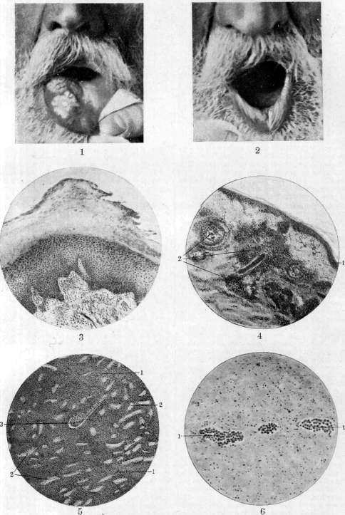
Figure 1. Leukoplakia of the tongue. On the leukoplakic spot occupying the right half of the anterior third of the tongue, a sharply delimited white shiny keratotic plaque 2nd/3 mm in height is visible. Figure 2. Leukoplakia of the lower lip (same patient). Figure 3. Histological preparation of leukoplakia of the penis; a marked hyperkeratosis is visible; a powerful multilayered granular layer; acanthosis. In the papillary and subpapillary layers of the dermis - round-cell infiltration. Figure 4. Leukemic infiltrates (2) in the skin (1). Figure 5. Leukemic. (i) infiltrates (from a large leukemic kidney) penetrate the entire parenchyma of the organ; the tubules (2) and glomeruli (3) of the kidney are greatly widened. Figure 6. Leukemic stases in the vessels (i) of the brain.
See also the article Leukemia, Leukoplakia. Of the 93 cases of L. of the urinary tract recorded in the literature before 1929, 51 cases were of the bladder and urethra, 29 of the renal pelvis, and 13 of the ureters. In relation to L. of the kidneys, the morbidity in women and men is the same. Morphologically, L. of the urinary tract does not differ from L. of the mouth and genital organs. Here, the lesion can be partial (focal) and more extensive, involving the entire surface of the renal pelvis or bladder or two adjacent organs. As etiological factors, stones, tumors of the bladder and kidneys, and irritation by abnormal urine are also indicated. Syphilis is not given great importance by urologists. Histologically, in L. of the bladder and upper urinary tract, metaplasia of the transitional (cuboidal) epithelium of the affected mucous membrane into the multilayered squamous epithelium of the external integuments is noted; in L. of the urethra, there is increased keratinization and desquamation of the epithelium. - Diagnosis of L. of the urethra and bladder is not difficult with the help of urethro- and cystoscopy. L. of the renal pelvis can occur with symptoms of pyelitis, with hematuria, and pollakiuria. The keratinized desquamating epithelium sometimes forms membranes which, passing through the ureter, can cause attacks of renal colic. The presence in the urine of such membranes and cell clusters from flat keratinized and columnar epithelium with a cystoscopically normal bladder suggests L. of the kidneys. - Treatment. Nephroureterectomy for unilateral L. of the upper urinary tract (Fedorov, Israel). For focal L. of the bladder, Varadulin obtained excellent results from diathermo-coagulation.
Related articles
Mentioned in
Cite this page
“Leukoplakia.” Soviet Medical Encyclopedia. English translation of Bolshaya Meditsinskaya Entsiklopediya, 1st ed. (Moscow, 1928–1936), ed. N. A. Semashko. https://sovietmedicalencyclopedia.pages.dev/article/leukoplakia/