Urogenital Organs
Historical document, translated for reference. It reflects medical knowledge of the 1920s–30s and is not medical advice.
Summary
This article describes the urogenital organs, which are closely connected in most vertebrates as part of the urogenital system. It covers the development and structure of excretory organs in invertebrates and vertebrates, including the embryonic and phylogenetic changes in kidney formation.
Encyclopedia article (1928–1936)
UROGENITAL ORGANS, Excretory organs, which are quite closely connected in most vertebrates together with the sexual organs into one urogenital, or urinogenital system. In invertebrate animals, such a connection usually does not exist—their urinary organs are represented by excretory tubes (nephridia of worms, Malpighian vessels of insects, etc.), while the sexual organs consist of gonads, their excretory ducts, and sometimes copulatory organs. The excretory organs serve to remove from the body the final products of metabolism dissolved in water—salts and nitrogenous compounds. Among the variously constructed excretory organs of invertebrates, the nephridia of worms deserve special attention. In lower, acoelomate worms, as well as in the larvae of higher worms, they are constructed according to the "protonephridia" type, consisting of a system of more or less branched tubes, the terminal branches of which are closed by special excretory, so-called flame cells, provided with an intracellular canal with a ciliary flagellum in it. Through the action of this flagellum, the fluid secreted by the cells is propelled through the nephridial canal to the excretory opening. In higher worms, the larval protonephridia are replaced by "metanephridia"—segmentally arranged paired tubules, opening with a ciliary funnel into the body cavity and leading directly to excretory pores on the lateral surface of the body. Such excretory organs are typically developed in earthworms and leeches. In vertebrates, the excretory organs are constructed according to the metanephridia type and consist of a series of successive excretory tubules, which initially open with a ciliary funnel into the body cavity and are connected to a common excretory duct. In the embryonic development of higher vertebrates, a sequential replacement of three different excretory organs is observed, which are called the pronephros, or head kidney (pronephros), the primary, or trunk kidney (mesonephros), and the permanent, or pelvic kidney (metanephros). This replacement corresponds to the phylogenetic replacement of excretory organs in the vertebrate series. In lampreys, the head kidney sometimes remains a functioning organ in the adult animal; in other cyclostomes, in fish, and in amphibians, the trunk kidney is the permanent excretory organ, while in reptiles, birds, and mammals, it is the pelvic kidney. However, upon closer examination, this replacement has the character of a gradual evolution of a single original organ, in which the anterior part develops earlier and has the simplest structure (pronephros), while the main mass of the organ achieves greater complexity with the gradual increase in the number of tubules (trunk kidney). Finally, in higher vertebrates, development in the posterior part of the organ goes even further and leads to the complete isolation of a complex of the most complex tubules, forming the permanent pelvic kidney. The excretory tubules of vertebrates develop from the epithelium of the body cavity (Figure 1), into which they open with a wide ciliary funnel. Initially, they have, like the metanephridia of worms, a regular metameric arrangement. The head
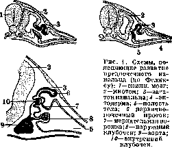
kidney consists of a few, only anterior tubules, which connect at their ends to form a common excretory duct, emptying into the cloaca. In addition to the excretory tubules, a special filtration apparatus develops, which is located either in the wall of the body cavity in direct proximity to the funnels of the pronephros (external chamber with a vascular glomerulus) or in the wall of the tubule itself (internal chamber with a vascular glomerulus or Malpighian corpuscle). The formation of the Malpighian corpuscle is especially characteristic of the trunk and pelvic kidneys. The tubules of the trunk kidney initially also have ciliary funnels. Later, however, these often become overgrown. With the development of the trunk kidney, the pronephric duct becomes the primary renal duct. The tubules of the pelvic kidney are more complexly constructed; they always begin only with a Malpighian corpuscle, and they never have funnels. The tubules of the pelvic kidney develop similarly to the secondary tubules of the trunk kidney from mesodermal nephrogenic tissue, however, they are not only devoid of funnels from the very beginning, but also connect not directly with the primary renal duct, but with a special outgrowth of it—the ureter (Figure 2). The latter becomes completely separated from the primary renal duct and then empties directly into the cloaca or (in mammals) directly into the urinary bladder. The tubules of the pelvic kidney differentiate into a series of successive sections. Beginning with the Malpighian corpuscle, the tubules form an initial convoluted section, then an elongated two-limbed loop of Henle, and finally a connecting tubule, emptying into the so-called straight collecting tubules of the kidney. The latter represent, in their development, the terminal branches of the ureter.
The permanent kidney of mammals is a relatively small compact organ of characteristic bean-shaped form with a concave inner wall, into the depression of which (hilus) the ureter and blood vessels enter. They are located not in the pelvic, but in the lumbar region, usually one somewhat in front of the other. Even macroscopically, in a cross-section of the kidney, one can notice the division into an outer—cortical and an inner, radially striated, medullary substance. In the cortical substance, the Malpighian corpuscles, convoluted and connecting tubules are located, while the medulla consists mainly of straight collecting tubules, but the loops of Henle also penetrate into it. For mammals, the fan-like arrangement of the collecting tubules, converging to a common excretory opening on the papilla, which protrudes into the expansion of the ureter, the so-called renal pelvis, is characteristic. Such a group of converging collecting tubules forms the so-called pyramid of the medullary substance. The number and arrangement of such pyramids can be very different (Figure 3). In many small mammals, the medulla consists of only a single pyramid ending with a single
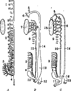
Fig. 2. Diagram of the renal tubules of amniotes and their relation to the sex glands: A—indifferent initial stage; B—female; C—male; 1-pronephros; 2-mesonephros; 3-metanephros; 4-ureter; 5-stalked hydatid; 6-funnel of the oviduct; 7-ovary; 8-epididymis and epoophoron; 9-paradidymis and paroophoron; 10-Wolffian duct; 11-urogenital sinus; 12-rectum; 13-uterus; 14-Müllerian duct; 15-testis; 16-uterus masculinus.
papilla. In others, a single papilla extends into a long ridge, carrying along its entire length the openings of the excretory ducts (carnivores, artiodactyls, monkeys). From the longitudinal ridge, lateral ridges may branch off, and finally such a complex papilla may break up into numerous independent papillae, as is also observed in the human kidney. A completely special type of kidney arises in the case where each papilla, with the corresponding branch of the renal pelvis and ureter, with the pyramid and part of the cortical substance, becomes isolated and forms a separate lobe. Thus, a multi-lobed form
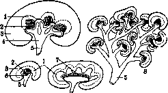
Figure 3. Diagrams of the structure of different types of kidneys in mammals (according to Gerhardt): 1-medulla; 2-papilla; 3-cortical substance; 4-branched renal pelvis; 5-ureter; 6-renal pelvis; 7-renal ridge; 8-multi-lobed kidney.
of the kidney arises, as is observed in bulls, bears, and cetaceans (Figure 3). Finally, in the horse and tapir, instead of a renal pelvis, a long passage develops, deeply penetrating forward and backward into the kidney parenchyma. The openings of the collecting tubules empty into this passage without the formation of any papillae. In all terrestrial vertebrates, a reservoir for urine—the urinary bladder—develops from the wall of the cloaca, into which, however, only in mammals do the ureters empty directly. The sex glands (see), or gonads, develop completely independently of the kidneys in the walls of the body cavity. Initially, in invertebrates, they have their own special excretory ducts. In higher worms, the sexual products are discharged into the body cavity and from there are expelled outward through the medium of special genital tubules. These, however, often fuse with the nephridial tubules. Thus, here already a connection between the sexual system and the excretory system is established, a connection that is so characteristic of vertebrates. In vertebrate animals, the sexual products are expelled exclusively through the ducts of the excretory system. The excretory duct of the primary
Fig. 4. Diagram of the primary kidney in the embryo of tailed amphibians: A-male; B-female; 1-testis; 2-vasa efferentia; 3-Müllerian duct; 4-sexual part of the kidney; 5-Wolffian duct; 6-ovary; 7-excretory part of the kidney.
sexual apparatus breaks up along into ducts, which receive the names Wolffian and Müllerian ducts (see Wolffian duct and Müllerian duct). The seminiferous tubules of the male sex gland connect with the primary renal tubules (usually with the anterior ones) through the medium of cords growing from them, forming the efferent ductules (vasa efferentia). Se
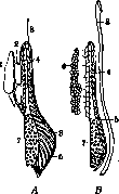
The flow proceeds through these canals into the Wolffian duct, which in lower vertebrates simultaneously serves as both the ureter and the vas deferens (fig. 2, 4). With the development of the pelvic kidney in higher vertebrates, the primary kidney loses its function as an excretory organ and partially regresses. Its anterior, sexual portion forms together with the highly coiled part of the Wolffian canal the epididymis, while the posterior, excretory portion sometimes remains as a small rudiment adjacent to the testis (paradidymis); in the female, both portions of the primary kidney regress, and their remnants sometimes persist in the peritoneal fold between the ovary and the oviduct (epoophoron, paroophoron).
The actual sex glands of mammals are rather compact organs of oval or bean-like shape. The ovaries are located in the posterior part of the abdominal cavity, while the testes move even further back and in most mammals protrude from the abdominal cavity into a special outgrowth of the latter, which is contained in a sac formed by the body wall with its muscles—the scrotum (fig. 5). Both the ureters and the genital ducts open
A
B
C
D
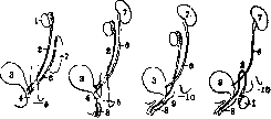
Figure 5. Schematic representation of the male urogenital apparatus of a turtle (A), a cloacal mammal (B), and viviparous mammals (C, D)—side view: 1-testis; 2-spermatic duct; 3-bladder; 4-urogenital sinus; 5-cloaca; 6-ureter; 7-kidney; 8-penis; 9-urogenital canal; 10-anus. directly into the common cloaca in reptiles and birds. In mammals, the anterior part of the cloaca, associated with the bladder, separates as the urogenital sinus (sinus uro-genitalis), into which the genital ducts open, and in monotremes, the ureters also open, while in viviparous mammals they open directly into the bladder. In viviparous mammals, the cloaca disappears, the urogenital sinus opens independently to the outside, forming in the female a small section, so-called vestibulum vaginae, and in the male elongating into a long urogenital canal. Mammals are also characterized by the process of fusion of the oviducts, leading to the formation of more or less significant unpaired sections. In all placental mammals, this results in a single vagina. In most cases, the process of fusion goes further, involving the next expanded section of the oviducts—the uterus, which is 'double' in many insectivores, rodents, and elephants, 'bicornuate' in other rodents, pigs, and some carnivores, 'simple' in some bats, monkeys, and humans, in which only the initial sections of the oviducts, so-called Fallopian tubes, retain their paired nature. - Connected with the urogenital excretory ducts are various accessory glands. In male mammals, at the end of the spermatic duct, there are large vesicular glands (gl. vesiculares), and in the walls of the urogenital canal, glands develop that differentiate into paired prostate glands (gl. prostaticae), which in primates fuse into a single unpaired gland, and also paired Cowper's glands (gl. Cowperi) at the base of the copulatory organ! In females, corresponding Bartholin's glands are present in the vestibulum vaginae. The copulatory organs of vertebrates may have different origins. In terrestrial vertebrates, they develop in connection with the cloacal wall (see). In mammals, this is a single organ (penis), developing in the anterior wall of the cloaca. Embryonically, its primordium arises from the anterior part of the cloacal tubercle and contains a portion of the urogenital sinus, which elongates along with the growth of the genital tubercle into a long canal. At the base of the genital tubercle, a paired ridge develops, which in the male forms the walls of the scrotum, and in the female monkeys and humans the so-called labia majora. In the female, the genital tubercle develops weakly and transforms into the clitoris and the labia minora. M. o. of humans—SEE Separate organs.
I. Shmal'gauzen. Statistics of diseases of M. o. In the department of diseases of M. o., according to the international nomenclature on which the following statistics is based, there are three groups: diseases of the urinary organs, diseases of the male sexual organs, and diseases of the female sexual organs. This department does not include venereal diseases, malignant neoplasms, and postpartum diseases, but includes benign neoplasms of the female sexual organs.- Mortality. In European countries, annually per 10,000 population, from 2.0 to 4.5 people die from diseases of M. organs, with more men than women (table 1). Table 1. Mortality from diseases of M. o. in European countries. Diseases of female organs. Countries Years Kidney diseases Italy . . 17,410 4.3 - As can be seen from the table, nephritis constitutes a significant portion of this figure, and in some countries (Italy) pyelitis is also included included. In any case, kidney diseases constitute the most frequent cause of death in the group of urogenital diseases. Next is mortality from diseases of the female sexual organs, which amounts to up to 1.4 persons per 10,000, while mortality from diseases of the male sexual organs is negligible and is usually not singled out in the data of most countries. Mortality rates from nephritis in the USA significantly exceed European ones: instead of the maximum figure of 4.0 per 10,000—from 8.5 to 9.6, and in recent years there is a tendency to increase, as can be seen from the following figures: 1920—8.94; 1921—8.54; 1922—8.85; 1923—9.01; 1924—8.96; 1925—9.63; 1926—9.83. In cities, mortality from urogenital diseases is higher than in rural areas. Thus, for Prussia (1927) with a coefficient of 3.7 male and 2.6 female per 10,000 population (table 2), in Berlin 4.8 males and 3.1 females died. Table 2. Mortality from urogenital diseases in Germany. Sex 1913 1925 1926 1927 Female...... 3.5 2.8 3.3 2.5 3.4 2.5 3.7 2.6 The years of war had their effect on increasing mortality from urogenital diseases: the pre-war figure in Germany of 3.5 (male) and 2.8 (female) steadily increased until 1917 (4.5 and 3.1); then followed a decrease until 1920, when it fell below the 1913 level (3.4 and 2.7). The years of military blockade thus had an impact in this area. For the city of Moscow, published data for 1925-28 are available. It amounted to 1.8 per 10,000 population (both sexes). By age, the largest number of deaths from diseases of the urinary organs occurs in the group from 22 to 44 years, from diseases of the male sexual organs—in the group from 50 years and older, and from diseases of the female sexual organs—in the prime age from 20 to 44 years. Mortality by individual professions is given in English statistics (1900-02). If we take mortality (of men) from all causes as 1,000, then from kidney diseases 35 died. Below this figure are such professions as agricultural workers (14), railway workers (17), teachers (23), clergy (27), etc., and above—butchers (46), musicians (54), day laborers (67), tavern workers (72), etc. Morbidity is most fully traced for the city of Moscow and province for 1926 (detailed development is timed to the census year). Per 1,000 population it amounted to 57.8; of which urinary—8.4, male sexual organs—2.5 and female sexual organs—46.9, and in the province respectively 35.9—6.2—1.4 and 28.3. In the group of urinary diseases in the city of Moscow, 2.3°/oo falls on inflammation of the kidneys (in the province—2.0°/oo). By sex, morbidity from urinary diseases (fig. 6) is higher among women, as can be seen from the following figures (table 3), Table 3. Diseases per 1,000 population. Men Women Urinary diseases .... Acute and chronic nephritis .... 4.5 1.6 2.9 7.7 2.4 5.3 Diseases of male sexual organs per 1,000 male population give an indicator of 5.2
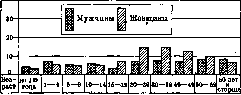
Figure 6. Urogenital diseases. (city) and 3.2 (province), and diseases of female sexual organs respectively—91.7 and 52.4. Sexual diseases among women occur 17 times more often than sexual
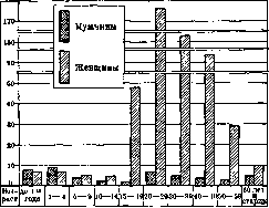
Figure 7. Sexual diseases.
Diseases in men. The character of the disease by age is illustrated by table 4 and figure 7. Table 4. Character of the population's morbidity from urogenital diseases by sex and age per 1,000 population of Moscow in 1926. Age Urogenital diseases Diseases of urinary organs Diseases of sexual organs m. f. m. f. m. f. Under 1 yr......... 11.0 9.5 3.0 2.7 8.0 6.8 From 1-4 yrs...... 15.6 12.5 6.2 5.4 9.4 7.1 » 5-9 yrs ..... 9.1 10.4 5.1 4.8 4.0 5.6 » 10-14 yrs ..... 7.2 9.3 5.6 4.4 1.6 5.0 » 15-19 yrs ..... 7.7 54.1 4.2 6.6 3.5 47.5 » 20-29 yrs ..... 12.9 188.4 6.4 13.3 6.5 175.1 » 30-39 yrs ..... 11.7 166.4 7.1 13.8 4.6 152.6 10.6 106.9 7.4 13.3 4.2 93.6 » 50-59 yrs ..... 11.2 38.8 7.9 10.0 3.3 28.8 60 yrs and older . . . 14.2 16.5 8.9 6.7 5.3 9.8 From this it is seen that morbidity of urinary organs in men increases from 20 yrs and reaches its highest development after 60 yrs, while in women the maximum falls in the age group 20-49 yrs. Diseases of male sexual organs are more common in the under 5 age group and among persons 20-29 yrs. Diseases of female sexual organs have a maximum at 20-29 yrs and 30-39 yrs, but they are also numerous in adjacent groups. In the productive group, sexual diseases among women occur 30 times more often than among men. Of individual diseases, one can demonstrate the age-specific morbidity from nephritis, diseases of the mammary gland, and metrorrhagias (table 5). Table 5. Morbidity from nephritis (acute and chronic), diseases of the mammary gland, and metrorrhagias (per 1,000 pop.). Age Under 1 yr..... 1-4 yrs ... 5-9 yrs ... . 10-14 yrs . . . . 15-19 yrs . . . . 20-29 yrs 2 30-39 yrs . . . . 40-49 yrs . . . . 50-59 yrs . . . . 60 yrs and older Nephritis Diseases of mam- Met- gland ror- rhagias f. f. >0.4 >0.2 ) 2.6 2.7 8.5 7.0 4.0 7.5 6.1 0.4 1.2 ' 0.2 0.2 In relation to nephritis, it can be noted that among men it is more common from 30 years and older, while among women-from 15 to 59 years, particularly more common in women than in men. Diseases of the mammary gland are noted mainly in the 20-39 age group (maximum 20-29 yrs), and metrorrhagias-from 20 to 49 years, i.e., in the productive age. Increased coefficients are found in the breast age for diseases of male sexual organs, in the 20-39 age group-for acute and chronic nephritis, diseases of male sexual organs and especially female sexual organs (especially mastitis and metrorrhagias), in the 40-59 age group-for acute and chronic nephritis and diseases of female sexual organs (metrorrhagias) and finally in the age of 60 years and older-for diseases of urinary organs (especially nephritis). When distributing urogenital diseases by month of the year, it appears that there is apparently no definite regularity. The role of profession in female morbidity according to research on factory and plant workers by S.M. Bogoslovsky is less significant than one might have assumed. Morbidity with loss of capacity to work in Moscow province for 1925-28 fluctuates within the following limits per 100 insured (table 6): for urogenital diseases (both sexes)-from 4.0 to 4.8; for diseases of urinary organs-from 0.6-0.7; for male sexual diseases-0.5; for female sexual diseases-from 6.9 to 8.6, and in recent years both the absolute number of cases of loss of capacity to work and the ratio to the total mass of insured show a tendency to increase. The average duration of one case of incapacity was for urogenital diseases in men from 22.4 to 28.8 days, in women from 17.1 to 25.5; for male sexual diseases-from 14.1 to 17.2, and for female from 11.8 to 18.3. Sex. As in the case of general morbidity, morbidity with loss of capacity to work in women is higher in all noted years almost twice, and for sexual diseases-17 times.-By individual industries, it is observed that men show increased morbidity in the sewing, food, and leather industries, while women, in addition to those mentioned, also in the metal industry (table 7). Table 7. Indicators of loss of capacity to work in industry of Moscow province in 1928 (per 100 insured). Uro- gen- ital dis- eases Urinary organs Sexual organs m. f. m. f. Metallurgical . . . 1.2 17.3 0.5 1.4 0.7 15.8 Woodworking . . . 1.2 9.0 0.6 0.7 0.6 8.3 0.8 7.3 0.4 0.9 0.4 6.4 1.3 0.5 23.4 6.3 0.7 0.2 1.6 0.9 0.6 0.3 21.8 5.4 Processing of min. substances 1.3 15.5 0.7 1.7 0.6 13.8 1.7 10.3 0.7 1.4 1.0 8.9 Processing of animal products 1.1 15.5 0.5 1.2 0.6 14.3 1.2 10.6 0.7 1.3 0.5 9.3 Printing . . . . . . . 1.1 17.6 0.5 1.2 0.6 16.4 Increased morbidity of men from sexual diseases can be noted in the leather industry, and of women-in the sewing, printing, metal, food, and in the processing of animal products. The influence of professional labor on the growth of diseases of female sexual organs with loss of capacity to work can be traced in tobacco production (table 8). Diseases of the uterus and appendages among production workers amounted to 3.6 per 100 insured, while among administrative-technical personnel only 0.6; Table 8. Morbidity of insured by professional groups in tobacco production (1927). Female dis- eases Number of cases of loss of capacity to work Production workers N rt И O o a в &g и я s S S a 2 и Я И щ *5 WIO И o Я га B л « H ^5 U и и «Й E- Л „ '. И K «я* Inflammation of uterus and appendages ..... Menstrual disorders .......... 3.6 0.8 2.0 4.6 0.2 3.1 6.4 1.6 3.2 4.4 0.9 2.5 0.6 Uterine bleeding at the same time among cigarette machine operators the indicator increased to 4.4, among cigarette makers to 4.6 and among machine packers of cigarettes to 6.4. Uterine bleeding is more common among packing, cigarette, and cigarette machine operators compared to production workers.-Disability. Of the entire mass examined at the Moscow Bureau of Medical Expertise (1925-1926), in 1925 1.2% were transferred to disability due to diseases of urogenital organs, and in 1926-1.1%. Thus in the total mass of causes of disability the proportion of diseases of urogenital organs is small. By groups of industries it is lower among employees than among workers, with the exception of medical-sanitary workers (2%) (table 9). By groups of disability due to diseases of urogenital organs (table 10) only about half received the right to a pension (1925), since 43.7% were assigned to groups IV-VI, while for all diseases-12.4%. Thus the severity of loss of capacity to work due to urogenital diseases is small; it cannot be placed alongside, for example, diseases of the cardiovascular system, where only in the first two groups were assigned disabled persons 60.1%. Table 9. Distribution by causes of disability in connection with professional group (1925-26). Years a б o ч и e Served i a в „ в з ч o ю «в a o ° B o n o "в в и н в к в W « оз Ю й л к a на- в к в s в X н в o a н o Ей «O И o i W o оз ю к ав к в в Piaj £ o) g B Ffb7 °K O B аз B o «в к 5 в в g И o и a 03 03 Ni r* ь в S3 8 И « B в Jfi 03 wg «a Ч и ело освидетельствованн ы'x 4 604 57 1 359 1 203 1 886 | 4 555 1 147 1 752 1 385 | 102 1 919 6 176 87 | 561 1 326 | 1 026 1 5 280 1 222 | 982 1 349 | 275 1 1200 Urogenital diseases (per 100 examined) 0.7 0.3 - 0.2 1.2 - 0.8 2.5 0.8 0.6 0.7 0.8 1.0 0.4 0.4 0.9 0.4 1.8 1.1 1.6 1.2 1.4 0.9 0.5 0.9 0.4 0.2 2.0 0.6 0.9 Table 10. Distribution of disabled workers by causes and groups of disability (1925-26). Causes of disability l o Number examined Abs. In% IV - VI For all 1 diseases. . . / Urogenital \ organs.. 1 1925 1926 1925 1926 10 472 12 603 126 140 100 100 1.2 1.1 8.2 8.3 7.1 2.1 50.4 51.5 22.2 35.8 29.0 31.1 27.0 32.1 12.4 9.1 43.7 30.0 Pathological involvement can be traced on the materials of the examination of industrial workers of Moscow (1925-28). The entire mass of workers and employees, divided by production, gives the following indicators of involvement of the urogenital apparatus (per 100 examined of each sex separately) (table 11): table 11. Professions Diseases of urinary organs йа Их S3 Z « и в o o *?» O o B оз и яЗ B o o ВЧ И m. f. Production workers ...... Auxiliary Staff..... 0.3 0.3 0.5 1.0 1.3 0.7 0.4 0.4 0.2 22.4 17.6 11.5 Women have greater involvement than men, and everywhere employees give lower indicators than workers, with the exception of diseases of urinary organs in men. Upon more detailed examination of the group of female diseases, this rule is almost never violated (table 12).
The combination of hazards of a particular type of work can have an effect on increasing the incidence of diseases of the female reproductive sphere (Table 13). Thus, in Table 14, it can be seen that with increasing length of service in wage labor, the indicators of inflammation of the appendages of the uterus also increase, both among auxiliary workers and among office workers, and to exclude the influence of age, a group of women in the productive period from 20 to 39 years was taken. The same ratios can also be traced for some other pathological conditions of the female sphere in the group of industrial workers. As can be seen from Table 14, the pattern of increasing indicators with increasing length of service is clear. Table 14. Influence of length of service in wage labor on the incidence of female diseases per 100 examined (Industrial workers; age 20-39 years). Diseases of the female genital organs Length of service Inflammation of the serous membrane of the uterus........ Inflammation of the muscular membrane of the uterus........ Inflammation of the mucous membrane of the uterus........ Inflammation of the appendages of the uterus............. Curvature of the uterus.... Change in position of the uterus............. Prolapse and prolapse of the vag-( ina........... 0,4 0,7 1,9 3,6 3,5 1,6 2,1 «5 0,9 2,1 3,7 4,9 2,2 2,3 1,0 1,3 3,8 4,8 6,6 3,5 3,8 1,3 1,1 3,3 4,5 6,0 3,1 3,3 1,2 Further, still unfinished studies of pathological incidence among individual professions show that in some cases, the incidence of female diseases can be associated both with the work process and with the surrounding production environment. For example, among tobacco workers who come into contact with tobacco, the coefficient of endometritis (5.9% compared to 4.5% in the production group of tobacco workers) and metritis (7.7% compared to 3.3%) is high. In other cases, socio-domestic factors also play a role, as is noted, for example, among teachers: the incidence of female diseases among unmarried women is 1.7%, while among married women (births not included) it is 12.1%.
Related articles
Mentioned in
Cite this page
“Urogenital Organs.” Soviet Medical Encyclopedia. English translation of Bolshaya Meditsinskaya Entsiklopediya, 1st ed. (Moscow, 1928–1936), ed. N. A. Semashko. https://sovietmedicalencyclopedia.pages.dev/article/urogenital-organs/