Wolffian Duct
Historical document, translated for reference. It reflects medical knowledge of the 1920s–30s and is not medical advice.
Summary
This article from the 1928–1936 Soviet Medical Encyclopedia describes the embryological development, structure, and fate of the Wolffian duct, explaining its role as the primary excretory duct and its transformation into the vas deferens in males or its regression into vestigial structures in females.
Encyclopedia entry (1928–1936)
WOLFFIAN DUCT, or Wolffian canal, represents, according to modern embryological nomenclature, the excretory duct of the primitive kidney, or primary ureter, which during further development enters into connection with the male gonad and functions as the vas deferens. The development of the Wolffian duct is closely connected with the development of the pronephric duct (pronephros, German Vorniere) and occurs in embryos at a very early stage, when the differentiation of the mesoderm begins (see). The latter, as is known, divides into somites, or primary vertebrae, arranged in a segmental order on the sides of the neural tube, and unsegmented mesodermal plates bounding the body cavity. Between these two formations lies the intermediate part (nephrotome), which gives rise to the excretory organs.
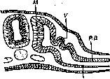
1-st
Figure 1. Diagram of the development of the excretory system: M-dermatome; PB-parietal layer of the mesoderm; VB-visceral layer of the mesoderm; V-rudiment of the pronephros. In it, correspondingly to the somites, pouch-like evaginations are formed, facing the dorsal side (see figure 1), which are then constricted off and turn into vesicles (pronephric vesicles, Vornierenbläschen); this differentiation proceeds from the head to the tail and in the human embryo occupies the region from 7 to 14 somites. Individual vesicles then begin to elongate and grow in a caudal direction; fusing with each other, they form a longitudinal canal lying under the ectoderm. In this way, the pronephric duct is formed, which grows without the participation of new vesicles towards the tail in the form of a continuous cellular cord, which subsequently acquires a lumen, reaches the hindgut or cloaca, and ends in it (see figure 2). The pronephros achieves significant development in fishes and some amphibians; in other vertebrates and man it does not function, but immediately behind it begins the development of the mesonephros, or Wolffian body (see above), the tubules of which flow into the same duct. From this moment on, it receives the name of the Wolffian duct and begins to function as the primary ureter. The second period in the development of the Wolffian duct is connected in male embryos with the development of the gonad, which enters into connection with the Wolffian body, whereupon the Wolffian duct also becomes the excretory duct for it. The fate of the Wolffian duct in adult individuals of various vertebrate classes is not uniform. Where the Wolffian body is preserved throughout life as a permanent kidney (fishes, amphibians), the Wolffian duct remains as the ureter, while in males it serves at the same time as the vas deferens, whereupon seminal vesicles are formed at its lower end in the form of diverticula. In reptiles, birds, and mammals (see figure 3), in connection with the development of the definitive kidney (metanephros), proceeding independently of the Wolffian duct, the latter, of course, loses the role of a ureter and in males passes entirely into the vas deferens, while in females it undergoes obliteration and disappears, being preserved only in the form of rudimentary remnants. There are two remnants in the female genital organs: 1) the epoophoron, where the Wolffian duct forms a longitudinal canal (ductus longitudinalis), and 2) Gartner's duct (ductus Gartneri), a canal that runs along the lateral surfaces of the uterus and vagina up to the vestibule of the vagina. Usually it is obliterated, but sometimes can be detected in newborns in the form of a short canal flowing into the vestibule of the vagina, and even persist in adults along its entire length as an anomaly.
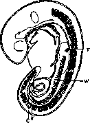
Figure 2. Wolffian canal of a human embryo 4.9 millimeters in length (after Ingalls): V-region of the pronephros with glomeruli; W-Wolffian canal with segmental tubules and glomeruli; C-caudal part of the Wolffian duct; K-cloaca; C1, Th1, L1-boundaries of individual parts of the spine: C1-cervical, Th1-thoracic, L1-lumbar vertebrae.
Figure 3. Relationship of the Wolffian body and the gonad. A-undifferentiated stage: U-Wolffian body; W-Wolffian duct; K-gonad with germinal epithelium. B-male type: W-Wolffian duct; G-glomeruli of the Wolffian body; U-Wolffian body; R-rete testis. C-female type: W-Wolffian duct; G-glomeruli of the Wolffian body; U-Wolffian body (parovarium); E-ovarian follicles. It is obliterated, but sometimes can be detected in newborns in the form of a short canal opening into the vestibule of the vagina, and even persist in adults throughout its entire length as an anomaly.
Related articles
Mentioned in
Cite this page
“Wolffian Duct.” Soviet Medical Encyclopedia. English translation of Bolshaya Meditsinskaya Entsiklopediya, 1st ed. (Moscow, 1928–1936), ed. N. A. Semashko. https://sovietmedicalencyclopedia.pages.dev/article/wolffian-duct/