Mullerian Duct
Historical document, translated for reference. It reflects medical knowledge of the 1920s–30s and is not medical advice.
Summary
This article describes the embryological development of the Mullerian ducts, which give rise to the female reproductive tract. It details their formation, fusion, and subsequent regression in male embryos, and explains how variations in this process lead to anatomical anomalies.
Encyclopedia article (1928–1936)
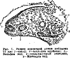
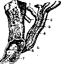
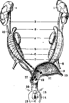
MULLERIAN DUCT, or paramesonephric duct; ductus, Mulleri (named after the famous physiologist Johannes Muller, who first described it), an embryonic formation that develops in the embryos of all vertebrates alongside the Wolffian duct (Fig. 1) and gives rise to the excretory pathways of the female genital apparatus: the tubes, the uterus, the vagina in mammals, and the oviducts in all other vertebrates. The Mullerian duct is formed in the embryos of both sexes: in selachians by the splitting of the Wolffian duct, and in all other vertebrates independently. The primordium of the Mullerian duct in the human embryo occurs at the end of the 1st month and the beginning of the 2nd month: the epithelium covering the Wolffian body in its cranial part deepens, forming a longitudinal groove; at the lower part of the edges of the groove they close, forming a tube lying outside the Wolffian duct (Fig. 2); the upper part remains as a slit; in total, a funnel is obtained, immersed in the mesenchyme of the Wolffian body and ending blindly. The further development of the Mullerian duct occurs by the growth of the blind end of the funnel in the caudal direction, at first it goes outside the Wolffian duct, and at the end of the 2nd month turns inward, crosses the Wolffian duct and continues further in the caudal direction, adjacent to the Mullerian duct of the opposite side. In this form, the Mullerian ducts reach the urogenital sinus and open into its posterior wall at the so-called Mullerian tubercle, located between the openings of the Wolffian ducts (Fig. 3). The mechanism of the closure of the Mullerian ducts and their transition to the inner side of the Wolffian ducts is quite complex and is associated with the growth of the urogenital folds of the Wolffian body and the formation of the so-called genital cord. In the 3rd month, the Mullerian ducts in male embryos begin to atrophy and dissolve; only two distal sections remain from them: the cranial and caudal. The first is preserved in the form of a Morgagni hydatid, appendix testis, the second, with further development, is surrounded by the prostate gland and forms the vesicula prostatica, or utriculus masculinus. In female embryos, at the end of the 3rd month, the adjacent sections of the Mullerian ducts begin to fuse into one common channel; fusion begins in the middle third and from there spreads forward and backward. The resulting uterovaginal canal in the 4th month becomes wider, and on it a constriction between the uterus and the vagina is outlined; the upper parts of the uterus become separated only at the end of the 4th month; the boundary between the uterine sections and the tubes is outlined earlier at the place of attachment of the round uterine ligament (ligamentum teres, in male embryos gubernaculum Hunteri). The various anomalies of the excretory genital tracts in women, starting with a bicornuate uterus and ending with a complete duplication of the uterus and vagina, are explained by the incomplete fusion of the Mullerian ducts. Each of these anomalies corresponds to the normal development of the genital tracts in various representatives of mammals, since the degree of fusion of the Mullerian ducts is extremely diverse. Fusion does not occur at all in monotremes and marsupials, which is why they have two uteri and two vaginas; in placental mammals, with one vagina there can be two uteri (uterus duplex in elephants, some rodents), one uterus divided into two parts (uterus bipartitus in carnivores, pig), or an elongated uterus in two horns (uterus bicornis in ungulates, insectivores). In birds, reptiles, and amphibians, fusion of the Mullerian duct does not occur, and each gives rise to an oviduct, with the right Mullerian duct remaining rudimentary in birds (see also Vagina, Uterus, development).
Related articles
Cite this page
“Mullerian Duct.” Soviet Medical Encyclopedia. English translation of Bolshaya Meditsinskaya Entsiklopediya, 1st ed. (Moscow, 1928–1936), ed. N. A. Semashko. https://sovietmedicalencyclopedia.pages.dev/article/mullerian-duct/