Sex
Historical document, translated for reference. It reflects medical knowledge of the 1920s–30s and is not medical advice.
Summary
This article discusses sexual reproduction and the separation of sexes in plants and animals, covering numerical sex ratios, sexual characteristics, and seasonal changes in appearance during mating periods.
Encyclopedia article (1928–1936)
174 Sexual reproduction and dioeciousness. Various forms of asexual reproduction in the course of phylogenetic development of plants and animals are replaced by sexual reproduction. The appearance of sexual reproduction is very often associated with dioeciousness, i.e., the development of sexual products subject to fusion during fertilization in two different individuals, male and female. The separation of sexes has significant evolutionary significance, ensuring division between two individuals of functions related to the appearance of offspring requiring care, protection, and upbringing. On the other hand, dioeciousness provides a wider range of variability, since during fertilization there is mixing of the hereditary plasma of father and mother, which also has great importance for the evolution of organisms. At various stages of the evolutionary ladder, one can discover various stages of separation of sex. In the filamentous alga Spirogyra sex, one can note the difference in the structure of cells of different individuals; filaments with large cells apparently correspond to female, and filaments with smaller cells to male individuals. During conjugation, the contents of male cells flow into female cells, as a result of which egg-shaped zygotes are formed, each of which is surrounded by a dense shell and gives rise to a new Spirogyra filament. However, Hartmann showed that individual specimens of Spirogyra behave simultaneously as male and as female individuals: during conjugation of three filaments with each other (Fig. 1), the middle filament B functions in relation to filament A as male, and in relation to filament C as female. In more highly organized plants and animals, dioeciousness manifests itself most clearly in the form and behavior of sex cells. The most general characteristic of gametes of both sexes is relative immobility and the large size of the egg compared to the sperm, which differs in mobility and is equipped with variously structured movement organelles (cilia, flagella, membranes). The appearance of differently structured gametes in different sexes is associated with the presence of multicellular organism organs that are the site of formation of sex cells. The sex glands, testes of males and ovaries of females, are sometimes called primary sexual characteristics. Numerical sex ratio. The most developed data on the numerical ratio of sex relate to humans. For every 100 girls born alive, on average 105 boys are born. Variations for different countries are very insignificant. Thus, according to Bodio, for the period 1887-95, for every 100 girls boys are born (live births): Spain.....105.8 Holland...100.2 For different animals the numerical sex ratio is as follows: J3, S. Conjugation of three Spirogyra filaments (from Hartmann). Fig. 1. Hungary France Sweden Belgium . . . 105.0 . . 101.6 . . 104.5 . . 101.5 England . . 104.6 Animal For every 100 females males are born Author Cattle . . Pigeons ..... Diptera . . . 101.0; 98.3* 104.6 115.4; 97.7 104.9; 111.8 105.0 94.7 115.0 105.5 105.8 Korvenen, Düzing Korvenen, Düzing Irwin Korvenen, Wilkens Keno Darwin Keno Standfus Keno * Two adjacent numbers refer to data from different authors. Thus, the numerical ratio of sex usually deviates little from equality. To obtain reliable results, it is necessary to count sex in embryos (if possible) or in recently born young. Otherwise, when determining the numerical ratio of sex in nature, one can encounter significant deviations from equality due to various biological peculiarities of one or another species. Thus, in the spider Latrodeotes mactans for every 100 females there are 819 males, in the fish Lophius piscatorius-385, in the fish Cottus gobio-183, in the black swift-400, in the beetle Maerodartylus-131; conversely, in the mollusk squid (Loligo) for every 100 females there are 16.6 males and in the octopus (Octopus)-13.3. Such violations of equal sex ratio do not reflect the picture of their birth in such a ratio, but the result or a higher percentage of death of one sex or the difficulty of detecting one sex due to its inconspicuousness (protective coloration) or hidden way of life or other biological conditions (for birds, for example, the non-simultaneity of the flight of males and females to wintering grounds or to nesting sites). In most cases, the reasons for deviation from the normal numerical sex ratio in nature can be established, and one can take it as a rule that at birth the number of males and females is the same. (Explanation of this circumstance see below-Determination of sex.) Sexual characteristics, In many animals the male differs from the female very insignificantly, and sometimes it is impossible to distinguish them by external appearance. However, by the structure of the sex organs in such monomorphic species sex is not difficult to determine. In addition to the difference in anatomical and histological structure of the sex glands, the male of monomorphic species usually differs from the female in the structure of the sex-conducting pathways and often in copulatory adaptations. The latter have especially complex structure in insects, particularly in butterflies. In addition to organs playing a direct role in the process of fertilization and ensuring the penetration of spermatozoa into the sex-conducting pathways of the female, many adaptations outside the sexual apparatus related to the act of fertilization are known. These include nuptial tubercles on the thumb of the male frog, serving him to grasp and hold the female during spawning. Special adaptations are found in some orthopterans. In crickets of the genus Oecanthus, the male has on its back 5 pits formed by the depression of the chitinous cover, into which the ducts of the so-called Hankokov's glands open. At the time of mating the pits are filled with the liquid secretion of the glands, and the female, sitting during copulation on the back of the male, drinks this liquid. After copulation the female opens the spermatophore attached to her sexual opening and eats it. The food bait-in the form of the secretion of Hankokov's glands apparently serves to keep the spermatophore longer at the sexual opening of the female, so that the sperm can pass into her sex pathways. Among the characteristics distinguishing the male from the female in insects are adaptations for producing sounds. Such adaptations are found in male crickets, grasshoppers, cicadas, etc. Their role in attracting females was attributed to them, but this position remains unproven. In the female of the silkworm (Bombyx mori) and many other butterflies at the end of the abdomen there are glands producing a fragrant substance, undoubtedly attracting males, since they gather around a piece of paper on which the contents of the fragrant glands is squeezed out, or around the cocoon with not yet emerged female. In many insects and vertebrate animals, males possess organs serving them as weapons in fights during the breeding period and for protection from enemies. These include the horns of deer, the tusks of boars and babirusas, the tusks of walruses, the tusks of elephants, etc. The characteristics described here constantly distinguish the male from the female; however, there are many examples of the appearance of distinguishing characteristics only during the non-breeding period, the so-called nuptial plumage, especially clearly noticeable in some fish. In the male salmon during spawning a curved process develops on the lower jaw and large pointed curved teeth grow. In Pacific salmon fish, pink salmon and chum salmon, during spawning (spawning) the silvery coloration darkens very strongly, reddish spots and stripes appear, the jaws greatly elongate and are provided with large teeth. All these characteristics disappear after the breeding period (Fig. 2). Bright coloration during spawning is acquired by some other fish, e.g., the stickleback (Gasterosteus), the minnow (Phoxinus) and others. The appearance of nuptial plumage also occurs in some birds. Here its preservation is not limited only to the breeding period, in the nuptial plumage the bird sometimes remains for more than half a year. Thus, the wild drake (Anas boschas) in winter and spring carries bright plumage, sharply distinguishing it from the female, and in summer and autumn has modest coloration, reminiscent of the coloration of the female. Nuptial tubercles of male frogs increase during spawning and thus should be partially attributed to seasonal sexual characteristics. As for the phenomenon of constant sexual dimorphism, its manifestations in the animal world are extremely diverse. Essentially the entire organism of the male differs from the female, but sexual differences are especially clearly manifested in a number of special characteristics, which have received the name secondary sexual characteristics (see). Of the anatomical features distinguishing the male from the female in constantly dimorphic species, one can note the following. In the worm Bonellia viridis the female has a body the size of a plum and a proboscis capable of stretching to 1 m in length, while the male, living inside the female, reaches several millimeters in length (Fig. 3). In many butterflies the coloration of the wings of the male is brighter than that of the female; sometimes females are completely wingless. In beetles the male is often significantly larger than the female and has various chitinous appendages in the form of unpaired
Fig. 3.
Fig. 4.

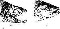
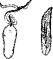

Figure 3. Bonellia viridis: a - female (reduced by 2 times); b - male (enlarged several times). Figure 4. The larynx of a duck (a) and a drake (b). or paired horns. In many viviparous fishes, there are forms where the male has bright coloring that is absent in the female, for example, guppy (Lebistes reticulatus), swordtail (Xiphophorus Helleri) and others. Sexual dimorphism is particularly diverse in birds. It is very clearly expressed in the coloring of chickens, ducks, pheasants, peacocks, African
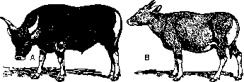
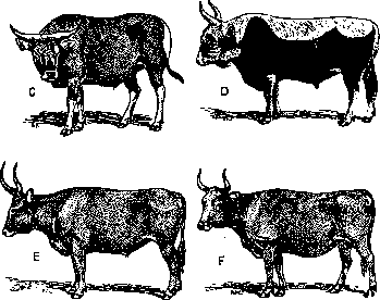
Figure 5. Bull (A), cow (B) and castrated bull (C) of the Hungarian Grey breed; bull (D), cow (F) and ox (E) of the Grey Ukrainian breed. (From Zavadovsky.)
ostriches, as well as in the bird-of-paradise, various species of hummingbirds, etc. One of the remarkable features of sexual dimorphism is the structure of the vocal apparatus and singing in many passerine birds. The lower larynx

-
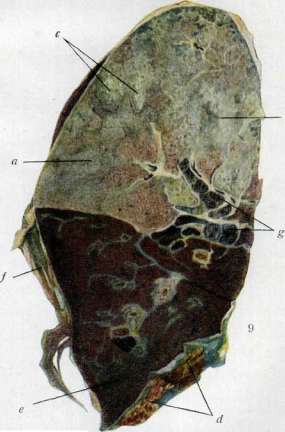
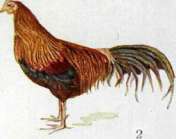
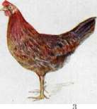
Fig. I. Normal comb. Fig. 2. Castrated comb. Fig. 3. Normal comb. Fig. 4. Castrated comb with regenerated tissue. Fig. 5. Castrated comb. Fig. 6. Castrated comb with regenerated tissue. Fig. 7. Fibrous exudate f d/ in the pleura during pleurisy; a - lung pleura, Fig. 8. Abundant fibrinous exudate f d/ and pneumonic exudate; a - sharp hyperemia of vessels, Fig. 9* Large-focal pneumonia (gray hepatization) with ]!:.! ! ti : i.::.....-: i. i - n punctate foci of purulent exudate, easily separated from the lung (a)\ d-pleural exudate and atelectasis of the lower lobe (c}\f- old pleural adhesions: л,г-anthracosis of bronchioles with ик жь'к.]. To art. Pneumonia, ffa,i. The syrinx in male songbirds and some other birds has a different structure than in females; sometimes it is equipped with expansions that act as resonators (fig. 4). The behavior of males during the breeding season (the display of capercaillies and some other birds, the poses assumed by peacocks and turkeys in the presence of females, pugnacity) characterize the manifestation of the male instinct, whereas in females of most birds the instinct of incubation and care for hatched chicks can be noted. Secondary sexual characteristics of mammals were partially mentioned above. In some species, distinct differences in the coloration of both sexes can be noted. In the nilgai antelope (Portax p ictus) the male is colored bluish-gray with a black mane on the neck, while the female is reddish with a white stripe on the chest; the gray Ukrainian cattle is characterized by a darker coloration of the bull; the same is observed in the Malayan cattle (Bos banteng) (fig. 5).- A distinct sexual dimorphism exists in humans. Man differs from woman on average by greater height, thicker bones with more intensely developed tubercles at the sites of muscle attachment. (For other differences in the skeleton of man and woman-see Identification in forensic medicine.) The subcutaneous fatty tissue is better developed in women, with fat deposits located near the mammary glands, on the mons veneris, on the thighs and buttocks. In Hottentot women steatopygia, according to Darwin, can reach an extraordinary degree that hinders movement.-Sexual dimorphism is also manifested in the structure of the larynx, which in men, in addition to greater volume and length of the vocal cords, is characterized by a sharper angle at which the plates of the thyroid cartilage converge, forming the so-called Adam's apple. Due to anatomical features of the larynx, the male voice is lower compared to that of a woman. The type of hair cover also belongs to the secondary sexual characteristics in humans. In many races, man is characterized by the presence of a beard, mustache and more intense development of hair cover on the entire body; hair is located mainly on the chest, back, forearms and shins. The hair cover on the pubis and around the external genitalia in men has the shape of a rhombus, in women the shape of a triangle.--Sex also leaves a certain imprint on human behavior, although the differences in psyche noted by bourgeois scientists mainly originate from distorting educational influences associated with the disenfranchised position of women in capitalist countries. Development of sexual characteristics. Research on the connection of secondary sexual characteristics with humoral influences spreading through the bloodstream began with the experiments of Berthold (Berthold,' 1849), who removed the testes of a cockerel and transplanted them to another location in the body. The cockerel retained all characteristic male signs. A number of subsequent studies led to the establishment of a connection between the development of sexual characteristics in vertebrates and the secretory activity of the sex glands. The regularity of this connection apparently does not extend to insects, since castration experiments on caterpillars of various butterflies (the gypsy moth, the cabbage white) did not lead to changes in sexual characteristics. Even simultaneous removal of wing primordia along with castration did not prevent the development of typical coloration in both males and females.-Removal of the sex glands in vertebrates affects their appearance, and males and females of different classes react to castration differently. The most complete data on the dependence of the development of sexual characteristics on the internal secretory activity of the sex glands have been obtained in chickens (see Castration). A castrated hen becomes indistinguishable from a castrated cockerel. On the basis of these data, M. Zavadovsky built his classification of secondary sexual characteristics (see Secondary sexual characteristics). The dependence of the development of some sexual characteristics on the sex hormone made possible the "transformation of sex", i.e., the imparting to a castrated male of female-dependent characteristics and to a castrated female of male-dependent characteristics by implantation of sex glands of the opposite sex. This experiment was first performed by Steinach, who coined the terms masculinization and feminization. The experiments of M. Zavadovsky, conducted on chickens, led to very demonstrative results. When testes are transplanted to a castrated hen, as early as 10 days later, one can observe enhanced growth of the comb, wattles and earlobes and the appearance of bright coloration of the head plumage (see separate table, fig. 5). The hen acquires a cockerel's voice and male sexual instinct, in short, those male characteristics that are lost by the cockerel as a result of castration ("dependent" or eusexual male characteristics, according to the terminology of M. Zavadovsky) (see separate table, fig. 6). Transplantation of an ovary to a castrated cockerel (see separate table, fig. 2) leads to the growth of hen plumage and the formation of a comb, in shape and color resembling that of a hen (see separate table, fig. У). The ovary, located in the abdominal cavity, forms a large number of yolks that stretch the abdominal wall and give the bird the appearance of a laying hen. The voice of such a feminized cockerel is close to the voice of a hen. This experiment also allows one to conclude that in the presence of an ovary in a castrated male, "dependent" characteristics develop. The general conclusion from the results of castration and transplantation of sex glands from male to female and vice versa is that the tissues of male and female are equally capable of developing both male and female characteristics. This circumstance M. Zavadovsky designates as the principle of equipotentiality of somatic tissues of male and female. The study of the influence of sex hormones on the development of sexual characteristics showed a peculiar interaction between male and female hormones in chickens. Transplantation of a testis to a normal or partially castrated hen did not cause the formation of male characteristics in it, whereas transplantation of an ovary to a normal cockerel, on the contrary, led to the formation of female characteristics. From this, M. Zavadovsky concluded the dominance of the female sex hormone over the male. This conclusion is consistent with the results of long-term observations of castrated chickens. As mentioned above, removal of the ovary in a hen leads to the formation of an individual with characteristics typical of a castrate. After a few months, without any additional intervention, a castrated hen begins to grow a cockerel's comb, manifests the male sexual instinct and cockerel's voice, as it were, spontaneously transforms into a male. Previous experiments suggest the presence of testicular tissue here. Dissection of such hens reveals in the abdominal cavity, in the region of the anterior part of the kidney, to the right of vena cava post., a round body, sometimes having a duct that opens into the ureter (fig. 6). Histological examination of this organ indicates that it is a male gland that can reach full development, up to the formation of spermatozoa (fig. 7). On the basis of these experiments, Zavadovsky comes to the assertion of the bisexual nature of the hen. According to his data, not only the right sex gland of the hen, which develops after castration into a testis, possesses bisexual potential, but also the left one. In the latter, this potential is revealed after incomplete castration, when the severely damaged gland begins to regenerate. In this case, ovarian tissue is often partially replaced by testicular tissue. A hen with such a regenerating left ovary often acquires male characteristics. The cockerel apparently does not possess bisexual potential, all the more so since the male hormone,
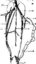
Fig.
Fig. 6. Position of the pre-gonad gland in a chicken: 1-posterior vena cava; 2-vein of Asab; 3-great vein; 4 and 5-arteries; 6-reduced oviduct; 7-rectum; 8-ureter; 9-sex duct; 10-upper part of the kidney; 11-right sex gland.(From Zavadovsky.) ,y.4«'JJ b Fig. 7. Histological structure of the right sex gland of a chicken. As shown above, it is suppressed by the female, and if a rooster has ovarian tissue, he would inevitably have to exhibit female characteristics. A certain deviation from this scheme is found in the so-called hen-feathered breeds of chickens, e.g., Sebright-Bantam, in which the rooster has neither sickle-shaped tail feathers nor lanceolate feathers on the neck and loins. The peculiarities of this breed are explained, however, not by the production of the female sex hormone by the male gland, as Morgan previously thought, but by the hereditary properties of this breed. This was determined by cross-transplantations of gonads from hen-feathered breeds to normal ones and vice versa. A castrated Sebright rooster with a transplanted testicle from a rooster of a hen-feathered breed retains its hen-feathered characteristics, while a castrate of a normal breed with a transplanted Sebright testicle retains its type of feathering (Danforth). The results of castration and transplantation of sex glands in mammals are somewhat different. In contrast to birds, the appearance of a castrate more closely resembles the female. A castrated male nilgau antelope acquires the reddish coloration of the female, a male deer after castration loses its horns; the same is true in breeds of sheep where the male is horned and the female is hornless. A castrated man is beardless and mustachioed, has a high voice, a horizontal hairline on the pubis, and often has fat deposits in the areas characteristic of women (breasts, thighs, buttocks) (see Castration). When transplanting sex glands from one sex to another in guinea pigs, Steinach, Lipschütz, etc., were able to obtain 'sex transformation' to the same extent as in similar experiments with chickens. There is reason to assume that in mammals, the male is bisexual, while the female is unisexual. This position, however, requires more serious proof. An indirect argument in its favor is the existence of the so-called 'free-martin'. This term refers to a developmental abnormality in calves, consisting in the fact that the external genital organs resemble those of a female, while the internal ones are closer to those of a male. Tandler and Keller (Tandler, Keller; 1911, 1916) and especially Lillie (Lillie, 1917), who used the enormous material from Chicago slaughterhouses, showed that a 'free-martin' is formed in the case of fraternal twins of different sexes with a common chorion and communicating blood vessels (fig. 8), provided that the second twin is male. The 'free-martin' is thus interpreted as a female undergoing transformation into a male under the influence of the humoral environment of her normal male twin. A similar transformation of a female into a male was obtained by Burns and Witsch by pairwise grafting amphibian embryos. In tailless amphibians, the phenomenon of sex transformation is also observed under natural conditions, which is why cases of hermaphroditic individuals are often found (see summary by Crew). Castration of the male leads to the loss of a number of secondary sexual characteristics and brings the appearance of the castrate closer to that of the female. By analogy with mammals, one can tentatively speak of the bisexuality of the male here. A particularly convincing argument in favor of this position is the existence in male toads of the so-called Bidder's organ (see Bidder's organ), which after removal of the testicles transforms into a normally functioning ovary. The research of Pfluger, Hertwig, and especially Witsch (Pfluger, Hertwig, Witsch) showed the existence in frogs of two so-called differentiated and undifferentiated races. In the first, the division into males and females occurs already during metamorphosis, which is why they always give a normal sex ratio. In the second, almost all tadpoles initially have the appearance of females, although they are essentially hermaphrodites, and then about half of them more or less gradually transform into males. Material collected from nature of this race at different times of the year gives different numerical sex ratios with a large predominance of females in young individuals that have not yet undergone transformation. The presence of hormonal influences on the development of sexual characteristics in vertebrates allowed M. Zavadovsky to construct a theory of morphogenesis. This theory comes down to the view of the developmental process as a reaction of developing tissues and organs to external influences relative to them. The formation of a 'dependent' sexual characteristic proceeds, according to Zavadovsky, according to the formula x+y→A, where x is the tissues that give the characteristic, y is the irritant that causes its development, in this case the hormone of the sex gland, and A is the ready characteristic (e.g., the rooster's comb). The sex hormone, participating in the formation of sexual characteristics, is arbitrarily isolated from the phenomenon and as such is opposed to the 'reacting tissues'. This reflects the formal-logical, mechanistic understanding of causality underlying the causally-analytical method used by Zavadovsky. This mechanistic understanding of causality is clearly formulated by Mill in the basic principle of inductive logic—the 'principle of the single difference'. The essence of this principle is as follows. If after the introduction of some factor a certain phenomenon appears (or after its removal, under otherwise equal conditions, disappears) [e.g., the disappearance or appearance of sexual characteristics after removal or transplantation of sex glands (Blyakher)], then in this case this factor that we are introducing or removing is the cause of the phenomenon. The impossibility of penetrating the essence of the phenomenon with the help of this principle depends, firstly, on the fact that in no case can one be sure that the 'single' difference that determines the nature of the process has been revealed, and secondly, that behind the solution of the question of the cause of this phenomenon there arises the question of the cause of this cause, and so on. In the end, one either comes to some primary cause, i.e., to God, or stops the inductive analysis at some arbitrary stage. After solving the question of the sex hormone as the cause of the development of sexual characteristics, Evans, Zavadovsky, and others were forced to raise the question of the cause of the origin of the sex hormone in the body. The corresponding cause is also sought outside, and vitamin E (vitamin X), or the 'reproduction vitamin', isolated from wheat germs, lettuce leaves, etc., is recognized as such. The necessity of vitamin E for the normal functioning of the sex gland is apparently indisputable, but the fact of this influence does not bring us closer to understanding the development of sexual characteristics. Instead of penetrating deeper into the phenomenon, this method of research takes us further and further away from the organism, from its specific peculiarities, the study of which alone can answer our questions about the essence of the phenomena of the development of sexual characteristics. After all, continuing the analysis further, we must determine the cause of the origin of vitamin E in plants, and we will undoubtedly be able to find it outside the plant, say in the soil. The importance and practical value of the facts of the dependence of sexual characteristics on endocrine influences is beyond doubt. It is only necessary to keep in mind that this kind of research is unable to penetrate the essence of the phenomenon, it does not reveal its driving forces, its motive, its self-movement. Work in this direction is a matter for the future.
Fig. 8. Communicating blood vessels of the chorion of twin embryos in a cow. (From Zavadovsky.)
and others succeeded in obtaining 'sex transformation' to the same extent as in similar experiments with chickens. There is reason to assume that in mammals, the male is bisexual, while the female is unisexual. This position, however, requires more serious proof. An indirect argument in its favor is the existence of the so-called 'free-martin'. This term refers to a developmental abnormality in calves, consisting in the fact that the external genital organs resemble those of a female, while the internal ones are closer to those of a male. Tandler and Keller (Tandler, Keller; 1911, 1916) and especially Lillie (Lillie, 1917), who used the enormous material from Chicago slaughterhouses, showed that a 'free-martin' is formed in the case of fraternal twins of different sexes with a common chorion and communicating blood vessels (fig. 8), provided that the second twin is male. The 'free-martin' is thus interpreted as a female undergoing transformation into a male under the influence of the humoral environment of her normal male twin. A similar transformation of a female into a male was obtained by Burns and Witsch by pairwise grafting amphibian embryos. In tailless amphibians, the phenomenon of sex transformation is also observed under natural conditions, which is why cases of hermaphroditic individuals are often found (see summary by Crew). Castration of the male leads to the loss of a number of secondary sexual characteristics and brings the appearance of the castrate closer to that of the female. By analogy with mammals, one can tentatively speak of the bisexuality of the male here. A particularly convincing argument in favor of this position is the existence in male toads of the so-called Bidder's organ (see Bidder's organ), which after removal of the testicles transforms into a normally functioning ovary. The research of Pfluger, Hertwig, and especially Witsch (Pfluger, Hertwig, Witsch) showed the existence in frogs of two so-called differentiated and undifferentiated races. In the first, the division into males and females occurs already during metamorphosis, which is why they always give a normal sex ratio. In the second, almost all tadpoles initially have the appearance of females, although they are essentially hermaphrodites, and then about half of them more or less gradually transform into males. Material collected from nature of this race at different times of the year gives different numerical sex ratios with a large predominance of females in young individuals that have not yet undergone transformation. The presence of hormonal influences on the development of sexual characteristics in vertebrates allowed M. Zavadovsky to construct a theory of morphogenesis. This theory comes down to the view of the developmental process as a reaction of developing tissues and organs to external influences relative to them. The formation of a 'dependent' sexual characteristic proceeds, according to Zavadovsky, according to the formula x+y→A, where x is the tissues that give the characteristic, y is the irritant that causes its development, in this case the hormone of the sex gland, and A is the ready characteristic (e.g., the rooster's comb). The sex hormone, participating in the formation of sexual characteristics, is arbitrarily isolated from the phenomenon and as such is opposed to the 'reacting tissues'. This reflects the formal-logical, mechanistic understanding of causality underlying the causally-analytical method used by Zavadovsky. This mechanistic understanding of causality is clearly formulated by Mill in the basic principle of inductive logic—the 'principle of the single difference'. The essence of this principle is as follows. If after the introduction of some factor a certain phenomenon appears (or after its removal, under otherwise equal conditions, disappears) [e.g., the disappearance or appearance of sexual characteristics after removal or transplantation of sex glands (Blyakher)], then in this case this factor that we are introducing or removing is the cause of the phenomenon. The impossibility of penetrating the essence of the phenomenon with the help of this principle depends, firstly, on the fact that in no case can one be sure that the 'single' difference that determines the nature of the process has been revealed, and secondly, that behind the solution of the question of the cause of this phenomenon there arises the question of the cause of this cause, and so on. In the end, one either comes to some primary cause, i.e., to God, or stops the inductive analysis at some arbitrary stage. After solving the question of the sex hormone as the cause of the development of sexual characteristics, Evans, Zavadovsky, and others were forced to raise the question of the cause of the origin of the sex hormone in the body. The corresponding cause is also sought outside, and vitamin E (vitamin X), or the 'reproduction vitamin', isolated from wheat germs, lettuce leaves, etc., is recognized as such. The necessity of vitamin E for the normal functioning of the sex gland is apparently indisputable, but the fact of this influence does not bring us closer to understanding the development of sexual characteristics. Instead of penetrating deeper into the phenomenon, this method of research takes us further and further away from the organism, from its specific peculiarities, the study of which alone can answer our questions about the essence of the phenomena of the development of sexual characteristics. After all, continuing the analysis further, we must determine the cause of the origin of vitamin E in plants, and we will undoubtedly be able to find it outside the plant, say in the soil. The importance and practical value of the facts of the dependence of sexual characteristics on endocrine influences is beyond doubt. It is only necessary to keep in mind that this kind of research is unable to penetrate the essence of the phenomenon, it does not reveal its driving forces, its motive, its self-movement. Work in this direction is a matter for the future.
L. Blyakher. Stimulation of sexual maturation. Sexual maturity in all animals does not coincide in time with birth or hatching from the egg. As a rule, it occurs significantly later. The period of infantile-immature state varies in different animals, but for each species it is strictly defined. Under normal conditions, the range of variation in the timing of sexual maturation is negligible, however, pathology knows cases of shifts toward significant shortening. The literature contains references to cases of sexual maturity occurring in boys and girls at the age of 5-6 years (see Pubertas praecox). In all such cases, premature sexual maturity was associated with early endocrine activity of the sex glands. Experimental studies of recent years on various animals have shown that premature sexual maturity can be caused by introducing the pituitary hormone into the body of an infantile animal. The works of Smith, Zondek, Aschheim and others have shown that the implantation of even a very small piece of tissue from the anterior pituitary gland (weighing up to 1/1000 g) can cause premature maturation of the sex gland. The pituitary gland, taken from a human or any of the male and female mammals, can in equal degree cause a reaction of premature sexual maturation in mice as early as 100 hours after implantation. At the same time, the pituitary gland of pigeons in Smith's experiments gave a negative result. The pituitary gland is active not only from sexually mature animals, but it is equally active in infantile animals and even in the fetus in the last months of pregnancy. Until recently, the pituitary gland was known as the only irritant for the maturation of the sex gland, and as Zondek showed, no other tissue or gland with internal secretion can give a similar reaction to sexual maturation. Under the influence of the pituitary hormone, the reaction of sexual maturation proceeds violently. In the sexual apparatus of an infantile animal, sharp changes occur both macroscopic and microscopic. The reaction of sexual maturation is manifested first in females in the growth and maturation of ovarian follicles, in the maturation of egg cells, in the premature appearance of ovulation, and in the premature formation of yellow bodies. However, in experiments, ovulation does not always occur due to the premature transformation of follicular cells into lutein cells. In these cases, so-called atretic yellow bodies are formed, containing the egg cell inside them. Due to the growth of follicles and the formation of a large number of yellow bodies, the ovary in experimental mice exceeds in volume and weight the ovary of normal animals of the same age by 10-15 times. Along with changes in the ovary, changes also occur in the uterus and vagina. The uterus and oviducts increase in volume, turning from pale-yellow to lilac-red due to the strong dilation of blood vessels; in the mucous membrane of the uterus and vagina, all the characteristic changes of estrus occur. The epithelium of the mucous membrane of the uterus and vagina changes from single-layered to multi-layered, the upper layer degenerates, flattens, loses nuclei, desquamates, and enters the vaginal mucus in the form of anucleated keratinized scales or masses. Estrus, which appears during sexual maturation and recurs each time during ovulation, is a characteristic sign of sexual maturity in all mammals. In humans, the analogous phenomenon is menstruation. Estrus in the process of sexual maturation is a secondary phenomenon; its appearance is determined not by the pituitary hormone, as Zondek showed, but by the hormone produced by the mature ovarian follicle. The transplantation of pituitary pieces to infantile or sexually mature castrates in double and even quadruple doses does not cause any changes in the genital apparatus, whereas under the influence of the follicular hormone, even in castrates, all the characteristic phenomena of estrus appear. These experiments showed that the pituitary hormone affects the sexual system only through the sex gland, and through the latter also on the other organs of the generative system. Maturation of the sex gland is connected with the hormone of the anterior pituitary. In experiments causing sexual maturation, the transplantation method is not the only one. The injection method of pituitary extracts is widely used. The anterior pituitary, as Evans showed, produces 2 substances with different effects. One of them stimulates growth, and the other excites sexual maturation (see Pituitary - chemistry of the pituitary). Due to their different physicochemical nature, they are easily separable and can be obtained from the anterior pituitary separately. According to their morphogenetic and physiological properties, these two hormones are antagonists, which is manifested in their combined action. With simultaneous introduction into the body of an infantile animal of these hormones, premature sexual maturation does not occur. Consequently, the growth hormone inhibits sexual maturation, because in its absence, sexual maturation occurs in all cases. The secretion of two different hormones by the anterior pituitary, Evans tries to associate with the presence of 2 types of glandular cells in the pituitary. Basophilic cells, located in the central part of the pituitary, according to Evans, produce the hormone that stimulates sexual maturation, while the periphery, rich in eosinophilic cells, secretes the growth hormone. In addition to the pituitary gland, the pituitary hormone can also be obtained from the placenta and the urine of pregnant women (see Prolan). The results of experiments in the field of premature sexual maturation have been verified by many researchers on various objects. For this purpose, amphibians, reptiles, fish, birds, and many representatives of mammals, up to anthropoid apes, were tried. In all cases, it was possible to stimulate sexual maturation in females and males with the help of the hormone from the anterior pituitary. The pituitary hormone has a stimulating effect not only on the immature sex gland, but also on the mature gland in a state of rest, which is observed, for example, during hibernation. The literature contains references to cases of disruption of the sexual rhythm with the help of injections of pituitary hormone. In experiments with frogs, fish, and snakes, it was possible to cause in winter months in females egg-laying, and in males sexual desire and a surge of spermatogenesis. Experiments with the removal of the pituitary in tailed amphibians also showed that the development and function of the sex glands are connected with the pituitary gland. In case of hypophysectomy (see also Pituitary, normal and path. physiology) in immature animals, the development of the sex glands in males and females was suspended, as a result of which the other organs of the generative apparatus did not develop. The oviducts remained three times narrower and shorter than normal, in males the cloacal swelling did not develop at all and the production of mature sexual products was inhibited. All the above facts indicate that the process of premature maturation under experimental conditions can be caused by the action of the pituitary hormone. However, from this one cannot conclude that in the organism there is exactly such a connection between the activity of the pituitary and sexual maturation. Zondek, considering the pituitary the 'motor of the sex gland', bases his judgment on a causal-analytical experiment, with the help of which the connections characterizing the organism as a whole cannot be revealed. The establishment by the analytical method of this or that 'cause' simplifies the true picture of interaction, arbitrarily dividing the organism into externally related components (see above).
N. Lintvarere. Determination of Sex. Previously it was believed that the determination of S. in relation to the moment of fertilization occurs at different times in different organisms, which is why progamous, syngamous, and metagamous determination of S. were distinguished. Progamous determination of S. occurs before fertilization, syngamous during fertilization, and metagamous after fertilization. However, it must be admitted that in most organisms the determination of S. occurs at the moment of fusion of the egg and sperm, i.e., syngamously. At present, it can be considered proven that the S. of each individual is determined by certain genes, depending on the distribution of the so-called sex chromosomes. (For a detailed description of the sex chromosome apparatus and the theory of sex determination based on it by McClung and Wilson, see Heredity.) Wilson's theory, according to which S. is determined by sex chromosomes, is supported by the overwhelming majority of biologists. The so-called index hypothesis, proposed by some authors (Hacker and others) in opposition to Wilson's theory, according to which heterochromosomes are not determinants of S., but on the contrary, are a manifestation, an index of the sexual character of the individual, must be recognized as unfounded. Its defenders usually refer to certain special cases of S. determination, when even before reduction the structure of the gametes predetermines the distribution of chromosomes and thus the determination of S. This argumentation is based on a metaphysical understanding of the determining factor as an 'absolute primary cause,' and a failure to understand that it in turn is subject to external influences both from the surrounding cells of the organism and from the external environment. In the question of the essence of the genetic action of sex chromosomes, there are also some disagreements. Individual authors (Win-ge) believe that the X-chromosome carries a recessive gene of female S. (in the case of male heterogamety), and the Y-chromosome a dominant gene of male S.; hence XX=♀, XY=♂. However, this contradicts both the existence of the XO type and the facts when male individuals due to malformation or experimental develop certain female characteristics and vice versa, from which it must be concluded that both males and females carry both male and female sex genes. Therefore, Correns suggested that both male and female genes are localized in autosomes, while in heterochromosomes there are 'realizers'—in the X-chromosome a gene that inhibits the manifestation of male S. or ensures the possibility of realization of female, in the Y-chromosome—a 'brake' of female or 'realizer' of male. Thus, the difficulty with the presence of female genes in males and vice versa is overcome; but the existence of the XO type also contradicts this theory. Moreover, it is completely unnecessary, and in methodological terms a metaphysical complication, to explain the impossibility of simultaneous full manifestation of male and female genes in dioecious organisms by the existence of special realizer genes, rather than by the nature of the interaction between these genes themselves. The most acceptable theory was proposed by Goldschmidt. According to his views, each haploid set of autosomes contains (in the case of male heterogamety) a male gene (or gene complex) M, each X-chromosome a female gene F, which is stronger than the M gene, and each Y-chromosome a qualitatively similar but weaker than M female gene f, which may also be completely absent. The ratio of the strength of these genes is such that 2F > 2M > F + f. Therefore, individuals having two X-chromosomes exhibit female S., while those having XY exhibit male (2nXX= MMFF=♀; 2nXY= MMFf=♂, where n is the haploid set of autosomes). In case of violation of the correct ratio between M and F (when crossing different races or due to the presence of extra sex chromosomes or extra sets of autosomes), the predominance of female factors over male or vice versa can become very small or disappear completely; in these cases intersexes develop. Intersexes of the first kind (violation of the balance of sex genes by crossing races having M and F of different strength) were studied by Goldschmidt in the silkworm, of the second kind (3nXX)—by Bridges and Dobzhansky in Drosophila. In both cases, the researchers came to basically similar views; the views of these authors are represented by a table taken from Bridges' article (p. 176). Considering that each hereditary factor or group of factors possesses a certain valence, Bridges assumes that the 'female' valence of the X-chromosome is 100, and the 'male' valence of the haploid set of autosomes is 80. Then a normal female, as well as tri- and tetraploid females will give an index (ratio of valences) =1.25; a male will give an index of 0.63, intersexes—0.82. Theoretically we—Number of X-chromosomes Number of haploid sets of autosomes Ratio of valences Sex type of the resulting individual 3 4 3 2 1 2 1 1 2 4 3 2 1 3 2 3 1.87 1.25 1.25 1.25 1.25 0.82 0.63 0.42 Superfemale ♀ tetraploid ♀ triploid ♀ normal (di-ploid) ♀ haploid ♀ intersex ♂ supermale ♂ According to Bridges, we can theoretically conceive individuals in which the number of X-chromosomes would be relatively too large or too small. The former, with an index of 1.87, Bridges calls 'superfemales,' and the latter with an index of 0.43—'supermales.' Superfemales and supermales externally differ little from normal females and males, being characterized by reduced viability and complete infertility. Bridges' views are based on cytological observations. It further turned out that the intersexual organism develops up to a certain moment as a male, and then as a female or vice versa. In this case, the predominance of genes of a certain S., not strong enough to lead to the formation of a normal male or female, affects the time of development of sexual characteristics: the greater the predominance of genes of a given S., the earlier the characteristics inherent to that sex begin to develop. The general character of this last rule received confirmation on plant objects in Wettstein's experiments on dioecious mosses, where experimentally obtained FM and FFM individuals were hermaphroditic, but the first developed antheridia in larger quantities and earlier than archegonia, while the second—the opposite. Besides dioecious organisms, as is known, various forms of normal hermaphroditism exist (in contrast to the hermaphroditism of humans and dioecious animals, which is an anomaly, most often intersexuality). The extreme form of normal hermaphroditism—so-called spatial monoecy, where male and female sexual systems function simultaneously in the same individual (annelids and flatworms, many mollusks, etc.). Goldschmidt and Vichi explain this case by a complete equilibrium of male and female genes (F=M). However, such subordination to the equilibrium formula in this case is hardly correct, all the more so that in spatial monoecists male and female gonads often develop very differently early in ontogenesis (see above on the significance of this moment). It is more probable that here there is simply a different interaction between sex genes, in which there is no antagonism between F and M characteristic of dioecious organisms; thus F does not prevent the manifestation of M and vice versa, and therefore the comparative strength of these genes plays no role. Another form of normal hermaphroditism—consecutive (sequential) monoecy, where sexual systems function in the same individual, but not simultaneously, but successively (many mollusks, lower crustaceans, etc.). Obviously here there is an antagonistic interaction of F and M, but the predominance of sex genes of one S. is not great enough to completely exclude the action of the opposite; the stronger pair of genes probably usually determines the preceding, and the weaker pair the subsequent sexual phase. Goldschmidt interprets consecutive monoecists by analogy with intersexes as forms in which the duration of life is great enough to cover both male and female phases. In dioecious organisms lacking internal secretion of sex glands, in his opinion natural death occurs before the 'turning point' from one sexual phase to another.—The third phase, closest to dioecy—rudimentary hermaphroditism, when only one sexual system functions, but alongside it a rudimentary system of organs of the second S. develops temporarily or for life. Thus, in male toads there is a rudimentary ovary (Bidders organ), which when the testes are removed can begin to function; in many races of frogs, the gonads of males initially develop as ovaries and only then transform into testes. According to Vichi, in rudimentary hermaphrodites there is a relatively strong female factor f of the Y-chromosome, due to which (MM-Ff) turns out to be too small a value to completely exclude the development of female characteristics; and, obviously, the greater f, the more significant the rudiments of hermaphroditism. In the type under consideration, unlike the two previous ones, heterochromosomes are already present, although morphologically it differs little from X (in the studied case—in tailless amphibians).
Wichi also believes that the excess of male genes over female in males should always be equal in magnitude to the reverse excess in females (FF - MM = MM - Ff). From this he deduces that the 'force differentiating the male', carried by spermatozoa with the Y-chromosome, must be three times greater than the 'force differentiating the female' in eggs and spermatozoa with the X-chromosome, since upon fertilization of the egg (with the X-chromosome) by an X-spermatozoon, the balance of sex genes will be 2 (F - M), and upon fertilization by a Y-spermatozoon (M - f) - (F - M); if (M-f)-(F-M)=2 (F-M), then M-f=3(F-M). However, this entire second part of Wichi's theory is an arbitrary attempt to fit all cases of sex determination into equilibrium formulas. The first part—the explanation of rudimentary hermaphroditism due to the relatively large magnitude of f—should be considered correct. A special place is occupied by two types of sex determination: unisexual monoecy and progamic sex determination. Unisexual monoecy (in some cases functional, in others rudimentary) consists of the fact that individuals genetically belonging to a particular sex nevertheless develop as hermaphrodites. Thus, in the roundworm Angiostomum nigro-venosum there is alternation of generations: the dioecious generation consists of females with 12 chromosomes (2nXX) and males with 11 (2nXY); the spermatozoa of males without the X-chromosome are non-viable, and all offspring are female; nevertheless, these individuals, genetically identical to females, develop as hermaphrodites. In their spermatogenesis, one X-chromosome is eliminated, but the spermatozoa of hermaphrodites without the X-chromosome are viable, and therefore half of their offspring receives 12, half receives 11 chromosomes; the dioecious generation is restored. Rudimentary unisexual monoecy is observed, for example, in the stonefly (Perla marginata), in which males have morphologically well-developed but non-functional ovaries. The difference of this latter case from the rudimentary hermaphroditism of amphibians lies in its undoubtedly secondary nature. The origin of unisexual monoecists from dioecious forms is beyond doubt, both due to the complete dioeciousness of almost all other representatives of the classes to which they belong, and due to the presence of a clearly developed heterochromosomal apparatus in them (in Angiostomum and in Perla). As mentioned above, in some cases (in some worms and insects), the distribution of sex chromosomes is predetermined by the structure of the sex cells even before reduction. Thus, in the grape phylloxera there are two categories of parthenogenetic females: from eggs of one category only males emerge, from the other only females; in the worm Dinophilus apatris, unfertilized eggs are of two sharply different varieties: large and small, with only females emerging from the first and only males from the second; in certain aphids belonging to the XO type, before the reduction division, the entire chondriome (see Mitochondria) accumulates at one pole, and the X-chromosome always goes to this pole. These are cases of so-called progamic sex determination. In all these cases, although the distribution of chromosomes is influenced by external factors, sex determination exactly follows this distribution. Thus, progamic sex determination does not refute but confirms the chromosomal theory of sex determination. As for the possibility of the influence of external factors on the chromosomal apparatus, such a possibility exists a priori in all other cases of syngamic sex determination; it is possible to influence the distribution of sex chromosomes and thereby the sex of the offspring experimentally (temperature, etc.; the most important work in this direction belongs to Seiler). This may seem to be an obstacle to the chromosomal theory only with a metaphysical misunderstanding of the role of the determining factor and the problem of interaction. The nature of the action of sex genes on the morphogenesis of sexual characters cannot yet be considered fully elucidated. According to Goldschmidt, all genes are pro-enzymes, under the influence of which hormones are formed in the developing organism, which in turn directly determine the formation of individual characters of the organism. However, this is only a hypothesis for now. Only the different nature of the action of sex factors in vertebrates and invertebrates has been firmly established. In the latter, the development of secondary sexual characters is completely independent of the sex glands. The phylogenetic evolution of sex and the mechanism determining it obviously went from hermaphroditism to dioeciousness. This is especially clear in plants, where dioecious in the usual sense are only some representatives of higher flowering plants (the dioeciousness of some mosses has a completely special character and it also represents a secondary phenomenon). But in animals, the vast majority of lower forms are hermaphroditic, and almost all higher forms are dioecious. The most primitive is obviously spatial monoecy, in which, according to our view, there are two pairs of genes (or gene complexes): FFMM, where one pair does not prevent the manifestation of the other, and therefore the comparative strength of them is indifferent. When antagonism arises between F and M, the male and female sexual systems can no longer develop simultaneously, and spatial monoecy turns into consecutive. F and M are generally not equal; if a mutation (transgenation) essentially weakening it occurs in one of the genes of the stronger pair (say F), then FFMM will turn into FfMM. Thus, along with individuals homozygous for both sex genes (females), there will be individuals heterozygous for the female factor (males). The chromosome carrying f, present in only one copy in one sex, is the Y-chromosome, thus we obtain a complete mechanism of sex determination in dioecious organisms. As long as f is relatively large, the organisms carrying it are rudimentary hermaphrodites; with its further decrease (or disappearance), they become fully dioecious.--A number of factors force us to accept that the Y-chromosome and the gene f degenerate during evolution; these include the established facts of the absence of genes in the Y-chromosome, so that the presence of several Y-chromosomes in one individual had no effect on its phenotype (including sexual characters—therefore here f = 0); the almost always smaller size of Y compared to X, and finally—the existence of the XO type. The fact that in a number of highly organized animals, in particular in humans, the Y-chromosome is present, while it is absent in many insects and worms, does not contradict the general degenerative course of its evolution, since dioeciousness undoubtedly arose independently many times at different points in the evolutionary process, and the degeneration of the Y-chromosome of course proceeded at different rates in different branches of the phylogenetic tree. The hypothesis presented differs in some essential points from the views of Wichi, who also recognizes the primitiveness of spatial monoecists but believes that the existence of male and female systems in them is explained by the equilibrium of sex genes (F = M). But since in dioecious organisms F > M > f (with female heterogamety), Wichi is forced to accept that F and f always change simultaneously and in opposite directions, with f always decreasing by an amount three times greater than the increase in F [since according to Wichi (M-f)=3(F-M)—see above]. Such a strained construction necessarily follows from subordinating the problem of spatial monoecy and the question of the comparative magnitude of the gene balance in males and females (FF - MM and MM - Ff) to the equilibrium scheme. At the same time, Wichi does not take into account the possibility of different interaction between F and M in dioecious and hermaphroditic organisms (the problem of antagonism). Goldschmidt pointed to this possibility as early as 1920, but did not draw the inevitable conclusion from this that there is no need to accept the equilibrium of F and M in spatial monoecists. The tendency to universalize the equilibrium scheme among the most prominent researchers of the problem of sex determination is a natural consequence of their lack of a consciously applied dialectical method. Sex dimorphism and morphism—see Dimorphism.
Related articles
Mentioned in
Cite this page
“Sex.” Soviet Medical Encyclopedia. English translation of Bolshaya Meditsinskaya Entsiklopediya, 1st ed. (Moscow, 1928–1936), ed. N. A. Semashko. https://sovietmedicalencyclopedia.pages.dev/article/sex/