Wolffian Body
Historical document, translated for reference. It reflects medical knowledge of the 1920s–30s and is not medical advice.
Summary
The Wolffian body, also known as the mesonephros, is a paired organ that develops in vertebrate embryos behind the pronephros. It serves as an excretory organ in lower vertebrates and contributes to the reproductive system in higher vertebrates.
Encyclopedia article (1928–1936)
WOLFFIAN BODY, otherwise known as the primary kidney, mesonephros, Germ. Urniere, named after the 18th-century embryologist Kaspar Friedrich Wolff, who first described it in a chick embryo; it is a paired organ that develops in the embryos of all vertebrates behind the head kidney (pronephros, Vorniere). In the human embryo, the formed Wolffian bodies are two longitudinal ridges located on both sides of the root of the mesentery (radix mesenterii); their extent varies in embryos of different ages, and as development proceeds, they appear to descend toward the caudal end, namely: in an embryo of 4.9 mm they extend from the VII cervical to the II lumbar segment (Ingall), in an embryo of 10.2 mm they begin from the II-III thoracic segment, in an embryo of 16 mm from the IX thoracic, in an embryo of 25 mm from the XI-XII thoracic, and finally in an embryo of 42 mm they extend only from the IV lumbar to the I sacral segment (Warren). In the outer part of the ridge is located the Wolffian duct (see); the inner surface, facing the mesentery, is covered with a high epithelium and subsequently becomes the rudiment of the sex glands, which is why the entire ridge is sometimes called the urogenital fold (plica urogenitalis). In humans, the Wolffian bodies do not reach great development; in other mammals they are sometimes weakly developed (guinea pig, mouse), and sometimes, conversely, extremely strongly developed, as, for example, in the pig (see Figure 1). The Wolffian bodies are well developed in fish, amphibians, and reptiles. The Wolffian bodies consist of: 1) primarily - renal tubules, 2) the vascular glomerulus (glomerulus), 3) the Wolffian duct. The tubules, similar to the tubules of the head kidney, develop from that part of the mesoderm that lies between the somites and the unsegmented along the length layers of mesoderm lining the body cavity. They represent the second generation of renal tubules and are located in the caudal direction from the tubules of the head kidney. When the somites finally separate from the common mass of mesoderm, the intermediate part (nephrotome) remains in connection with the mesoderm of the body cavity, along the boundary between the parietal and visceral layers, and forms a funnel-shaped depression opening into the body cavity - the rudiment of a tubule (see Figures 2 and 3). It further grows in the dorsal direction as a solid, bending cord and enters into connection with the previously formed duct of the head kidney, which thus becomes the Wolffian duct (see Figure 4). The formed tubule that has obtained a lumen remains in communication with the excretory system: with the body cavity through the funnel, which receives a ciliated covering, and enters into a new connection with the vascular glomerulus by means of Bowman's capsule - a one-sided outpouching of the tubule wall immediately behind the funnel. The beginning of the vascular glomerulus is given by segmental branches of the descending aorta (vasa afferentia), which form a wonderful network (rete mirabile), i.e., they break up into capillaries and again unite into efferent vessels (v. efferentia). From the connection of the vascular glomerulus with Bowman's capsule, the Malpighian glomerulus is formed, as in the definitive kidney (see Figure 5). Ordinarily, two tubules are formed per somite, but the correct segmental arrangement in higher vertebrates is soon disrupted due to the formation of new tubules and branching of the old ones. The ciliated funnels of the Wolffian body, analogous to similar funnels of the nephridia of annelid worms, should drive the fluid of the body cavity through the tubule, through the vascular glomerulus fluid from the blood is filtered into them, and the walls of the convoluted tubule secrete products of reverse metamorphosis; through the Wolffian duct this embryonic urine is poured into the cloaca. But this is the case only in lower vertebrates; in mammals the funnels soon close, the tubules lose their connection with the body cavity, and their beginning remains the Malpighian glomeruli. Generally, in mammals the Wolffian bodies function for a limited time depending on the rate of development of the definitive kidney, which takes over their function; in humans they begin to atrophy very quickly, in the mouse vascular glomeruli do not form at all, and the excretory function is performed, apparently, by other organs. On the inner surface of the ridge of the Wolffian body, where the high rudimentary epithelium is located, the sex glands are formed very early, which at the time of their formation are closely connected with the Wolffian body. Then the tubules of the Wolffian body give offshoots that penetrate into the parenchyma of the sex glands, and in the case of the testis, they enter into connection with its tubules, forming the sperm-conducting ducts. The fate of the Wolffian bodies varies in different classes of vertebrates. In Anamnia (fish, amphibians) the Wolffian body is the final kidney, functioning throughout life, and even retains communication with the body cavity by means of funnels; in addition, it remains in connection with the testis. Therefore in males it can be divided into two parts, different in appearance (some amphibians): that part which adjoins the testis - narrower, and the other - purely renal. In the first, the renal tubules take on the role of sperm ducts; in this case, in some forms the tubules even retain their glomeruli, continuing to function as renal, while in others the glomeruli disappear and the tubules turn into sperm-conducting ducts. In Amniota (reptiles, birds, mammals), in connection with the development of the definitive kidney, the Wolffian bodies lose their excretory function and as such disappear; in males they enter into the composition of the sexual apparatus, forming sperm-conducting ducts (ductuli efferentes) and tubules of the epididymis (epididymis), and in addition, in both sexes some remnants of them may remain. In particular, in the male, remnants of the Wolffian body in the form of small, blindly ending tubules are described in the epididymis under the name of vasa aberrantia Halleri; in females they form two rudimentary organs: epoophoron (organ of Rosenmüller), in which remnants of the Wolffian body form ductuli transversi (see below), and paroophoron. The first is located in the broad ligament of the uterus, between the tube and the ovary, and consists of a series of transversely arranged tubules (ductuli transversi), which partly penetrate into the parenchyma of the ovary, partly end in the ligament and are connected by a longitudinally running canal (ductus longitudinalis), formed by the remainder of the Wolffian duct (see); paroophoron consists of short blind tubules, also scattered in the broad ligament of the uterus, but without a longitudinal duct.
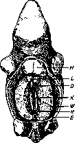
Figure 1. Wolffian body of a 19 mm long pig embryo (according to Corning): H - heart; L - lungs; D - intestine (cut off); K - sex gland; W - Wolffian body with Wolffian duct; R - root of the mesentery; E - hindgut (cut off).
Figure 2. Diagram of the development of the excretory system: M - myotome; W - Wolffian duct; UT - funnel of the Wolffian body; U - tubule of the Wolffian body; C - body cavity.
Figure 3. Diagram of the development of the excretory system: M - myotome; W - Wolffian duct; UT - funnel of the Wolffian body; U - tubule of the Wolffian body; C - body cavity.
Figure 4. Diagram of the development of the excretory system: M - myotome; W - Wolffian duct; U - tubule of the Wolffian body; UT - funnel of the Wolffian body; G - glomerulus; C - body cavity.
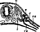
Figure 5. Tubule of the Wolffian body of a 10.2 mm human embryo (semi-schematic): UG - Wolffian duct; UK - tubule of the Wolffian body; PB - parietal layer of the glomerulus; VB - visceral layer; G - glomerulus; VA - afferent vessel; VE - efferent vessel.
Figure 5. Tubule of the Wolffian body of a 10.2 mm human embryo (semi-schematic): UG - Wolffian duct; UK - tubule of the Wolffian body; PB - parietal layer of the glomerulus; VB - visceral layer; G - glomerulus; VA - afferent vessel; VE - efferent vessel.
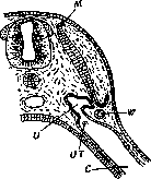
Figure 5. Tubule of the Wolffian body of a 10.2 mm human embryo (semi-schematic): UG - Wolffian duct; UK - tubule of the Wolffian body; PB - parietal layer of the glomerulus; VB - visceral layer; G - glomerulus; VA - afferent vessel; VE - efferent vessel.
Figure 5. Tubule of the Wolffian body of a 10.2 mm human embryo (semi-schematic): UG - Wolffian duct; UK - tubule of the Wolffian body; PB - parietal layer of the glomerulus; VB - visceral layer; G - glomerulus; VA - afferent vessel; VE - efferent vessel.
Figure 5. Tubule of the Wolffian body of a 10.2 mm human embryo (semi-schematic): UG - Wolffian duct; UK - tubule of the Wolffian body; PB - parietal layer of the glomerulus; VB - visceral layer; G - glomerulus; VA - afferent vessel; VE - efferent vessel.
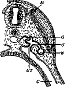
Figure 5. Tubule of the Wolffian body of a 10.2 mm human embryo (semi-schematic): UG - Wolffian duct; UK - tubule of the Wolffian body; PB - parietal layer of the glomerulus; VB - visceral layer; G - glomerulus; VA - afferent vessel; VE - efferent vessel.
Figure 5. Tubule of the Wolffian body of a 10.2 mm human embryo (semi-schematic): UG - Wolffian duct; UK - tubule of the Wolffian body; PB - parietal layer of the glomerulus; VB - visceral layer; G - glomerulus; VA - afferent vessel; VE - efferent vessel.
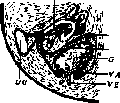
Figure 5. Tubule of the Wolffian body of a 10.2 mm human embryo (semi-schematic): UG - Wolffian duct; UK - tubule of the Wolffian body; PB - parietal layer of the glomerulus; VB - visceral layer; G - glomerulus; VA - afferent vessel; VE - efferent vessel.
Figure 5. Tubule of the Wolffian body of a 10.2 mm human embryo (semi-schematic): UG - Wolffian duct; UK - tubule of the Wolffian body; PB - parietal layer of the glomerulus; VB - visceral layer; G - glomerulus; VA - afferent vessel; VE - efferent vessel.
Related articles
Mentioned in
Cite this page
“Wolffian Body.” Soviet Medical Encyclopedia. English translation of Bolshaya Meditsinskaya Entsiklopediya, 1st ed. (Moscow, 1928–1936), ed. N. A. Semashko. https://sovietmedicalencyclopedia.pages.dev/article/wolffian-body/