Retroperitoneal Space
Historical document, translated for reference. It reflects medical knowledge of the 1920s–30s and is not medical advice.
Summary
This article describes the anatomical structure of the retroperitoneal space, detailing the complex arrangement of fasciae and connective tissue layers that house the retroperitoneal organs. It explains the relationships between the endoabdominal fascia, the retroperitoneal fascia, and the renal capsules, providing a detailed topographical overview as understood in the early 20th century.
Encyclopedia article (1928–1936)
RETROPERITONEAL SPACE (cavum, s. spatium retroperitonaeale) represents the dorsal section of the abdominal cavity (cavum abdominis) and is located between its posterior wall and the posterior parietal layer of the peritoneum. Its upper boundaries are determined by the place where the peritoneum transitions onto the liver, stomach, and spleen, and its lower boundary is at the level of the promontory. A cavity as such does not exist, because this entire space is filled with adipose and loose connective tissue, in which the so-called "retroperitoneal organs" (organa extra saccum peritonaei) are embedded, and which is distributed in several layers separated by fascial sheets. The outermost of these, lining the posterior and lateral muscular walls of the retroperitoneal space from the inside, bears the name fascia endoabdominalis (Fig. 1). It appears as a complex formation and consists of: 1) fascia transversalis, lining the lateral walls up to the level of the outer edge of the quadratus lumborum muscle; 2) fascia quadrata, which is a direct continuation of the previous one onto the quadratus lumborum; and 3) fascia psoatis and iliaca—a continuation of the fascia quadrata onto the muscles of the same name; these sheets from both sides meet in front of the spine. Upward, the endoabdominal fascia extends to the diaphragm, where it receives the name fascia diaphragmatica; downward, it transitions into the fascia pelvis. The next (more deeply located) fascial sheet, called fascia retroperitonaealis Langenbecki, is a derivative of a thin layer of subperitoneal adipose tissue, which is absent in some places (on the diaphragm, where the peritoneum lies against the endoabdominal fascia) and in other places acquires the characteristic features of a fascia. As such, it begins from the sides at the level of the peritoneal fold at the place where the latter transitions from the outer wall of the abdomen to the posterior one (at the outer edges of the ascending and descending colon) and reaches the midline, weaving into the sheaths of the vessels located in front of the spine (aorta, inferior vena cava, and renal vessels). Throughout this extent, the retroperitoneal fascia passes behind the ascending (descending) colon, kidneys, and ureters and attaches—in places quite firmly—to the fascia quadrata and fascia psoatis. Upward, it reaches the diaphragm,
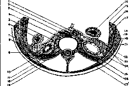
Figure 1. Cross-section through the kidney region (schematic): 1-v. cava; 2-crus med. diaphragm.; 3-aorta and its sheath; 4 and 22-fasc. Toldti; 5-ureter; 6-colon desc.; 7-paracolon; 8 and 16-textus cellulosus retroperiton.; 9-paranephron; 10-fasc. psoatis; 11-m. psoas; 12-aponeur. m. transversi; 13-fasc. lumbo-dors.; 14-colon ascend.; 15-periton.; 17-fasc. transversa; 18-m. transversus; 19-m. obl. int.; 20-m. obl. ext.; 21-fasc. superfic.; 23-fasc. praerenalis; 24-fasc. retroperiton.; 25-fasc. quadrata; 26-m. quadratus; 27-m. latissim. dorsi. (From Stromberg.)
having previously joined with the fascia praerenalis, and is lost in the adipose tissue located between the liver and the diaphragm (Stromberg, Shevkunenko). Downward, being located above the iliac vessels, it extends to the promontory and merges with the adipose tissue of the small pelvis. Between the fascia endoabdominalis and the fascia retroperitonaealis lies a layer of adipose tissue (textus cellulosus retroperitonaealis), which is not equally developed in different sections. In the region of the quadratus lumborum muscle, the connection of these sheets is intimate, but starting from the place where the psoas muscle merges with the iliacus muscle, the connection of the fascia retroperitonaealis of this area with the fascia endoabdominalis (fascia iliaca) becomes looser, the adipose layer is more pronounced here, and along the posterior-lateral walls of the abdomen (especially in the region of the iliac fossa) it reaches its greatest development. In the direction of the small pelvis cavity, there is less adipose tissue, especially at the place where the peritoneum folds over the inner edge of the psoas muscle, and it gradually transitions into the adipose tissue of the small pelvis. Anterior to the outer edge of the quadratus lumborum or psoas muscle, the adipose layer in the form of tela subserosa extends to the linea alba. Upward, gradually thinning, the textus cellulosus penetrates to the diaphragm, with the upper section of this layer known as the massa adiposa pararenalis (Gerota). The fascia retroperitonaealis thus divides the entire retroperitoneal space into two sections: the posterior—textus cellulosus retroperitonaealis, in which the aorta, inferior vena cava, thoracic duct, and lymph nodes are embedded, and the anterior, in which the "retroperitoneal organs" with their surrounding adipose tissue are located. Figure 2. Sagittal section through the region of the right kidney and pelvis (schematic): 1-pleura; 2-XI rib; 3-fasc. lumbo-dorsal.; 4-XII rib; 5-aponeurosis m. transversi; 6-arcus lumbo-cost. lat.; 7-m. quadrat. lumb.; 8-fasc. quadrata; 9-m. erector trunci; 10-fasc. retroperiton.; 11-crista os. ilii; 12-fasc. iliaca; 13-m. iliac. int.; 14-fascia surrounding vasa iliac. com.; 15-pleura diaphr.; 16-diaphr.; 17-fasc. diaphr.; 18-hepar; 19-gl. supraren.; 20-duplicatura fasc. praeren.; 21-ren dex.; 22-periton.; 23-fasc. praerenal.; 24-fasc. retroperitonaealis; 25-fasc. praeuret.; 26-appendix. (From Stromberg.)
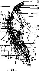
The section of the fascia passing behind the kidney and the adipose tissue surrounding it is called the fascia retrorenalis, s. fascia renalis posterior (Zuckerkandl, Gerota). At the outer edge of the kidney, corresponding to its convexity, along an arcuate line, a thin plate separates from the fascia retroperitonaealis (Fig. 2), which passes in front of the kidney, weaves along its inner edge into the sheaths of the aorta, vena cava, and renal vessels, and is called the fascia praerenalis, s. fascia renalis ant. Upward, it reaches the diaphragm, where it merges with the fascia retrorenalis, which usually occurs at the lower edge of the space devoid of peritoneum—along the posterior edge of the liver. Downward, both sheets also merge below the lower pole of the kidney, corresponding to its convexity.
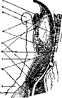
Figure 3. Sagittal section through the region of the left kidney and pelvis (schematic): 1-pancreas; 2-flex. coli sin.; 3-arcus lumbo-cost. lat.; 4-paranephron (capsula adiposa); 5-paracolon; 6-textus cellulosus retroperiton.; 7-colon descend.; 8-paraureterium; 9-flex. sigmoid.; 10-mesosigmoid. (From Stromberg.)
Thus, the kidney is enclosed in a fascial capsule—capsula renis externa, s. fascia renalis (Gerota). There is no complete closure of its sheets at the lower pole of the kidney, because, corresponding to the ureter, it extends downward in the form of a sheath—a sleeve surrounding the ureter. The fascia praerenalis along the ureter bears the name fascia praeureterica and, just like the first, splits off from the fascia retroperitonaealis at some distance from the ureter; inward, it weaves into the sheath of the large vessels of the abdominal cavity, below the bifurcation of which it transitions into the fascia of the same name on the other side, and in the small pelvis, it is lost in the adipose tissue. The fascia praerenalis and fascia praeureterica are reinforced by the fascia Toldti (Fig. 2), which represents a degenerated mesoperitoneal layer of connective tissue. The capsula renis externa is filled with adipose tissue surrounding the kidney, which bears the name capsula adiposa renis, s. paranephron. Continuously extending downward along the course of the ureter and surrounding it, it reaches the urinary bladder and, being located between the fascia praeureterica in front and the fascia retroperitonaealis behind, has received the name paraureterium. Since below the bifurcation of the aorta the fascia praeureterica of one side...
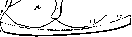
fascia endoabdominalis, peritoneum, fascia retroperitonealis, fascia praerenalis, paracolon, paranephron, textus cellulosus retroperitonealis. Mil Pnc. 4. Cross-section at the level of the lower anterior edge of the III lumbar vertebra: 1-musculus obliquus externus; 2-musculus obliquus internus; 3-musculus transversus; 4-sheath of musculus rectus; 5-ligamentum teres; 6-peritoneum; 7-colon descendens; 8-XI rib; 9-fascia transversalis; 10-XII rib; 11-site of cannula insertion into fascia transversalis; 12-fascia superficialis; 13-fascia lumbo-dorsalis; 14-aponeurosis musculi transversi; 15-fascia quadrata; 16-fascia psoatis; 17-sheath of musculus psoatis; 18-ureter; 19-sheath of musculus quadratus lumborum; 20-sheath of musculus erector trunci; 21-site of cannula insertion into fascia retroperitonealis; 22-jejunum; 23 and 24-duodenum; 25-colon ascendens; 26-crus mediale diaphragmatis; 27-vena cava; 28-aorta; 29-liver. (From Stromberg.) ...side passes into that of the other, then in the lesser pelvis the right and left paraureterium connect and reach the urinary bladder as a common mass, separating, however, by special laminae from the vas deferens located above the ureter and the prevesical cellular tissue (Stromberg, Kupriyanov). Between the fascia praerenalis-praeureterica posteriorly and the peritoneum and colon ascendens-descendens anteriorly, there is also located fatty tissue-paracolon. More precisely, posteriorly this space is bounded by the fascia retroperitonealis-praerenalis-praeureterica (fig. 3), reinforced in the medial sections by the fascia of Toldt; anteriorly it is bounded by the parietal peritoneum of the posterior wall from the point of reflection to the outer edge of the colon ascendens-descendens and a section of fascia of Toldt, not fused with the fascia praerenalis, connected here with the parietal peritoneum, representing a differentiated anterior leaf of the mesocolon; laterally-by the site of connection of the peritoneum with the fascia retroperitonealis (figure 4), medially-by the radix mesenterii; superiorly-by the root of the mesocolon transversum, with the right paracolon passing into the retroduodenal and retropancreatic masses; inferiorly-by the cecum on the right and the site of transition of the colon descendens into the S-Romanum on the left. Between the posterior wall of the colon ascendens-descendens anteriorly and the fascia of Toldt posteriorly along the extent of these intestinal sections, there is also embedded fatty tissue-retrocolon; however, in view of the weak development of the fascia of Toldt here, both spaces: paracolon and retrocolon are practically considered as one-paracolon. The right and left paracolon are separated by a sagittal partition, composed of structures located along the left edge of the aorta: superiorly-bridges from the posterior surface of the duodenum to the sheath of the aorta and vena cava, then sympathetic plexuses, lymph nodes, remnants of paraganglia, vasa mesenterica inferiora, vasa haemorrhoidalia, vasa spermatica; inferiorly-a bridge between the peritoneum and the ligamenta lutea of the spine at the promontorium, sometimes-mesosigmoideum. The location of the partition more to the left of the midline causes the right paracolon to be larger than the left (Shevkunenko, Bobrov). The location, shape, and boundaries of the paracolonic masses change according to the various forms of the position of the large intestines in accordance with the shape of the abdomen, conditioned by irregularities of its development (mobility, incomplete rotation of the intestine or absence of rotation, dystopia, sinistro-dextropositio, etc.). An idea of the limiting boundaries of the fatty cellular masses is provided by injections of liquid suspensions (gypsum, zinc chloride with dyes, gelatin, India ink, oil mixtures, etc.) into the corresponding layers. Upon injection into the textus cellulosus, the mass spreads superiorly to the diaphragm, inferiorly-into the lesser pelvis to the urinary bladder and rectum, penetrating here more often from the right from the iliac region, from where it also reaches the internal opening of the canalis femoralis; anteriorly-to the linea alba (tela subserosa); through the ligamentum hepato-duodenale it penetrates to the porta hepatis and sometimes gets between the leaves of the mesocolon transversum. Upon injection into the paranephron and paraureterium, the mass spreads over the upper pole of the kidney and the adrenal gland, with the latter being displaced anteriorly by the fluid accumulating behind it and separated from the paranephron by a lamina of fascia praerenalis; it fills the entire capsula renis externa and along the ureter, gradually narrowing, reaches the level of its flexure and then spreads along the cellular tissue to the urinary bladder. Upon injection into the paracolon on the right-the mass reaches the flexura hepatica and duodenum; inferiorly-to the level of the fundus of the cecum or sometimes to the linea innominata; laterally-to the site of connection of the fascia retroperitonealis with the peritoneum at the site of the latter's reflection ("Umschlagsfalte"); medially-to the sagittal partition along the left edge of the aorta. Upon injection into the paracolon on the left-superiorly the mass reaches the flexura lienalis and the level of the pancreas, inferiorly-along the root of the mesosigmoideum and further along the lateral and sacral wall of the lesser pelvis it merges with the mesorectum and reaches the urinary bladder; the lateral and medial boundaries-are the same as on the right. A connection between the retroperitoneal cellular tissue and the extrapleural tissue has been established. However, the penetration of injection masses, resp. purulent accumulations, from the retroperitoneal space into the extrapleural space does not occur, apparently due to the fact that there is sufficient space in the retroperitoneal cellular tissue for the spread of accumulations within it, whereas spread toward the diaphragm is hindered by the presence of relatively dense bridges between the peritoneum and the fascia endoabdominalis. The penetration of masses in the opposite direction-from the extrapleural space into the retroperitoneal-is possible and occurs through the hiatus lumbo-costalis diaphragmatis-a slit-like gap above the XII rib above the arcus Halleri, covered only by the fascia endoabdominalis, which is loosened here and is the weakest spot in the diaphragm. This area almost corresponds in its position to the upper pole of the kidney, separated from the pleura only by loose connective tissue laminae-fascia retroperitonealis and fascia endoabdominalis (Stromberg). The circulatory system of all layers of cellular tissue is in close connection; fascial partitions-frontal and sagittal-do not hinder the anastomosing of vessels of different layers. 7 groups of blood vessels are described. 1. Renal group: primarily this includes the arteriae perforantes internae (from the arteriae renis interlobares), perforating from the inside outward the superficial layer of the kidney cortex and entering the renal capsule; the fatty capsule is nourished by branches departing from the renal arteries, forming an anastomotic network along the anterior and posterior surface of the kidney and connecting with the arteria suprarenalis inferior and arteriae capsulae adiposae from the spermatic and lumbar groups. The veins of the fatty capsule anastomose with the veins of the renal parenchyma. Some of them, having started in the fatty capsule, penetrate into the substance of the kidney (venulae stellatae), others accompany the arteriae perforantes internae and flow into the veins of the fatty capsule.-2. Mesenteric group: arteria mesenterica superior and arteria mesenterica inferior, supplying the colon ascendens and colon descendens, passing under the parietal peritoneum, also supply the cellular masses located corresponding to these intestinal sections. The same applies to the venae mesentericae superiores et inferiores. From the paracolonic layers, vascular branches penetrate into the paranephron and from there into the textus cellulosus. In 75% there are well-defined anastomoses between the vena colica dextra and vena spermatica (vena porto-spermatica dextra), vena colica sinistra and vena renalis (vena porto-renalis sinistra), vena colica dextra and vena suprarenalis dextra (see Portal vein).-3. Adrenal group: branches of the arteria suprarenalis inferior (from the arteria renalis) pass along the anterior surface of the fatty capsule and anastomose with the arteries of the renal and spermatic systems; arteria suprarenalis media (from the aorta) gives branches to the internal and posterior surfaces of the paranephron, anastomosing with the arteries of the spermatic and lumbar groups; arteria suprarenalis superior (from the arteria diaphragmatica inferior) gives branches to the superior-posterior section. The veins flow into the vena suprarenalis, vena spermatica and arcus venosus perirenalis.-4. Spermatic group: at the level of the lower pole of the kidney, a branch departs from the arteria spermatica, ascending along the lateral edge of the kidney and anastomosing with the arteria suprarenalis and with other (see above) vessels of the fatty capsule. Corresponding to the course of the artery is the vein (arcus venosus perirenalis), anastomosing with the venous plexus of the ureter.-5. Lumbar group: arteriae lumbales I, II and III penetrate with their branches into the textus cellulosus, paranephron and anastomose with the arteries of the paracolon; correspondingly, veins from all cellular layers flow into the venae lumbales I and II.-6. Diaphragmatic group.-7. Aortic. Both of them are composed of inconstant branches of the arteria diaphragmatica inferior and the aorta.-The veins of the entire retroperitoneal cellular tissue belong to three systems: vena cava inferior, vena cava superior and vena portae due to the anastomoses indicated above, as well as the anastomoses of the vena azygos with the vena cava inferior and hemiazygos with the vena renalis sinistra and furthermore-the constant anastomosis vena azygos-venae oesophageae-venae cardiacae and vena coronaria ventriculi-vena portae. The lymph vessels of all sections of the retroperitoneal cellular tissue are in close connection. The textus cellulosus is connected primarily with the nodes located along the aorta; partly into these same nodes, partly into the renal ones, flow vessels from the genitalia.
The paranephron and paraureterium have two lymphatic networks: one is located superficially in the adipose capsule under the peritoneum and drains into the nodes near the kidney; the other is in the fibrous capsule and is connected with the lymphatic vessels of the cortex. The common vessels of both networks drain into groups of nodes (lumbar lymph nodes) located at the bifurcation of the large vessels, on the right side predominantly at the site where the renal vein enters the inferior vena cava; in this case, the glands of the right and left sides communicate via small trunks passing in front of and behind the aorta (Stahr). In addition, Krymov described 3 lymph nodes embedded in the adipose capsule: one in front of and slightly above the middle of the kidney (encountered rarely) and two behind (at the upper and lower poles; the upper one more often). The lymphatic pathways of the paracolon drain predominantly into the lumbar lymphatic plexus, with communications existing on the right side between the vessels of the vermiform appendix, the cecum, the ascending colon, and the anterior section of the paracolon, paranephron, and hepatoduodenal ligament. The retroperitoneal space contains the kidneys, adrenal glands, and ureters; the abdominal aorta and the inferior vena cava with its roots (Fig. 5); the lumbar part of the sympathetic trunk, lumbar lymph nodes, and the cisterna chyli. Pathology. Diseases of the retroperitoneal space concern the organs located within it and inflammatory-purulent processes of its connective tissue. Tumors, in addition to those originating from the kidney, adrenal gland, and pancreas, develop from the lymph nodes (tuberculosis, leukemia, pseudoleukemia). Among benign tumors, retroperitoneal lipomas are encountered, sometimes reaching very significant sizes—"soft tumors" that give a picture of false fluctuation, which can lead to an erroneous diagnosis of ascites (Raug). Among malignant ones, sarcomas and carcinomas are observed, both primary and metastatic (more often from testicular tumors). Primary sarcomas (more often in women) have as their starting point the retroperitoneal lymph nodes, the renal capsule, the connective tissue near the spine, and very rarely the trunk of the sympathetic nerve (proper neurocytomas). The source of a retroperitoneal tumor can also be the remnants of the primary kidney—the Wolffian body (see). In the retroperitoneal connective tissue (more often in the paranephron), echinococcal cysts are observed, reaching large sizes; in this case, the sacs of the parasites (delicate fibrous membrane) are easily torn, which causes the ease of seeding of the retroperitoneal space. Recognition
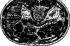
Figure 5. Horizontal section through the abdomen at the level of the 1st lumbar vertebra: 1-descending part of the duodenum; 2-pleural cavity; 3-inferior vena cava; 4-crura of the diaphragm and abdominal aorta; 5-spleen. (According to Cunningham.)
of retroperitoneal tumors, especially those reaching large sizes, from intraperitoneal ones presents significant difficulties. Given the low mobility of retroperitoneal tumors in general, their recognition is sometimes facilitated by the following technique: with one hand placed from the side of the lumbar region, one attempts to impart a rotational movement to the tumor, then the other hand, examining the tumor through the abdominal wall from the front, catches this movement; this sign is inconstant. Likewise, the displacement of the hepatic or splenic flexures of the colon in retroperitoneal tumors cannot be considered a constant and reliable sign, since even under normal conditions the flexures, especially the hepatic one, can be positioned relatively low. Retroperitoneal tumors usually grow in the direction of least resistance, i.e., toward the peritoneal cavity, which causes the difficulties in distinguishing them from tumors of intraperitoneal organs; in this case, due to the compression of intraperitoneal organs, phenomena from the side of the latter may be noted. A clinical diagnosis is often possible only by exclusion, while laboratory methods of examination, functional tests, cystoscopy, etc., and X-ray examination (contrast masses and inflation of the intestine, pneumoperitoneum, pneumoren) provide essential services in differential diagnosis. Even greater difficulties arise when determining the nature of the tumor. Some help in this regard is provided by taking into account the general condition of the patient (e.g., tuberculosis of other organs in tuberculosis of retroperitoneal nodes, cachexia and metastases in malignant neoplasms) and special laboratory studies, for example, examination of white blood cells, the complement fixation reaction (Ghedini-Weinberg), the intradermal anaphylaxis reaction (Casoni) in echinococcosis, etc. However, it should be noted that under all these conditions, the diagnosis is often established only during the operation. Recently, Hesse drew attention to the fact that in tumors originating from the lumbar sympathetic trunk, there is an important diagnostic sign, expressed in an increase in the temperature of the limb on the affected side by 4-8°. Inflammatory diseases of the veins of the retroperitoneal connective tissue are either the result of the spread of a purulent process from the neighborhood, or are caused by the introduction of an embolus from a purulently disintegrating thrombus, or by spread per continuitatem. A certain peculiarity of the process compared with inflammatory diseases of veins in general is explained by the abundance of anastomoses and the length of the azygos and hemiazygos veins, as a result of which the sources can be processes in the retroperitoneal space, as well as in the cavity of the middle ear, the mastoid process, the cerebral sinuses (Napalkov's case), and the pelvic connective tissue. In addition, in view of the presence of anastomoses of the system of the vena cava and the portal vein, it is possible for purulent emboli to be carried into the latter and from there—the occurrence of pylephlebitis, liver abscesses, etc. (see Portal vein). Inflammatory-purulent processes of the connective tissue, in accordance with anatomical data, are considered as paranephritis (see), paracolitis, and retroperitonitis (in the retroperitoneal connective tissue). In accordance with the anatomical isolation of these sections of the retroperitoneal connective tissue, inflammatory-purulent processes in them also remain delimited for some time, as long as the fascial layers are not yet involved in the process. With the violation of the integrity of the fascial sheets, pus finds access to the posterior section of the retroperitoneal space—into the retroperitoneal space. In this layer of connective tissue, purulent processes develop, having as their sources diseases of various organs of the retroperitoneal space, the small pelvis, and the parapleural connective tissue (through the lumbocostal hiatus), known by various names: retroperitonitis, retroperitoneal phlegmon, retroperitoneal abscess. By the latter, one should understand the actual accumulation of pus, whereas retroperitonitis represents a widespread inflammatory-purulent process in the connective tissue. To this name (depending on the starting point of the disease) is added a definition establishing the etiological moment: retroperitonitis ex paranephritide, ex paratyphlitide, etc., or retrorenal phlegmon, retrocecal, etc. To provide an idea of the relationship of the connective tissue spaces among themselves and with neighboring organs, Stromberg provides the following illustrative table. A. Paracolic connective tissue (paracolon) B. Pararenal connective tissue (paranephron) Paraureteral connective tissue (paraureterium) 4.
4- Connective tissue of the broad ligaments
Pararectal tissue. C. Retroperitoneal tissue (Textus cellulosus retroperitonaealis). Subdiaphragmatic space (cavum hypophrenicum). Cavum hypophrenicum proprium (above lig. coronarium hep.). Cavum phreno-lumbale (below lig. coronarium h.). Regio retrorenalis. Regio retrocolica. Regio retrocaecalis. Regio inguinalis. Regio lumbalis. Regio fossae iliacae. Paravesical tissue (paracyston). Pararectal tissue (pararectum). Parauterine tissue (parametrium). Accordingly, the classification of suppurative processes of the retroperitoneal tissue is presented in the following form: A. Paracolitis, abscessus paracolicus (paracolon). B. Perinephritis, absc. perirenalis (capsula propria renis). C. Paranephritis, absc. pararenalis (paranephron). D. Periureteritis, absc. periuretericus (adventitia ureteris). E. Paraureteritis, absc. parauretericus (paraureterium). F. Retroperitonitis, phlegmona retroperitonaealis, absc. retroperitonaealis (textus cellulosus retroperitonaealis): 1. Abscessus hypophrenicus, s. subphrenicus. a) Absc. hypophrenicus superior, s. retrohepaticus (cavum hypophrenicum proprium). b) Absc. hypophrenicus inf., s. phreno-lumbalis (cavum phreno-lumbale). 2. e paranephritide, s. retrorenalis. 3. e paraureteritide, s. retroureterica. 4. e paracolitide, s. retrocolica. 5. e paratyphlitide, s. retrocaecalis. 8. phlegmona inguinalis, absc. inguinalis. U. Paracystitis, absc. paravesicalis (paracyston). H. Parametritis, absc. pelvi-cellulitis, absc. para-uterinalis (parametrium). I. Periproctitis, absc. perirectalis (fasc. propria recti). K. Paraproctitis, absc. pararectalis, s. pelvi-rectalis (pararectum). Infection can penetrate the retroperitoneal space primarily and secondarily. Primary infection is introduced into the retroperitoneal space by the instrument causing the wound (trauma); secondary infection is possible via two paths: metastatic and per continuitatem. The metastatic path is hematogenous and lymphogenous infection. The former is conditioned by the presence of well-developed connections of the venous system of all three sections of the tissue among themselves and with distant systems (anastomoses of the cava-portae and azygo-renalis are especially pronounced). The sources in this case are furuncles, panaritiums, eczema, angina, etc., whereby the infection can be direct (e.g., metastasis directly into the paranephron) or first an intraorgan (e.g., intrarenal) abscess forms, which then ruptures into the tissue surrounding the organ. Lymphogenous infection is possible from the kidneys and ureters, intestine, liver, organs of the small pelvis, urethra, and likewise from the thoracic cavity (Küttner, Stromberg). The spread of processes to the retroperitoneal tissue per continuitatem is noted following diseases of the kidneys and ureters (pyelonephritis, nephritis, stones of the kidneys and ureters), extra- and intraperitoneal organs (osteomyelitis of the ribs, diseases of the spine, psoitis, perforations of the ascending and descending colon, periduodenitis, perityphlitis, appendicitis, purulent lymphadenitis, abscesses of the liver, pancreas, cholelithiasis), organs of the thoracic cavity (pleurisy, empyema, lung abscesses), and organs of the small pelvis. Lit.: Bobrov V., Topographic-anatomical studies of the human duodenum and the retroperitoneal tissue in its vicinity, Proceedings of the Voronezh State University, vol. II, Voronezh, 1925; Volkov M. and Delitsin S., Pathogenesis of mobile kidney, SPB, 1897; Inflammatory diseases of the retroperitoneal tissue, XX Congress of Russian Surgeons, M., 1929; Stromberg G., Anatomical-clinical study of the retroperitoneal tissue and suppurative processes in it, diss., SPB, 1909 (lit.); Shevkunenko V., On the retroperitoneal tissue and fasciae, New Surgical Archive, vol. XVI, book 2, 1928; Gerota D., Beitrage zur Kenntnis des Befestigungsapparates der Niere, Archiv f. Anatomie und Entwicklungsgeschichte, Anatomische Abteilung, 1895.
Related articles
Mentioned in
Cite this page
“Retroperitoneal Space.” Soviet Medical Encyclopedia. English translation of Bolshaya Meditsinskaya Entsiklopediya, 1st ed. (Moscow, 1928–1936), ed. N. A. Semashko. https://sovietmedicalencyclopedia.pages.dev/article/retroperitoneal-space/