Hymen
Historical document, translated for reference. It reflects medical knowledge of the 1920s–30s and is not medical advice.
Summary
This article describes the anatomy, embryological development, and morphological variations of the hymen. It details the different types of hymenal structures, such as annular, semilunar, and labial forms, and discusses their histological characteristics.
Encyclopedia article (1928–1936)
HYMEN (hymen), a fold of the mucous membrane at the entrance to the vagina, separating the latter from the vulva. It lies (especially in children and towards old age) deep in the genital slit. If the thighs are spread wide and the labia majora and minora are separated, the hymen becomes taut and is visible in the depth of the vestibule. On the sides, it borders on the labia minora, below with the fossa navicularis, and above with the external opening of the urethra, above which the clitoris protrudes (see figure in Vol. III, p. 42). Usually, the hymen resembles a membrane that partially closes the vaginal lumen, narrowing the entrance to it, but often it only seems to terminate the vagina, being a continuation of its mucosa. Then its appearance will resemble a cuff, rising somewhat above the level of the vaginal vestibule. Externally, the hymen begins directly from the surface of the vestibule, the mucosa of which imperceptibly transitions onto the hymen; occasionally, transitional folds are encountered here. Internally, the mucosa of the hymen is a continuation of the vaginal mucosa. Its folds, which are always sharply defined, also continue onto the hymen, often branching at the transition. This leads to a certain asymmetry and irregularity. The hymen originates from the Müllerian tubercle, i.e., the place of connection of the distal end of the Müllerian ducts, which form the vagina, with the urogenital canal. In embryos with a body length of 7 cm, the epithelium of the vagina (Müllerian ducts) already emerges onto the surface of the urogenital canal, or sinus urogenitalis, and in an embryo of 7-9 cm, it begins to multiply intensively over the Müllerian tubercle and, accumulating, produces an expansion that compresses the Müllerian tubercle, causing the latter to turn into a disc covered from the inside by the vaginal epithelium and from the outside by the epithelium of the sinus urogenitalis, between which lies a layer of mesenchyme. This disc is the future hymen; its lumen remains filled for a long time; only in fetuses of 20-22 cm does the vaginal cavity connect with the outside world, and consequently, the morphological formation of the hymen is completed. The hymen is absent only in some congenital malformations.
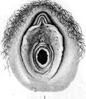
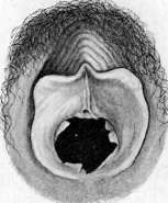
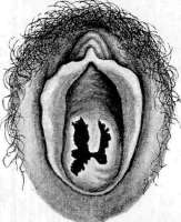
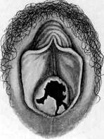
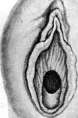
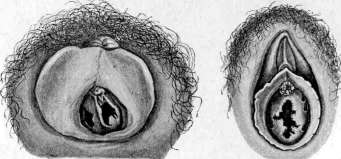
Figure 1. Annular hymen. Figure 2. Carunculae myrtiformes after childbirth. Figure 3. Hymen with a rudimentary septum. Figure 4. Hymen defloratus; a series of tears extending to the base of the hymen. Figure 5. Semilunar hymen. Figure 6. Hymen septus; openings of unequal size. Figure 7. Hymen fimbriatus.
To the article Hymen. The structure of the hymen is fundamentally identical to the structure of the folds of the vaginal mucosa. On the outer and inner surface, the hymen is covered by a stratified squamous epithelium with papillae. Between the epithelium is located a layer of connective tissue with elastic fibers, in which vessels pass. It is sometimes delicate and thin, sometimes thick and fleshy, sometimes containing much fibrous tissue. A pronounced hymen exists only in humans, but in many mammalian animals (monkeys, elephants, horses, donkeys, cows, goats, pigs), a clear narrowing at the transition of the vagina into the sinus urogenitalis is noticeable (Hunterian Museum in London), and in some, there is also a fold here running horizontally (Nagel). Hymens are extremely diverse. If one takes their external form, three main types can be outlined. Annular, or h. annularis, when the hymen forms a fold-ring, more or less uniform along the entire circumference of the vagina; inside the ring is a hymenal opening of various sizes and shapes, leading into the vagina (see figure 1). Semilunar, or h. semilunaris, when the hymen is well developed in the lower-posterior part of the entrance, gradually becomes smaller, and fades away in the direction upward and forward. The fold forming it sometimes exactly reproduces a crescent. If the height of such a hymen is the same everywhere, it resembles a horseshoe. A semilunar hymen always leaves the upper-anterior part of the vaginal entrance free. This is an eccentrically lying opening of the hymen (see figure 5). Labial, or h. labialis, when the hymen is expressed on the right and left, and in the lower-posterior and upper-anterior sections is little noticeable or in the upper-anterior even absent. Then, as it were, two lips and a slit-like opening between them are visible. Occasionally, the labial form gives the impression of a third pair of labia. The three mentioned types are valuable for classification. In life, transitional forms are more common. Among them, the semilunar type dominates, followed by the annular, and as a rarity - the labial; the latter is more frequent in children (Brouardel, Thoinot). Besides the external form, hymens differ in thickness, density, extent, size and shape of the opening, and properties of the free edge. The thickness and density of the hymen are extremely unequal. There are thin and delicate hymens, but more often they are more or less thick and fleshy, usually soft, less often dense, sometimes even seemingly tendinous. The extent of the hymen and the hymenal opening is determined by direct measurement. Sometimes the opening is small, barely allowing a thin probe to pass; but congenital wide openings are encountered - up to 10 mm and more; this usually happens with low hymens and cuff-like ones. The free edge of the hymen shows the greatest variety. When examining the hymen, it is usually stretched and forms an even surface; in some, it nevertheless retains folding; the edge also remains folded; then the hymen resembles the beginning of a drawstring bag with folds (or even praeputium penis). Such an appearance is often had by a cuff-shaped hymen. The edge of the hymen, bounding the opening, is often (but far from always) smooth and even. Occasionally it is covered with more or less delicate fringing, h. fimbriatus (see figure 7). Sometimes individual small depressions are encountered along the edge, which give it a serrated appearance, h. denticulatus. In a number of cases, the marginal notches go deep, whereby they occasionally reach the very base and divide the hymen into separate lobules, of which there may be 3, 4, or more - h. lobatus. Cases are known where the hymen has not one, but several openings - 3, 4, or more - h. cribriformis. Occasionally, a bridge passes through the hymenal opening: from top to bottom, less often obliquely or even across; a hymen with two openings is obtained, usually called h. septus (see figure 6). A hymen is encountered from the free edge of which a thin process extends (see figure 3). Finally, there is a hymen without an opening - h. imperforatus, which coincides with the concept of atresia vaginae hymenalis (see Gynatresia). The described appearance and properties of the hymen exist with widely spread thighs and separated labia. In the usual position of a woman - vertical or horizontal - the hymen, like the vagina, will be in a collapsed state. If the hymen is of a semilunar form, then, collapsing, it, especially in children or with a relatively large extent, gives a fold looking forward, which resembles the keel of a boat. A trace of such a fold is sometimes noticeable even on the flattened hymen, and then it is as if a continuation of the raphe perinei. An annular hymen often folds into a cone. If it is voluminous, the cone may protrude into the genital slit. With a labial hymen, both its parts either connect at an angle, forming some semblance of a roof, or simply lie on top of each other. The size of the hymenal opening with spread thighs and separated labia is not something final and constant. It is known that the hymen can stretch, causing its opening to increase.
The extensibility of the hymen depends on its structure. A thin, delicate hymen does not stretch much; it tears easily and quickly. A dense, fibrous hymen also stretches poorly (the latter type is sometimes an insurmountable obstacle to sexual intercourse). With age, the elasticity and extensibility of the hymen also decrease. But fleshy, soft hymens, especially low or cuff-like ones, stretch easily and strongly (e.g., during a routine examination, the hymenal opening seems to be 8 or 6 mm in diameter, but if one tries to insert a finger, it often passes into the vagina with almost no resistance; sometimes, with an intact hymen, it is possible to insert two fingers, a vaginal speculum, etc.). Digital examination must be performed with great caution, especially in virgins. A finger can be inserted only if it passes freely and if there are no fresh injuries; otherwise, one can tear the hymen or enlarge existing tears of the hymen (Maschka, Salles, and others). The first coitus of a woman violates her so-called "virginity" and damages the hymen. Therefore, the beginning of sexual life and the violation of the integrity of the hymen in women as a rule coincide. True, besides sexual intercourse, the hymen can be damaged in other ways. One should also mention the changes in the hymen during some local diseases (noma, diphtheria, smallpox), in which, in cases of deep necrosis with subsequent development of a scar, nothing may remain of the hymen. During coitus, the appearance of abrasions of the hymen is observed, sometimes hemorrhage into its thickness, and, most importantly, more or less deep tears. Abrasions and hemorrhages pass quickly, disappearing without a trace. As for tears, it is extremely rare to note the healing of wound surfaces, and consequently, the restoration of the original appearance of the hymen. Usually, a tear heals in such a way that the epithelium of the hymen at the border with the tear begins to proliferate, passes onto the tear, and gradually covers it along its entire length. Surfaces covered with epithelium can no longer fuse, and in place of the intact hymen, a series of lobules or flaps is obtained. A hymen with tears is called h. defloratus (see figure 4), and the act itself that caused the tears, whether it be coitus or something else, is called defloration (defloratio). Tears of the hymen begin from the free edge. Depending on the properties of the hymen, the relationship with the male penis, and the intensity of the sexual act, there may be one, two, or more tears. They are located in different ways. With a semilunar hymen, there are usually two tears, one on each side, so that 3 flaps are formed; an annular hymen gives two or more tears, but there may be only one; with a labial one, the bridges usually tear, especially the lower one, while the lateral parts more often remain intact. The depth of the tears is also unequal: often the tears reach the base. These will be "complete" tears ("incomplete" tears are those that capture only a part of the hymen). Injuries to the hymen, especially tears, are accompanied by pain and bleeding. Is the hymen always torn during coitus? A negative answer must be given to this. A thin and delicate hymen and a thicker one, forming a semilunar or annular septum at the entrance to the vagina with a relatively small opening, and especially with an opening that does not yield well to stretching, are damaged relatively easily—the sexual act is accompanied by their tearing. It is otherwise with a low, fleshy, soft hymen with folds and generally easily stretching; in such cases, a woman can have sexual intercourse often and for a long time, and the hymen will remain intact and undamaged. The latter is not so rarely observed in life. Forensic medical casuistry knows examples of 14-year-old girls who had multiple sexual intercourses and kept the hymen intact; an undamaged hymen has been described many times in prostitutes, in primiparas, etc. With such a hymen, the first sexual intercourse does not cause pain or blood. All this emphasizes the importance of the property of the hymen to stretch. The normal cycle of changes in the hymen in women does not end with defloration. The further and final stage depends on the onset of motherhood. The birth act has the main significance here. It causes sharp stretching and tears in the birth canal. The entrance to the vagina and the hymen also stretch, are compressed, and torn. After childbirth, when involution ends, only its remnants are visible in place of the hymen; these are the so-called carunculae myrtiformes, i.e., myrtiform papillae, located along the circumference of the vaginal entrance singly with more or less wide gaps (in the place of tears; see figure 2). Thus, the hymen, its properties, and condition represent a valuable and extremely important sign for characterizing the sexual life of a woman. It can be considered a general rule: 1) Hymen intacta testifies that the woman has not had sexual intercourse; 2) Hymen deflorata, on the contrary, establishes that sexual life has already begun; 3) the presence of carunculae myrtiformes in place of the hymen says that the woman has been a mother. The cited provisions concern the norm. Of course, one must take into account all the diversity in the structure and properties of the hymen and the frequent deviations resulting from this, but at the same time, it should be remembered that these are precisely deviations and they can be with a certain type of hymen or with a certain impact on it.
M. Raisky. Pathology of the Hymen. In the overwhelming majority of cases, the hymen is traumatized during the first normal act of intercourse (defloratio). Sometimes, if the first coitus is performed roughly and violently (especially if the man is in a state of intoxication), the rupture of the hymen can have the character of a serious injury with a breach in the integrity of the mucosa of the vestibule or even the perineum and be accompanied by significant bleeding. With such ruptures of the hymen, in the opinion of Veit, one must always keep in mind that the injury can occur not only during the insertion of the penis, but also due to manipulations with a finger or some object in cases of sexual neurasthenia or sexual perversions. Physiological trauma to the hymen does not require special treatment; usually, ruptures heal on their own in 3-5 days. It is necessary to recommend, for the purpose of hygiene during this period, washing the external genitalia, the vaginal entrance, and the perineum with warm water several times a day (especially after urination and defecation) and to prohibit sexual intercourse. Attempts at sexual intercourse in the presence of unhealed wounds are very painful and often reflect very painfully on the woman's psyche, causing a feeling of fear of the sexual act and even disgust for it. Significant injuries to the hymen sometimes require more serious intervention, such as tamponing or even suturing of bleeding vessels. More often, cases are encountered where the hymen, due to the peculiarities of its structure, is an obstacle to the insertion of the penis or to the discharge of the natural secretions of the female genital apparatus (menstrual blood). Correct coitus is impossible with an imperforate hymen (hymen imperforatus s. atresia hymenalis). In this case, menstrual blood accumulates in the vagina, uterus, and tubes (see Gynatresia). Therapy consists of incision or, to avoid re-occlusion, excision of the hymen. With various kinds of anomalies of the hymen, the insertion of the penis may prove impossible, and attempts at coitus extremely painful. If, despite the impossibility of normal coitus, pregnancy still occurs (coitus ante portas), which is not observed so rarely, then the hymen may serve during childbirth as an obstacle to the crowning of the presenting part. The hymen, especially in elderly women, can be so dense and unyielding that the performance of the sexual act becomes impossible. Therapy consists of incising the hymen in a posterior direction, at least in two places. In the case of a septate hymen, it is necessary to incise the septum. If the septum is an obstacle to the birth of the fetus, it must be cut between two ligatures during pregnancy or during labor. In the case of rigidity of the hymen, simple incision of it is usually insufficient, as a result of which excision of a crescent-shaped section from its posterior part down to the base is recommended. The mucosa of the vestibule is sutured to the mucosa of the vagina with interrupted sutures. When deciding the question of the significance of one or another feature of the hymen as a cause of the impossibility of the sexual act and when choosing therapy, it is necessary first of all to take into account the husband's health in the sense of potentia coeundi, since the obstacle on the part of the hymen may be only apparent and depend on insufficient erection of the penis. In these cases, treatment of the woman cannot yield a positive result. As for other diseases of the hymen (inflammation, neoplasms), they do not occur in isolation and usually proceed together with diseases of the vestibule or vagina (see Vagina). It should be mentioned, however, that cystic formations are sometimes found in the hymen in newborns and adults. They are usually small. Kuntzsch described a case of a cyst of the hymen the size of a plum. The etiology of congenital cysts is unclear. Gruzdev admits the possibility of the origin of some hymenal cysts from the remnants of the Gartner's duct (Wolffian duct), which in individual cases were found in the thickness of the hymen. Cystic formations in the hymen in adult women are considered by Veit to be retention cysts of the sebaceous glands. E. Shvartsman. The Hymen in Forensic Medicine. Recognition of ruptures of the hymen is very important in forensic medical cases. They must be distinguished from natural notches (fissures) of the hymen, which are quite common. A congenital notch is a matter of nature, while a rupture is the result of trauma and, as a rule, from the insertion of a penis or finger (in children), usually associated with violence. Often the entire outcome of a case depends on the examination. If the rupture occurred recently and healing has not finished, then diagnosis is not difficult. Bleeding, or more often reddened and swollen edges, accurately establish the location of the rupture and its origin from trauma. But if healing has occurred (and it can finish in 3-5 days) and enough time has passed, then the question of what the depressions present in the hymen depend on becomes difficult, and sometimes even insoluble. Usually, one is guided by the following signs: 1) congenital notches are symmetrical; 2) they have an edge of uniform thickness, the same at the beginning of the notch and in the depth; 3) their surface in color, density, and uniformity of the epithelial cover is identical to the surrounding mucosa, without a trace of a scar. It can also be added that congenital notches are often located in such a way that they correspond to the folds in the vaginal mucosa. The cited signs help to sort things out, but not always, since the position of ruptures can fully correspond to the position of congenital notches, and the surface of ruptures, as lying in the mucosa, generally quickly acquires the properties of congenital notches. Ruptures are better determined when they involve at least slightly the vaginal mucosa. The question of the integrity of the hymen is of significant interest from the point of view of forensic medicine, since cases of violent encroachment by a man on the sexual inviolability of a woman are always possible. And if one can still sometimes doubt that a completely healthy adult woman under normal conditions could easily be a victim of sexual violence, then there is no reason to doubt the possibility of such in relation to girls and adolescent girls with all the severe consequences of this (in the sense of gross sexual, physical, and psychic trauma, and often also infection with venereal diseases). On the other hand, cases are always possible where an adult woman (and sometimes an adolescent) will more or less sincerely consider herself a victim (sometimes indeed not realizing that the sexual act occurred not without the participation of her conscious or subconscious sphere), and sometimes intentionally pass herself off as a victim in the hope of material benefits and compensation. All these cases force the expert physician (forensic medical expert or gynecologist) quite often to give a precise, exhaustive answer to a specific question of the court about the presence or absence of signs of virginity. Experience shows that it is not always easy for an expert physician to give the answer required of him; in individual cases, the resolution of the task turns out to be extremely difficult and always responsible, since in such cases the examination, in essence, alone decides the fate of the accused. The precise establishment of objective data on which the forensic medical examination is based when establishing the fact of virginity (or, conversely, establishing the fact of defloration) can sometimes, due to the variety of variants in the properties of the hymen, encounter great difficulties and doubts. For their resolution, a joint consultation of several forensic medical experts and gynecologists possessing great special experience in this regard may be required. The examination itself must be performed extremely carefully, with the observance of a number of rules and conditions (proper lighting, exposure of the external genitalia with the help of assistants, sometimes under anesthesia, proper instruments, etc.), but this complex setting and, in essence, humiliating procedure for the woman may in individual cases still not lead to the desired goal in the sense of an absolutely precise answer to the questions of the court.
Related articles
Mentioned in
Cite this page
“Hymen.” Soviet Medical Encyclopedia. English translation of Bolshaya Meditsinskaya Entsiklopediya, 1st ed. (Moscow, 1928–1936), ed. N. A. Semashko. https://sovietmedicalencyclopedia.pages.dev/article/hymen/