Fetus
Historical document, translated for reference. It reflects medical knowledge of the 1920s–30s and is not medical advice.
Summary
This article from the 1st edition of the Great Medical Encyclopedia (1928–1936) details the intrauterine development, anatomical growth, and physiological changes of the human fetus from the second month until birth. It discusses month-by-month measurements of length and weight, viability at different gestational ages, and the influence of maternal nutrition and external factors on fetal development.
Encyclopedia article (1928–1936)
Fetus (fetus), the intrauterine infant from the end of the 2nd month until birth. His and others distinguish two periods in human intrauterine development: the embryonic (see Embryo) up to the end of the 2nd month, and the fetal from the 3ኅ month until birth. Ballantyne, on the other hand, divides intrauterine life into 3 periods: 1) germinalis—the moment of fertilization, 2) embryonalis—until the beginning of the differentiation of the blastodermic layers into tissues and organs, and 3) fetalis—in the literal sense of the word. From this moment, human-specific features begin to manifest in the structure of the fetus, distinguishing it from other mammals. By this same time, the initial stage of embryo development (see) ends, and the stage of intrauterine development and maturation of the fetus begins, lasting until its birth. This maturation, if considered month by month, is manifested by a whole series of anatomical and functional changes, on the basis of which the age of the fetus is determined in obstetrics, and in connection with it, the duration of pregnancy itself.
Length, weight, and development of the fetus. The following changes deserve the most attention in practical terms. By the end of the 2nd lunar month, the length of the fetus reaches 3-4 cm, the branchial arches disappear, and the division of the limbs into their constituent parts becomes noticeable: on the arms—the shoulder, forearm, hand; on the legs—the lower leg, thigh, and foot. By the end of the 3rd month, the length of the fetus = 7-9 cm, weight 20-30 g, the fingers and toes begin to be distinguished (nail rudiments are noticeable) and the external


genitals differentiate (the formation of the scrotum, labia begins; the clitoris cannot yet be distinguished from the penis); the first ossification centers appear and are clearly visible (Fig. 1). By the end of the 4th month, the length of the fetus = 10-17 cm, weight—about 120 g, facial formation begins, hair appears, and the skull ossifies. The sex of the fetus is differentiated. The skin of the fetus up to 4 months is smooth and so thin and transparent due to the complete absence of subcutaneous fat that Figure 1. Fetus at the end of the 3rd month.
pregnancy (according to Dietrich). muscles and blood vessels shine through (Fig. 2). In a fetus born at the 4th month, weak limb movements and respiratory movements are noticeable (when placed in warm water). In the 5th month, fat deposition and the formation of subcutaneous tissue are observed, starting from the neck and buttocks. Lanugo hair appears on the more developed skin, and it begins to be covered with a cheesy vernix (see Vernix caseosa). The eyelids separate. Nails grow on the fingers and toes. Bile secretion begins, as a result of which the intestinal contents acquire a dark Figure 2. Four-month-old fetus (according to Dietrich). color (meconium). The length of the fetus by the end of the 5th month reaches 18-27 cm, and the weight on average is 280 g. Movements intensify and begin to be felt by the pregnant woman; likewise, the heartbeat is auscultated at this time. By the end of the 6th month, the fetus, with a length of 28 to 33 cm, reaches 670-680 g in weight. The amount of fat in the subcutaneous tissue and primordial vernix on the skin increases. A born fetus of this age can breathe and move its limbs for some time, but soon dies. By the end of the 7th month, the fetus reaches 35-38 cm in length with a weight of 1,100-1,200 g. The subcutaneous tissue is still weakly developed; the skin is red and has a wrinkled, senile appearance. At this age, the fetus rarely survives; in most cases, it is non-viable and dies from weakness. In the 8th month, the length of the fetus = 39-42 cm with an average weight of 1,800 g. The skin becomes smooth due to the sufficient development of subcutaneous tissue, covered with abundant down. The fetus at this term is viable, but requires special attention and care (see Prematurity). By the end of the 9th month, the length of the fetus reaches 43-45-47 cm, and the weight 2,200-2,500 g. The fetus has a well-nourished appearance, the face becomes smooth, and the redness of the skin disappears. A 10-month-old fetus is considered completely mature and full-term (see Newborn, Full-term and Premature). The absolute maximum increase in the length of the fetus, according to Bedu, occurs between 4½-5½ solar months; other authors consider the 5th or 6th lunar months. The relative growth of the fetus during pregnancy is characterized by unevenness 46. (Fig. 3). The ovum by the end of the 1st month reaches 8 mm in diameter, i.e., becomes 200 times larger than its original size; during the 2nd month, the ovum increases only 2 times,
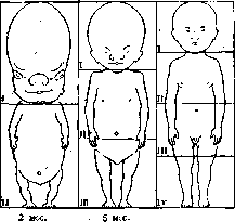
10 m:s. Fig. 3. Proportions of the head and torso at various stages of pregnancy (according to Stratz). while the fetus during the 10th month increases by 0.06. Axel Key establishes the position that the increase in the fetus extends first to its growth and then to its weight. The relative profit in weight, if considered by months, is also distinguished by its unevenness (Figs. 4-7). The intrauterine development of the fetus, its growth, and weight depend on a whole series of external factors—social conditions, the quantity and quality of nutritional material coming from the mother's blood through the placenta, and the ability of the fetal cells to assimilate and synthesize them. The question of the mother's dietary regimen, which most favorably affects the growth and weight of the fetus, has been little studied. Bartels also points to a possible relationship between fetal development and maternal nutrition. On the other hand, the years of the imperialist war, accompanied by significantly reduced nutrition and undernutrition in many countries, show that with insufficient maternal nutrition, fetal development can proceed normally, and the necessary nutritional materials are received by the fetus from the mobilized reserves of the maternal organism. Thus, the study of the weight and length of newborns of the so-called "wartime" showed that "war nutrition" had little effect on them in this regard and that their weight and length were not lower than those of pre-war newborns (Troitskaya, Dietrich, Tschirch, Richter, and others); only a decrease in the number of large fetuses is noted, while underdevelopment and malformations during these years were much rarer (Selitsky). Only some authors noted a decrease in weight and an insignificant decrease in length or even a drop in average weight (Lichkus, Valitsky). The amount of nutrients entering the fetal organism and the degree of their assimilation is closely connected with the conditions of the initial stage of implantation and growth of the fertilized ovum, the quality of the germ cells themselves, the site of attachment of the egg cell in the uterus, the state of the uterine mucosa, the architecture of the placental blood vessels, and a number of other factors. In women whose sexual maturity occurs earlier, larger fetuses are born. According to Wernich, in primiparas with the usual time of onset of sexual maturity, the fetal weight is 3,174 g; in primigravidas with the onset of menstruation in their 19th year—3,138 g; in women with the onset
45
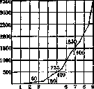
Figure 5. Fetal weight curve by months, compiled by Vignes according to Bedu's data. Figure 4. Embryo weight curve in the first weeks of pregnancy (according to Zangemeister). of menstruation earlier, the weight is 3,215 g. The amount of amniotic fluid also has a certain significance for the development and weight of the fetus. The weight of the fetus is also influenced by the weight and height of the parents (especially the mother), constitutional characteristics, race, nationality, and sex (male fetuses are heavier than female ones).
Figure 7. Fetal weight curve in primipara (according to
fetal weight curve in primipara (according to Vignes).
vignes). The average weight of boys is usually greater than that of girls. Nikolaev, based on his material, found an average weight of 3,289 g for Russian newborn boys and 3,260 g for girls. Figurnov and Shmerling arrive at the same conclusions. Alphant lists 383 boys and 161 girls among fetuses born with a weight exceeding 4,000 g. Fetuses of multiparous women weigh more than fetuses of primiparas. According to Tarnier's observations, boys in primigravidae had an average weight of 3,164 g, and girls 3,101 g; in multigravidae: boys 3,372 g, girls 3,120 g. Alphant cites statistics of 537 cases of large fetuses weighing 4,000–5,000 g; of these, 457 were born to multiparous women and 80 to primiparas, which indicates the dependence of the fetus's weight on the number of pregnancies. In women with a large number of births (9–10), fetuses begin to be born with a lesser weight, which Vignes associates with the unfavorable influence of short intervals between births. The age of the mother also has an effect on the weight of the fetus. According to Haeckel, the weight of the fetus increases in proportion to the mother's age until she reaches 29 years, after which there is a decrease in the fetus's weight. The height of the parents can also have an influence on the size of the fetus; the greater the height of the latter, the larger the fetuses are born. In the case of Gilford, a fetus born of giant parents weighed 10,700 g and had a length of 76 cm. All other things being equal, social, hygienic, economic, and domestic conditions and the external environment surrounding the pregnant woman have a significant influence on the weight and length of the fetus. In women belonging to more affluent social groups, employed in less hazardous industries and in better living conditions, fetuses are of greater weight (Pismenny, Figurnov, Nikolaev, etc.). These factors acquire particular importance in the last months of pregnancy. Rest before childbirth and the release of the pregnant woman from work in accordance with the Soviet Labor Code are factors aimed at protecting not only the health of working women, but also at the antenatal protection of the health of the fetus. Pinard establishes that thanks to pre-natal rest, the fetus gains in weight on average by 220 g compared to those fetuses that were born of mothers who did not have this rest. The works of Merlino-Ferrari and Figurnov established the dependence of the fetus's weight gain on the number of days of the mother's rest before childbirth; the longer the prenatal leave, the more significant the dimensions of the newborn. The weight and length of the fetus are different in various nationalities. According to Geller-Levitova, Georgian children are born with a greater weight than children of other nationalities. The average weight of newborns in Belarusians is 3,370 g, in Jews 3,223 g. Dietrich's data on the average weight of newborns in various countries also show that the greatest weight (3,523–3,527 g) is noted in Sweden, Norway, and America. In some cases, a large fetus weighing more than 4,000 g is the result of one or another disease of the mother. A large fetus in the presence of a large placenta and a large amount of amniotic fluid, according to some (Wallich, Bruhinsholz), is a sign on the basis of which congenital syphilis of the parents can be suspected. Nine children weighing more than 4,500 g, according to Sellet's statistics, had signs of congenital syphilis. When the mother suffers from diabetes, fetuses (according to M. Bary) can also be large, with large fat deposition. In a number of maternal diseases (both chronic and directly related to the pregnancy itself) that impair the nutrition of the fetus, the latter, although they may be quite mature, are distinguished by low weight and leanness. Such fetuses Pinard calls enfants-araignees (spider-children). On the other hand, in some cases, fetuses are born with a very small weight, but quite viable from a completely healthy mother; they were studied by Rouvier and given the name pseudo-prematures (false prematurity). Chemical composition of the fetus. The embryo is built of embryonic tissue, which contains 97.54% water. At 2½ months in the fetus, water makes up 93.82%. As the fetus matures, the amount of solid constituents increases and water decreases, as the following table (Michel and Perret) shows; the decrease in the amount of water is due to the increase in solid parts, the amount of which by the end of pregnancy reaches 30.84%. At the beginning of pregnancy, the amount of protein in the body of the fetus increases rapidly, and by its end, the fat content. The content of lime and iron also increases strongly by the end of pregnancy, the latter in a mature fetus, according to Leenhardt, being 38–42% in the blood and 60% in the liver as a reserve material. Physiology of the fetus. For the fetus, the mother's organism is the external world, from which it draws everything necessary for its development and growth. By the end of the 2nd month, placental circulation begins to form in the fetus, replacing the yolk-sac and allantoic circulation (see Placenta). Oxygenated and nutrient-laden blood is collected by the capillaries of the vena umbilicalis (Figure 8), through which it goes to the fetus, and pours into the inferior vena cava partly directly through the ductus venosus Arantii, partly after passing through the vascular system of the liver. From the inferior vena cava, the blood flows into the right atrium, then through the foramen ovale present in the septum between the atria with the help of a valve protruding along the right edge of the mouth of the inferior vena cava (valvula Eustachii) is directed into the left atrium, from where it enters the left ventricle. Thus, during diastole, the left ventricle is filled predominantly with arterial blood of the inferior vena cava with a small admixture of venous blood from the poorly developed pulmonary veins, and the right ventricle with venous blood from the superior vena cava. During systole, blood from the ventricles is directed to the large vessels of the fetus; from the left ventricle through the aorta ascendens into the large vascular trunks of the upper half of the body; only an insignificant part of this blood enters the descending aorta; the contents of the right ventricle in its smaller part are poured into the poorly developed branches of the a. pulmonalis, and in the greater part through the ductus Botalli it goes into the descending part of the aorta (Figure 9). In po
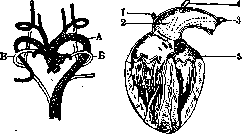
Figure 9. Heart and large vessels of the fetus: A—aorta (absence of arch); B—Botallian duct; V—arch; 1—a. subclavia sin.; 2—a. carotis; 3—a. subclavia dext.; 4—right Botallian duct; 5—initial part of truncus aortae (after Akimova-Voronkova).
Thus, the blood is strongly mixed with venous blood, part of it going to nourish the lower half of the fetus's body, and part through the umbilical artery going again for renewal in the placenta. A feature of the fetal circulation compared to the adult organism is the fact that in the fetus, arterial blood is everywhere mixed with venous blood, but to varying degrees: thus, the liver is in the most favorable conditions in this respect, since in it arterial blood is mixed only with the venous blood of the portal vein; the upper half of the fetus's body receives arterial blood with an admixture of venous blood coming from the lower half of the fetus's body and the veins of the liver and lungs; the lower half is in the worst nutritional conditions, since blood that is almost exclusively venous goes to it from the right ventricle. By virtue of these conditions, the liver and the upper half of the fetus's body are nourished much better than the lower half, and therefore develop faster. In the second half of pregnancy, this difference is smoothed out due to the fact that the Eustachian valve lags behind in its development by this time, which is why a greater mixing of the blood brought by the lower and upper venae cavae occurs in the right atrium, and the formation by this time of the valve of the oval foramen and the narrowing of the Botallo duct create conditions preventing the free passage of blood, which puts the circulation of all parts of the fetus's body in more equal conditions compared to the first half of pregnancy. The blood of the fetus differs in composition from the mother's blood by a greater number of red blood cells and platelets, a greater hemoglobin content (especially at the end of pregnancy) and other biological features. According to Karnitsky, it contains: red blood cells 6,000,000, hemoglobin 14.27%, white blood cells 18,000, of them lymphocytes 48%, neutrophils 40%, transitional forms 9%, eosinophils 2.8%. Specific gravity of blood 1.0616, of plasma 1.0285. Dry residue of blood 23.43%. Views on the amount of salts and the related question of osmotic pressure vary: some authors find the amount of salts in the fetal blood and its specific gravity to be greater, with a lower freezing point, others believe that the blood of the mother and the fetus has the same freezing point; consequently, the blood of the fetus and the mother will be isotonic. The difference in the blood of the fetus and the mother in terms of biochemical reactions is that in the serum of the fetal blood, the hemolytic substance (hemolysins and agglutinins) is expressed more weakly both in quantitative and qualitative terms. Polano establishes that the blood serum of the mother perfectly dissolves pigeon erythrocytes, while the serum of the fetus does not dissolve them. The agglutinating ability of the blood serum of the fetus and the mother is independent of each other: it can be expressed sharply in the blood of the fetus and insignificantly in the mother and vice versa (Schumacher, Muller, Schenk). In addition, the blood serum of the mother contains an agglutinating substance for the erythrocytes of the fetus. Fetal serum (experiments on cats) acts hemolytically on the mother's erythrocytes. This proves that the blood of the fetus and the blood of the mother relate to each other like the blood of two different individuals, and serves as indirect proof that very complex chemical processes take place in the placenta. As for enzymes in the blood of the fetus, they are also present, but in smaller quantities than in the mother, partly in an inactive state (zymogenic), and are used by it as needed. Shepetinskaya notes very low diastase figures in the blood of the umbilical vein—4–8 times less than in the mother's blood. In addition, the blood of the fetus contains a number of hormones produced by the endocrine glands of the fetus, as well as maternal hormones passing through the placenta, which has been proved by experiments (Mertz, Lüttge). Respiration of the fetus. The blood of the fetus that has entered the placenta from the umbilical arteries gives off carbon dioxide to the maternal blood in the capillaries of the villi and in exchange receives the oxygen necessary for the fetus. The placenta thus serves as an organ of respiration for the fetus, replacing its lungs. The transition of oxygen from the maternal blood to the vessels of the villi is proved both spectroscopically by the finding of oxyhemoglobin in the blood of the umbilical vein and by the difference in the color of the blood in the umbilical vein and umbilical arteries, as well as by the fact that a prolonged cessation of placental circulation due to certain causes leads to the death of the fetus from asphyxia. Nutrition of the fetus. During its intrauterine life, the fetus increases 800 times or more. With such colossal growth, it needs a large amount of nutrients, especially proteins. All these substances the fetus receives from the mother's blood through the placenta (see). In addition, the fetus receives water from the amniotic fluid, which is absorbed in its intestine after it swallows it. However, there are other assumptions. Kolosov believes that the process of entry of amniotic fluid into the stomach occurs (without the participation of the swallowing apparatus of the fetus) according to certain physical laws due to the pressure difference in the nasopharynx of the fetus and in the egg cavity. Kolosov calls this process intra-ovular water circulation and considers it a very important adaptation.
.
Metabolism of the fetus. The peculiarities of circulation, respiration, and nutrition in the fetus indicate that a constant exchange of substances occurs between the fetus and the mother, but the fetus, as an independent organism, also has its own metabolism. Proof of this is the characteristic temperature of the fetus, which is 0.3-0.5° higher than the temperature of the mother. In the fetus's own metabolism, assimilation processes prevail over dissimilation processes, which is evidenced by the rapid growth of the fetus, its relatively small oxygen consumption, and the small temperature difference with the mother. As a result of metabolism in the fetal organism, a whole series of degradation products is formed: CO2, water, urea, etc. For the excretion of these products of incomplete combustion, there are special devices in the form of the same chorionic villi through which all these products are excreted into the mother's blood. In addition, part of the water and urea is excreted through the fetal kidneys into the amniotic fluid. Besides the kidneys, the intestine functions in the fetus, albeit weakly. It usually contains meconium in the form of a resinous, black, odorless mass consisting of bile components, swallowed and unabsorbed parts from the amniotic fluid (hairs, epidermis scales), and mucus of the intestine itself. Meconium accumulates first in the small intestine, but as the fetus matures, it moves by means of peristalsis into the large intestine. Owing to optimal nutrition and growth, the liver of the fetus begins to produce bile from the 3rd month and contains glycogen and iron in abundant quantities. The gastric juice contains pepsin and rennet. The skin glands of the fetus begin to secrete vernix caseosa from the 5th month, mixed with epithelial cells and lanugo, which protect the skin from maceration. There is, however, a view of the vernix caseosa as a product of the activity of the amniotic epithelium. Muscle work in the fetus begins from the earliest stages of intrauterine life. The musculature is more elastic than that of an adult and is easily excitable. The most active in this regard is the heart muscle, which by the 5th month of pregnancy becomes so strong that heart tones can be auscultated with the ear through the abdominal wall of the pregnant woman. The heart rate in the fetus becomes slower as pregnancy progresses and reaches 120-140 beats per minute by the end of pregnancy. Likewise, the smooth musculature of the urinary bladder and intestines works in the fetus, as evidenced by the presence of intestinal peristalsis and the ability of the fetus to urinate. Skeletal muscles begin to contract at the 4th month and even earlier, and from the middle of the 5th month, contractions become so energetic that they are felt by the pregnant woman herself. The activity of the nervous system and especially the sense organs is poorly expressed in the fetus. Skin sensitivity appears later than motor ability. Of the other senses, taste appears first. Smell and hearing appear after birth. Sensitivity of the retina to light is detected relatively early, starting from 8 months. The intensification of fetal movements under the influence of heat applied to the abdominal wall of the pregnant woman or palpation indicates the development of skin sensitivity and skin reflexes, while the change in heart rate under the influence of carbonic acid during asphyxia indicates the work of the center regulating cardiac activity. Other parts of the nervous system, especially those governing vegetative life, also begin to function relatively early in the fetus. Internal secretion in the fetus has not yet been sufficiently studied, but a number of observations and theoretical premises based on the data we currently have in endocrinology indicate that it begins to manifest from the very early stages of life. The thyroid gland is formed by the 4 months of life and consists of follicles with a richly developed capillary network containing colloidal substance. The formation of this gland coincides precisely with that period of fetal development when its water-rich embryonic tissue begins to be replaced by tissues of a higher type -- epithelial, connective, and muscular. Thus, the very development of the embryo and the improvement of its protoplasm through the transition from a simplest form to a more complex one is in direct connection with the formation and function of the thyroid gland. Its normal development also determines the normal development of the fetus. The loss of its function causes a delay in fetal growth and various deviations in its formation. The parathyroid glands (glandulae parathyreoideae) can be detected in the 3rd week of embryonic development; they apparently possess a diverse function, normally supplementing the thyroid and sex glands. The thymus gland appears in the human embryo early and apparently exerts an influence on the growth and formation of the skeleton and sex glands, and also plays a role in nucleic metabolism. Likewise, the pituitary body as a glandular formation appears in the fetus in the 3rd week of intrauterine life. One can think that a whole series of pathological changes in the fetus, e.g., giant growth or, conversely, too small a size of full-term fetuses, is in direct dependence on one or another functional state of this very gland, as well as a number of other glands that have an influence on growth. Experiments by Drennan and Carlson on pregnant dogs show that the pancreas performs a diastatic function in the fetus, since by extirpating the pancreas in pregnant animals, they did not see a disruption of sugar metabolism, whereas after the birth of the fetus, glycosuria was observed in the mother. The sex glands arise in the embryo at the very early stage of development. Initially, they have an indifferent character, but in an embryo of 13 mm, signs of the male sex are already revealed, and the transformation of the indifferent sex gland into an ovary occurs somewhat later, when the embryo reaches 18-20 mm. The influence of the sex glands on the entire organism of the fetus is enormous. The hormone of these glands begins to manifest its participation in the structure of the embryo very early, and the first result of its influence must be considered the formation of secondary sexual characteristics -- external genitalia (3 months), and subsequently the correct development of the fetus with all the features of this development for each sex. Membranes. During its development in the uterus, the fetus is surrounded by three membranes, of which one -- of maternal origin -- is the decidual membrane, which is a modified mucous membrane of the uterus, and two -- of fetal origin -- are the amnion and chorion. All these membranes together with the placenta and amniotic fluid bear the name of the fetal egg, wherein the amnion, or water membrane, covers this egg from the inside and passes to the umbilical cord, while the chorion, or villous membrane, at the site of implantation of the egg in the uterus enters into the composition of the placenta, and in the remaining part forms the outer membrane of the egg in the form of a thin layer, which merges with the decidua at the 5th month of pregnancy. Between the fetus and the amnion is the amniotic fluid, in which the fetus is located. Position of the fetus. The fetus can assume various positions relative to the long axis of the uterus, which is of great practical importance in obstetrics, since incorrect positions complicate labor and can pose a danger to both the mother and the fetus. The normal and most frequently encountered is the so-called longitudinal position of the fetus, in which the longitudinal axis of the fetus coincides with the longitudinal axis of the uterus and when large parts can be felt above the pelvic inlet and in the fundus of the uterus (see Obstetric examination). In approximately 1% of all births, the fetus occupies a transverse or oblique position, in which the longitudinal axis of the fetus does not coincide with the longitudinal axis of the uterus, but is in a transverse or oblique position relative to it, in the vast majority of cases requiring artificial correction for a favorable termination of labor. Position (Situs). In the longitudinal position, the fetus can have its back turned to one side or the other relative to the uterine walls. If the back is turned to the left, then it is called the first position; if to the right, the second position. In some cases, the back may be turned directly forward or backward -- middle position. In transverse and oblique positions, the position is determined by the relation of the fetal head to one or the other side of the uterus: head to the left -- first position, to the right -- second. During its development in the uterus, the fetus tries to adapt to the size and shape of the uterine cavity, which is achieved not only by position, but also by the arrangement of its individual parts, and is called the attitude of the fetus. The normal attitude of the fetus is when the back is slightly arched, the head is flexed forward so that the chin is pressed against the chest, the legs are flexed at the hip joints and pressed against the abdomen, and the arms are crossed on the chest. Thanks to such an attitude, the fetus assumes an ovoid shape corresponding to the shape of the uterine cavity and occupies the smallest space in it. In obstetrics, to determine the position of the fetus, large and small parts are distinguished, referring the head and buttocks to the large parts, and the extremities to the small parts. Both are determined by palpation: the head and buttocks in the form of dense and rounded parts, the back in the form of a flat surface, and the small parts in the form of individual tubers. For the normal development of the fetus, the amount of amniotic fluid is of importance, with a normal amount of which (about 1 liter) the fetus, being in a suspended state, receives favorable conditions for the development and growth of all its parts.
While an excessive increase in amniotic fluid (see Hydramnios) has no direct effect on the fetus, its decrease (oligohydramnios, oligamnion) can lead to various malformations of the fetus due to creating purely mechanical conditions that hinder the growth of the fetus and the proper formation of its individual parts. The thread-like bands formed in the process between the walls of the amniotic sac, known as Simon's bands, can lead, through wrapping around and compression of the umbilical cord, to the death of the fetus as a result of asphyxia, as well as, in some cases, to the complete detachment of the fetus or to so-called spontaneous amputations of the arms, legs, or fingers of the fetus.
A. Sofoterov. Overterm Fetus. Prolonged pregnancy is spoken of in cases where pregnancy lasts longer than the established average norms. Practically, determining an overterm fetus in most cases is very difficult, since one has to solve an equation with many unknowns: 1) we do not know the exact time of conception, since ovulation can occur throughout the intermenstrual period; 2) in the overwhelming majority of cases, the time of fruitful sexual intercourse is unknown to us; 3) the duration of pregnancy in humans, as in animals, is not a constant value, but is subject to significant fluctuations depending on many factors (occupational and living conditions, hereditary and constitutional characteristics of the mother and father, nutrition are of the greatest importance; sex and weight of the fetus, mother's age, number of previous pregnancies, type of menstruation, etc. are of relative importance); 4) the cause of the onset of labor is unknown to us, and 5) we have no absolute signs to determine the overterm fetus. Thus, the question of an overterm fetus can be solved only by the totality of all data. Some authors consider an overterm fetus to be an extremely rare phenomenon. C. Ruge found only 4 cases of undoubted fetal post-maturity in the literature. Depending on whether the ovum was fertilized immediately after the past menstruation or before the expected one, the difference in the duration of pregnancy will be 26 days; therefore, one can speak of an overterm fetus in cases where pregnancy lasted 302 days or more without these 26 days; if, however, we obtained 302 days already after adding 26 days, then there can be no talk of post-maturity (Ruge). With such a calculation, Siegel found 2-3.3% of overterm fetuses in 125 of his own cases and 800 from the literature (only wives of men on leave during the war were taken into account, since the days of past sexual intercourse could be established for them). Jäger and Nürnberger out of 31 similar cases and Ruge out of 22 could never establish an overterm fetus; Veit found the duration of pregnancy from 302 to 351 days in 0.22%, Stavskaya in 6.3%, considering a pregnancy duration of over 290 days as an overterm fetus, Issmer in 5.8%. Large material from the State Institute of Obstetrics and Gynecology, processed using the method of variational statistics by Bogorov, shows that the average duration of pregnancy in humans is 280.9 days, or 40 weeks from the first day of the last menstruation or 286.8 days from the last day (Fig. 10). [Standard deviation (σ) ±13.9 and ±13.8; 90% lies within 3σ, i.e., 272-322 days.] The highest number of births in animals as well falls on the middle figures of the duration of pregnancy; the longer the latter is in a given animal species, the more significant the fluctuations (Teissier) (see Pregnancy—pregnancy in animals). Based on the foregoing, all cases in which pregnancy lasts more than 293 days after menstruation could be considered as an overterm fetus. Winckel proposed to refer to an overterm fetus all cases when the fetus is in the uterus for more than 41 weeks. In Russian literature, there are also isolated cases of overterm fetuses from 328 to 367 days. The shortest duration is 221-239 days; individual casuistic data speak of 210-216 days (Kehrer, Opitz, Sellheim). Social and living conditions have a great influence on the duration of pregnancy. It has been established that in women of physical labor, pregnancy is shorter than in office workers or those engaged in mental labor. Pinard as early as 1888 noted that in women who rested 6-8 weeks before childbirth, pregnancy lasted more than 280 days in 60%, while in those who worked until the last days, only in 48.2%. These observations are confirmed by other authors: the longer the maternity leave, the longer the pregnancy lasts and the larger the fetus (Belugin and Luria, Libov and Figurnov). Pismenny found among multiparous textile workers the average fetal weight before the revolution to be 3,451 g and after to be 3,526 g. There are indications that with complete rest, the duration of pregnancy is on average 20 days longer than with physical exercise. Hirsch and Kharitonov note a decrease in the duration of pregnancy in textile workers, Libov and Shmerling in female tobacco workers and tram conductors (however, the number of examined persons in these groups is small, and the conclusions require verification). Taking into account that in the USSR all women employed in production receive an 8-week maternity leave before childbirth, it is difficult to establish the influence of profession on the shortening of pregnancy; it is more correct to attribute the shortening to living conditions (heavy housework, frequent and rough coitus, etc.). Nutrition, poor and good, can equally contribute to both an overterm fetus and premature birth. The works of Rudolsky from Pashutin's laboratory showed that in starved animals, pregnancy is not interrupted ahead of time, but the amount of offspring and the sum of its weight are significantly less than usual. Lichkus and Valitsky also did not find a shortening of the gestational age in 1918, but in 1919 the number of premature infants increased by 1.5%; a similar phenomenon was noted by Kellog in silkworms, in which starvation for 2 generations increases the number of premature infants in the third. The lack of yolk in the mother's egg is of importance here. On the other hand, the slow development of the intrauterine fetus (Wachstumstendenz of Zangemeister) under poor nutrition is compensated by a longer intrauterine life. During the war and the blockade in Germany, more frequent cases of fetal post-maturity were noted. Good nutrition in obese women leading a sedentary lifestyle also contributes to the prolongation of pregnancy, apparently due to poor metabolism in the mother and reduced excitability of her neuromuscular apparatus. Constitutional and hereditary factors were noted by old authors; early termination of pregnancy and post-maturity are observed in some families through generations, as well as repeatedly in the same woman. Ciulla attaches importance to the size of the pelvis—with a wide pelvis, the duration of pregnancy is greater; however, post-maturity is also observed with a narrow pelvis, since in the latter case the presenting part does not descend into the pelvic cavity and does not irritate the lower segment of the uterus (Winge). Large women, blondes give birth later; the season of the year is not without influence—in winter pregnancy is longer, in summer shorter (in the plant and animal world, heat accelerates development). The racial factor has also been noted in animals: Cornevin observed generations of cows in which pregnancy lasted 10-11 months. It is also necessary to take into account the racial characteristics of the father: Sanger cites a case in which the duration of pregnancy in the same woman was unequal with the first and second husband. It is known to all obstetricians that even from the same husband a woman can alternately have either small, sometimes premature children, or large ones. This explains the fact that with a relatively insufficient pelvis, spontaneous births are sometimes observed, while at other times one has to resort to fetal perforation and even embryotomy due to the very large size of the fetus. Constitutional factors (A. Mayer, Rech) must include the size of the surface of the villous apparatus, which is proportional to the weight of the fetus and depends on the special property of the circulatory system of both the mother and the fetus. Sex of the Fetus. There is no consensus in the literature regarding the influence of the sex of the fetus on the duration of pregnancy. Ahlfeld does not recognize the influence of sex; Veit and Simpson, by analogy with the animal world, believe that pregnancy with boys lasts longer (cows carrying more than 286 days gave 152 males and 90 females). Idelson obtained a prolongation of the gestational age with girls by 3 days, Zangemeister by 1-2 days. According to A. Mayer, Ruge, and Nürnberger, pregnancy with boys is longer. Obviously, the divergence of authors on this issue must be explained by the fact that the sex factor is random, since upon verification on large material, the estimate of its reliability is 4 instead of 9. Fetus Weight. A correspondence between the weight and length of the fetus and the duration of pregnancy is not always observed; with the same duration of pregnancy, the fetus can weigh 3-4 kg or more. Sellheim cites a case where 214 days after the last menstruation, a woman gave birth to a child weighing 3,500 g with a length of 51 cm. A large fetus, weighing over 4,500 g, is still encountered with a duration of pregnancy of at least 288 days. The material of the State Institute of Obstetrics and Gynecology in Leningrad confirms that there is only a small direct correlation between fetal weight and the duration of pregnancy (r = +0.16 ± 0.029).
The age of the mother is of no essential significance, although in very young mothers, pregnancy may be shorter, and in older primiparae, it may be longer. Primigravidae usually give birth earlier than multiparae (276.3:279.9 days), although older authors (Ahlfeld, Horowitz) believed that pregnancy is longer in primiparae. The type of menstruation—the duration of menses and the length of the menstrual period—according to some authors, affects the duration of pregnancy. Vinay gives the following ratio: an intermenstrual interval of more than 30 days gives a pregnancy duration of 288 days, 30 days gives 281 days, 28 days gives 279 days, and less than 28 gives 269 days. Agabekov, Chernoyarova, and some foreign authors point out that women with prolonged menses give birth to larger infants. However, Schröder has already pointed out that the duration of pregnancy does not depend on the individual type of menstruation, which moreover is inconstant. Not all of the enumerated factors exert an identical influence on the over-term fetus. Each of them individually contributes to an insignificant fluctuation in one direction or the other, but when they are summed up in a single woman, the difference may reach up to 7 weeks (Nürnberger). Internal secretory glands exert an influence on the development of the fetus and possibly on the duration of pregnancy. In Switzerland, where goiter is widespread, in 50% the length of the children lags behind the norm, and in Berlin in 27%; ossification is delayed in 23% of them versus 3% in Berlin (Guggisberg, Riddle). In hyperthyroidism, the duration of pregnancy is shortened. Lehmann succeeded in shortening pregnancy by injections of thyroidin, on the basis of which Mayer regards post-term pregnancy as a dysfunction of the thyroid gland. Mandelstamm obtained an elongation of pregnancy by 2 days in a mouse by injections of corpus luteum extract. There are indications of enhanced growth in young animals upon injection of the anterior pituitary hormone (Long, Evans); likewise, the influence of the posterior pituitary on the onset of labor cannot be denied. The question of the influence of glands of internal secretion and the metabolism associated with them on the post-term fetus is in the period of study; a final conclusion cannot yet be made. The cause of the over-term fetus may be insufficient excitability of the neuromuscular apparatus, as well as weakness of the uterine muscles and insufficient development of the lower segment of the uterus (Fraenkel). Decreased excitability of the uterus is observed in its underdevelopment; infantilism can also be a cause of premature interruption of pregnancy; tumors, inflammatory processes, etc., also contribute to the early interruption of pregnancy. The diagnosis of an over-term fetus during pregnancy is based on data concerning the last menstruation, the appearance of the fetal heartbeat, the measurement of the length of the fetus and the dimensions of the head; in multiparae, also on the anamnesis. A large post-term fetus must be differentiated from multiple pregnancy, hydramnios, and transverse lie; in doubtful cases, the diagnosis is clarified by radiography (see below). Post-term development of the fetus during pregnancy does not cause complications. The latter may occur during labor if the post-term fetus is very large (see Labor). In the case of an over-term fetus in primiparae, trial labor should be recommended so that, in case of necessity, it may be terminated by Cesarean section; in multiparae with a corresponding anamnesis, Cesarean section at the onset of labor. Post-term retention of a dead fetus usually does not cause complications; sometimes, however, a deterioration in the woman's well-being and a subfebrile temperature of 37.2-37.4° are observed. In such cases, the question of terminating the post-term pregnancy may arise (see Labor). After labor, the diagnosis of a post-term fetus must be made on the basis of an analysis of all the indicated points: length and weight of the fetus, dimensions of the head (the transverse diameter of the shoulders is significantly greater than the circumference of the head), the degree of ossification of the cranial bones, the presence and size of the ossification center of the cuboid bone of the foot and the proximal ossification point of the tibia (established by radiography). A post-term fetus is difficult to distinguish from a large fetus; the duration of pregnancy from the last menstruation or from a definite sexual intercourse will help recognize a post-term fetus. Stavskaya performed a histological study of placentas in post-term pregnancies that ended in the birth of a live fetus, and gives valuable indications which, apart from biological interest, have the significance of an auxiliary diagnostic method. Mayer already pointed out that at the end of pregnancy, thrombosis of the placental vessels occurs with their subsequent obliteration. Stavskaya discovered significant degeneration and fibrous transformation (in particular, areas of calcareous degeneration), as well as villous atrophy and vascular obliteration in post-term fetuses; syncytium is absent; these changes were found even in small and medium post-term states. It must be thought that in prolonged post-term pregnancies, they are the cause of intrauterine death of the post-term fetus. A. Mayer sees the cause of fetal death in internal secretory disorders, in particular on the part of the mother's thyroid gland; other authors consider insufficient supply of Fe, Ca, and P to the fetus as the main cause. Male fetuses perish intrauterinely more often, which is explained by their more intensive growth and greater need for nutritive material. The over-term fetus is frequently the subject of judgment in judicial instances, in paternity and affiliation suits. The legislation of Western European countries establishes a maximum duration of pregnancy of 300 days (France), 302 days (Germany), 311 (England), 317 (America). Zangemeister proposes to increase the period adopted in Germany on the basis of the fact that in 4.2% of women pregnancy lasts from 320 to 340 days from the beginning of the last menstruation, and in 0.84% labor occurred 320-333 days after the established fertilizing intercourse. The old Russian code recognized 306 days. This question is resolved more correctly by the Code of the RSFSR, which does not establish a limit for the duration of pregnancy, but the question in each case is decided by the people's court with the assistance of expert testimony and witness statements. In insurance medicine, the question of post-term or pre-term development of the fetus has great practical significance, as it is closely connected with the question of "underruns" and "overruns" in maternity leave for pregnant women. If overruns burden the financial position of social insurance organs, then underruns undermine the target setting of the Mother and Child Protection organs by shortening the period of prenatal rest. Therefore, insurance physicians have particularly sharpened their attention on determining a 32-34 week pregnancy. The date of the last menstruation for determining the duration of pregnancy has only indicative significance under the condition that the woman gives correct information (pregnancy can also occur with amenorrhea). Early recognition of pregnancy and its duration, as well as a record of the first auscultation of the fetal heartbeat, can introduce a significant correction during subsequent observation. For this, it is necessary that women come to the consultation after the very first missed menstrual period. The reference points for determining a 32-34 week pregnancy are the height of the uterine fundus, the length of the child, and the size of its head. The height of the fundus is measured with a caliper or centimeter tape. In evaluating this value, it is necessary to take into account the constitution of the woman, the relation of the head to the pelvic inlet, and the size of the fetus. According to Libov and Figurnov, 3 types are encountered: 1) height of the fundus from the upper edge of the pubic symphysis—23-25 cm, abdominal circumference—80 cm, transverse diameter of the head—9 cm; 2) fundus—28 cm, abdominal circumference—90 cm, head—10 cm, stands tightly in the inlet; 3) fundus—30-32 cm, abdominal circumference—95-100 cm, head—10-11 cm. Baksht considers a fixed head to be a sufficient indicator of a 32-week pregnancy, which cannot be agreed with, since the head is sometimes fixed earlier and sometimes later. He measures the height of the fundus with a caliper and establishes the following three types: 1) height of the fundus—23 cm, transverse diameter of the head—9-11 cm, head pressed against the pelvic inlet; 2) head balloting, the same transverse diameter, height of the fundus—24-25 cm, and 3) head fixed. In breech presentation, the height of the uterine fundus at 32 weeks should be 24-25 cm, the transverse diameter of the head—10-11 cm. In transverse lies, the height of the fundus has no significance; the main thing here is the measurement of the length of the fetus and the transverse diameter of the head. At 34 weeks of pregnancy, the height of the fundus is 24-26 cm (75%), more often 25 cm (41%), transverse diameter of the head—11.5 cm (Baksht). The Skulsky formula is roughly applicable: x = (2L - 5) / 5, where L is the length of the fetus measured with a pelvimeter, multiplied by 2; 5 is subtracted from it for the difference in thickness of the uterus and abdominal wall in the lower and upper sections, and the resulting number is divided by 5, the coefficient of fetal length after 5 months. Despite the refinement of objective signs for establishing the term of a 32-34 week pregnancy, we are still far from having reached the ideal.
The accuracy of the determination undoubtedly depends on the experience and qualification of the physician, but it can be said that we will not achieve 100% agreement, since the duration of pregnancy—just like other periodic biological processes (teething, maturation, menstruation)—is not a constant value, but fluctuates not only in different women, but even in the same woman depending on various conditions.
R. Lurie. Fetus radiography is one of the valuable diagnostic methods in obstetrics. The radiogram of the fetus can serve as absolute proof of the presence of pregnancy in general, makes it possible to differentiate single and multiple pregnancies, and at the end of pregnancy gives an exact picture of the presentation of the fetus. In addition, serial images make it possible to form a clear idea of the mechanism of labor. The first attempts at radiography of the fetus date back to 1896 and belong to Varnier, who initially took pictures of an extirpated pregnant uterus and obtained a clear image of the fetus. In the same years, Davis first undertook studies on the living, but without result. Investigations in this direction were continued by a number of authors, but only in 1910 (Fabre, Barjon, Trillat) was it possible to obtain an exact image of the fetal skeleton. The success of radiography was greatly facilitated by the gradual improvement of the technique and in particular the new method proposed in 1904 by Albers-Schönberg. Subsequently, radiography of the fetus was successfully used by many authors, and studies were also directed towards obtaining images in earlier months (for the first time, images of a 5-month fetus were presented by Potocki, Delherm, and Laquerrière). With the help of radiography, the mechanism of labor began to be studied (Warnekros), characteristic signs and an X-ray syndrome of intrauterine fetal death were established (Spalding, 1922; Szendrö), and it also became possible to determine the age of the fetus. Thanks to valuable investigations in this field (especially the works of Bouchacourt, Blanche, 1924; Spalding, 1922; Barnes, Weill, Portes, Ponzio, Bermann, and others should be mentioned), radiography of the fetus has now become one of the valuable auxiliary diagnostic methods in the modern clinic. The technique of photographing the fetus in earlier stages of pregnancy continues to be a subject of study up to the very last time, and individual authors consider it possible, thanks to the improvements achieved in the technique, to obtain a clear image of the fetus already in the initial stages of ossification. Most authors, however, believe that a clear image can be obtained only from the 4th month (Bouchacourt indicates that from 4½ months the possibility of obtaining a radiographic image is the rule), while many (Reeb, Potocki, and others) advise in practical life for diagnostic purposes not to undertake radiography earlier than the 5th month. Mikeladze (1931), based on 68 exposures from 3½ months of pregnancy and later, believes that an image of the fetus can sometimes be obtained starting from the 4th month of pregnancy; after 7 months, obtaining a picture is the rule. Thus, at present, clear images of the fetus can already be obtained at 4 months of pregnancy (see separate table, pp. 483-484, Fig. 1-3 and 9). First of all, the femurs, clavicles, and ribs become visible on the radiogram, later the spine in the form of symmetrically arranged points along the midline, and finally, last of all, the skull bones. The technique of fetal radiography is quite difficult, especially in the early stages of pregnancy, due to the insignificant lime content in the fetal bones, the abundance of soft tissues, and the presence of amniotic fluid, which has a large capacity for ray absorption and secondary radiation, the respiratory movements of the mother and the movements of the fetus itself; all this combined significantly reduces the contrast of the radiogram. Accordingly, the following technical conditions are required for fetal radiography: 1) The use of compression, due to which, by pushing the amniotic fluid to the side, a flattening of the photographed object (the gestational sac) is created. 2) The use of maximally soft rays, making it possible to obtain the pattern of the fetal skeleton. When using harder rays, the fetal pattern is not obtained due to the insufficient lime content in the fetal bones. 3) The use of a very short exposure, within 1–1½ seconds, to exclude the influence of the mother's respiratory movements and the fetus's own movement. 4) The use of a high load of 100-150 mA, ensuring, with a short exposure, sufficiently intense radiation and obtaining a sufficiently sharp pattern of the fetus. 5) The use of a Bucky diaphragm, which eliminates secondary radiation and ensures maximum contrast of the image. 6) The use of high-quality and high-sensitivity films. The technique of the picture is as follows. The pregnant woman receives a mild laxative the day before and an enema on the day of the picture. Immediately before radiography, the pregnant woman urinates. Pictures can be taken in 3 different positions of the pregnant woman: 1) on the abdomen, 2) on the back, and 3) on the side. The position on the abdomen apparently gives the best pictures. In the position on the abdomen, the pregnant woman is forced to press her abdomen and symphysis as tightly as possible against the plate, which is easily achieved with the upper part of the trunk slightly raised, when the pregnant woman rests her elbows on the table. In this position, natural compression is achieved. The picture is taken at the moment of deep expiration. Equally successful pictures can be obtained in the position of the pregnant woman on her back. In this case, compression is produced by a special cylinder applied to the abdominal region between the navel and the pubis. The least successful pictures are obtained when the pregnant woman is on her side, due to the different thickness of the abdomen from the side of the navel and from the side of the sacrum; in this case, the contrast of the image in the part adjacent to the sacrum is insufficient. The picture itself, when using the radiotransverter apparatus of the Koch and Sterzel firm, is produced under the following most advantageous conditions: focal length—60 cm, diaphragm diameter of the aperture—10 cm, maximum softness of rays (3-3½ We), load—120 mA, exposure—0.8-0.9 sec. (in obese women up to 1½ sec.). Fetal radiography in the early stages of pregnancy can also be performed by Arkhangelsky's method. A light-sensitive plate is placed between two thin aluminum plates and covered with a rubber condom. Such a vaginal cassette is inserted with two fingers into the vagina and advanced into the posterior fornix. With the external hand, the uterus is shifted into a position of sharp anteversion and pressed by the cylinder against the cassette, which is supported from below by fingers inserted into the vagina. The conditions of the picture are the same as for a full-term pregnancy. The value of fetal radiography for the diagnosis of fetal position and multiple pregnancy lies in the objectivity and absolute accuracy of the method. In individual cases, the diagnosis of hydrocephalus and other forms of malformation is possible (anencephaly [Portes and others]). Cases of X-ray diagnosis of ectopic pregnancy have also been described (Knaff, 1922 and others). Fetal radiography can also be used to diagnose intrauterine fetal death. The pathognomonic signs established in 1922 by Spalding were subsequently confirmed by other authors (Kehrer and others), who believe that in radiography we have a valuable auxiliary tool for the diagnosis of an intrauterine dead fetus. Szendrö, already based on the study of images, came to the conclusion that with this complication there is also a characteristic X-ray syndrome. The most typical for an intrauterine dead fetus are the arrangement of the cranial bones, flattening of the skull and its wrinkling, elongation of the head in the direction of the occiput, and strong flexion of the spine (in the form of lordosis, kyphosis, or scoliosis). Kehrer considers the presence of flexion of the spine in the distal half or in the sacral part of the spine to be especially indicative (see separate table, Fig. 5-8 and 10-15). How relief-like the fetal pattern is obtained can be seen from the two attached radiograms of a multiple pregnancy and a 3½-4-month fetus taken according to Arkhangelsky's method (see separate table, Fig. 9). As can be seen from the attached radiogram (Fig. 2), the contours of the fetal skeleton and skull are obtained with sufficient relief even in the 4th month of pregnancy. Finally, the fetal radiogram makes it possible to directly study the mechanism of labor. Based on serial images, it turned out that the position of the fetus during pregnancy is completely unconstrained, while in the period of dilation and expulsion, the position of the fetus acquires characteristic features. In the period of dilation, the head and spine turn out to be strongly flexed and the arms tightly pressed to the trunk. After the discharge of the waters, the back of the fetus straightens out, and at the moment of a contraction, the spine straightens out completely, and the fetus takes on the form of a pyramid with the apex facing the pelvic cavity and the base toward the fundus of the uterus. The base of the pyramid is formed in cephalic presentation by the buttocks and thighs, in breech presentation by the shoulders and the head deflected to the side. In transverse presentations, it turned out that the child often lies with its back to the fundus of the uterus (radiograms 10-14). Fetal radiography, lasting only 1 second, presents no harm to the fetus.
V. Arkhangelsky. Fetal diseases are characterized by peculiarities arising from the environmental conditions and the very development of the fetus. Firstly, the fetus, isolated from the external environment by the maternal organism, can become ill either independently or through the transmission of pathogenic influence from the mother; secondly, the embryo bears enhanced functions of development, and the pathogenic agent, striking the latter, frequently causes the emergence of deformations, underdevelopment, heterotopias, and other monstrosities and anomalies, the more severe the earlier the damage occurred; thirdly, the course of diseases is under the influence of special conditions: temperature, humid environment, constant biochemical influences from the mother and the placental barrier, in some cases exerting favorable effects, and in others accelerating the death of the fetus. The causes of fetal disease are as follows. A. Damage to germ cells. I. Pathological heredity. A pathogenic germ, pre-existing in the germ cells of the paternal or maternal organisms, is transmitted to the offspring according to Mendel's laws. Those lesions that are accompanied by manifest deformations are recognized in the fetus. These include monstrosities: hemicephalia, cerebral hernias, harelip, cleft palate, clefts along the midline of the abdomen and spine, umbilicus...
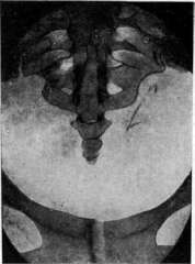
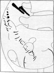
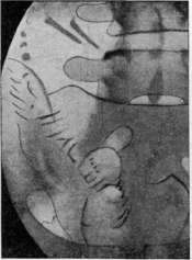
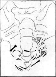
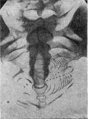
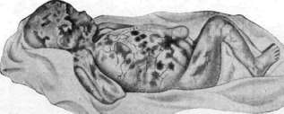
Figure 1. X-ray of a 3-month fetus. Figure 2. The same of a 4-month fetus. Figure 3. The same of a 41/2-month fetus. (Figures 1-3 after E. Bergman.) Figure 4. Skin changes in intrauterine streptococcal infection. (After Lebedev.)
Figure 5. X-ray of a dead fetus; lordosis and kyphosis of the spine. Figure 6. X-ray of a dead fetus; deformation and elongation of the skull. Figure 7. X-ray of a dead fetus; marked lordosis of the thoracic spine; displacement of the skull bones. Figure 8. Hydrocephalus. (After J. Granzow.)
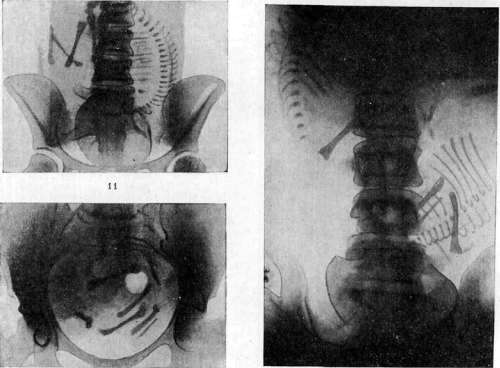
Figure 9. X-ray of a 3.5–4-month fetus. (According to Arkhangelsky.) Figures 10 and 11. Occipital presentation. Figure 12. Breech presentation. Figure 13. Transverse lie. Figures 14 and 15. Twins. (Figures 10–15 after A. Blanche.) Hernia of the umbilical cord, ectopia of the bladder, epi- and hypospadias, atresia of orifices, polydactyly, splitting of the foot and hand, congenital tumors (of the kidneys, genital sphere, etc.), naevi, etc. Other diseases, such as hemophilia, daltonism, deaf-mutism, metabolic, endocrine, and constitutional anomalies, predispositions to tumors, and a tendency to mental illnesses (epilepsy, idiocy, feeblemindedness, etc.), manifest themselves only later in extrauterine life. 2. Blastophthora. Hereditarily healthy germ elements can be affected by accidental agents in the bodies of their carriers. Such agents include: alcohol, syphilitic poison, certain industrial poisons (e.g., lead, mercury), X-rays and radium, ultraviolet rays; other intoxications (infectious, biological, and industrial), very high temperatures, trauma, a lack of certain substances (vitamins, lipoids) in nutrition, etc., may apparently act in the same way. Affected cells may perish, resulting in temporary sterility. If the damage is not so significant, the damaged elements recover, become capable of fertilization, but first produce unviable and pathological forms (e.g., Down syndrome), and then offspring that appear healthy. Thus, for example, after irradiation of the ovaries, the immediate pregnancy often (in almost 25%) ends in the death of the fetus and miscarriage, while in those born alive, pathological changes are observed in 5–20% of cases. Subsequent pregnancies prove to be more favorable. Once contracted, blastophthora can spread with a diminishing effect to 2–3 generations (alcoholism, syphilis). Recently, Rautmann described a new type of fetal disease under the name of erythroblastosis—general congenital hydrops (it consists in a significant proliferation of erythroblasts and to a certain extent resembles severe jaundice of newborns). Its immediate cause has not been clarified; some (von Gierke, 1931) classify it among degenerative changes of a germinative nature. Erythroblastosis usually has a familial character [thus, incidentally, Salomonsen recently reported on a family where 3 children were born with significant edema and erythroleukoblastosis; two of them were born dead, and the third died immediately after birth]. B. Lesions transmitted from the mother. Here we can distinguish the action of 1) pathological factors penetrating from the external environment into the mother's body, 2) biochemical influences from the mother, and 3) mechanical factors. 1. External pathogenic agents, in order to affect the fetus, must circulate in the maternal blood, from whence they can pass through the intervillous spaces and placental tissue into the fetal blood or amniotic fluid and affect the integument, mucous membranes, and internal organs of the fetus. Thus, the "barrier" significance of the placenta (retention of formed elements, neutralization of poisons, enzymatic effects) along the pathways from the mother to the fetus is far from absolute and leads only to the fact that the fetus turns out to be somewhat more resilient than the mother with respect to certain agents (chloroform, ether, CO, morphine, scopolamine, and other alkaloids). Moreover, a damaged placenta (inflammation, edema, hemorrhage, infarctions, etc.) can itself become a source of new fetal injuries. Infectious diseases appear to be the most frequent affliction of the fetus. Pathogenic microorganisms, having entered the intervillous spaces during maternal bacteremia, settle on the walls of the septa and on the outer surface of the villi, from whence they penetrate both into the uterine mucosa and into the syncytium and further deep into the interstitial tissue of the villi. The placenta can detain bacteria, but the latter, given significant invasion, nevertheless advance further (which is sometimes facilitated by placental edema) and reach the fetal vessels and the fetal surface of the placenta, from whence they infect the fetus through the blood and amniotic fluid. Such a pathway has been proven for the spirochetes of syphilis and for tubercle bacilli; apparently it is the same for acute infections. Bacterial toxins penetrate from the maternal blood into the fetal blood through the placenta (diphtheria and tubercle toxins); immune bodies produced by the mother can also be transmitted to the fetus in the same way (their transmission by a germinative pathway, i.e., from the germ cells of the parents, is denied), as well as antitoxins (e.g., of tetanus and diphtheria; Polano, Ehrlich), agglutinins (of typhoid; Chambrelent, Philippe, Yurevich), hemolysins, precipitins, opsonins, and anaphylactic reactive bodies. Intoxications. Poisonous substances in gaseous and liquid states, having entered the blood of the maternal organism, easily penetrate from the intervillous spaces by diffusion and osmosis into the fetal blood. In addition to those mentioned above, the passage into the fetal blood of very many other substances has been proven, such as: soluble salts of heavy metals (Pb, Hg, Cu, Sb, etc.), compounds of As and P, further NaI, KBr, KCN, salicylic, oxalic, benzoic acids, chloral hydrate, turpentine, camphor, naphthalene, aniline and its derivatives (aniline dyes), certain plant dyes (madder, indigo, crocus), alkaloids (quinine, strychnine, nicotine, curare, santonin), animal poisons (snake venom, cantharidin), hormones, etc. Highly toxic poisons cause fetal death and abortion in acute poisoning; chronic poisoning (e.g., in industrial production) can cause malformations in the embryonic stage of fetal development, and later various morphological and functional irregularities. Infectious toxins (syphilis) may apparently act in the same way. 2. The situation is similar with respect to toxins produced in the body of the mother herself during diseases of her organs and systems, in particular during disorders of the activity of endocrine organs and during pregnancy toxicosis. The placenta almost always suffers in this case. As a result of placental damage, the fetus easily perishes or suffers in development and is born weak, of low weight, and often prematurely. In nephritis, premature fetal death occurs in 60% of cases, in diabetes in 41%; in eclampsia, 50% of fetuses perish from asphyxia or as a result of reduced viability; premature birth takes place in acute yellow atrophy of the liver and other pregnancy toxicoses. Deficiencies in the mother's nutrition and labor regime—lack of vitamins, heavy labor, irrational use of maternity leave—can also affect fetal development. 3. Mechanical influences. The narrowness of the gestational sac, compression of the uterus from the outside or a reduction of its cavity by tumors, lack of amniotic fluid, etc., can produce fetal deformations—flattening of the head, curvature of the limbs, cause the formation of pes equino-varus, etc.; pressure can cause intrauterine bone fractures, be the cause of congenital dislocation, and so on. These deformations arise especially frequently in ectopic pregnancies and pregnancies in a rudimentary horn due to the aforementioned tightness of the underdeveloped gestational sac. A narrow pelvis and its tumors (exostoses) cause damage to the fetus during childbirth (skull indentations, fissures and fractures of bones, hemorrhages, edemas). Compressions, deformations, and even strangulations of limbs can be caused by umbilical cord entanglements (see Umbilical cord); other kinds of constrictions, disfigurements, and amputations are produced when there is a small amount of amniotic fluid by adhesive inflammatory processes between the membranes and the skin of the fetus, forming the so-called Simonart's bands (see Amniotic bands, constrictions, adhesions). The infliction of external injuries to the mother affects the placenta more than directly the fetus (hemorrhages, detachment of the placenta, etc.). Individual forms of fetal disease. A fully satisfactory classification of fetal diseases does not yet exist. The classification based on the origin of the disease, proposed by Ballantyne, deserves attention. The author divides fetal diseases into 4 groups: 1) idiopathic, 2) transmitted to the fetus from the parents (father or mother), 3) intoxications and infections transmitted from the mother, and 4) diseases resulting from traumatic influences. The cause does not determine the form, since the latter depends on the stage of fetal development in which the disease manifested itself: in the germinal stage we get not disease forms, but a predisposition to them; in the embryonic stage, diseases manifest themselves as malformations, and only in the lethal stage do they approach the forms of childhood diseases. The group of idiopathic diseases is not entirely defined and with further study of the genesis of intrauterine diseases will undoubtedly undergo significant changes. Ballantyne includes in this group: 1) general fetal dropsy (anasarca universalis), accompanied by premature closure of the foramen ovale, inflammatory processes, and cysts in the kidneys; the fetus is not adapted to extrauterine life; 2) congenital cystic elephantiasis with marked hyperplasia of the subcutaneous tissue and the formation of cysts with gelatinous or semi-solid contents;
The fetus is also non-viable; 3) elephantiasis congenita (simplex) with localized indurations in the subcutaneous tissue; a harmless and curable condition; 4) atrophia congenita panniculi adiposi, giving newborns an extremely emaciated appearance; 5) ichthyosis fetalis gravis and ichthyosis fetalis benigna (fish scale disease) with the formation of horny scales on the skin, giving the child's body a mottled appearance (f. arlequin of French authors); severe forms rapidly lead to death, mild ones may be cured; 6) naevus neuroticus congenitus - papillomatous formations located along the nerves (a variety of fish scale disease); 7) hypertrichosis congenita; 8) osteopathia foetalis (rachitis congenita, chondrodystrophia fetalis, achondroplasia) - a diverse skeletal disease manifested by developmental defects of cartilage and bone tissue and accompanied by micromelia, fractures with callus formation, often hydrocephalus, etc.; 9) inflammation of the peritoneum (peritonitis fetalis), characterized by the accumulation of fluid in the abdomen, often of significant size (serous, serous-fibrinous, or hemorrhagic effusion), sometimes with the presence of true inflammatory adhesions; gives a very poor prognosis; occasionally, so-called meconium peritonitis is observed; 10) congenital tumors should also be referred here, most often in the urogenital organs, liver, along the course of the sympathetic nervous system, and finally certain malformations. Diseases of the skeletal system deserve special mention. According to Ballantyne, they appear to be very diverse and arise in the embryonic and fetal periods. The causes of these bone system anomalies are also very diverse; they can be regarded as a consequence of improper differentiation of germ elements, disproportionate development of individual parts of the body due to impaired nutrition and blood circulation. (The most frequent "congenital anomalies" of the pelvis are rickets and so-called assimilation pelves; for details see Pelvis - contracted.) To the second group belongs primarily syphilis - the most frequent disease of the fetus, giving very diverse forms. All parts and tissues of the ovum can be affected by syphilis. Polyhydramnios or, conversely, oligohydramnios with inflammatory lesions of the membranes, formation of adhesions, etc., is often noted. The fetus itself is often affected by malformations (underdevelopment, midline clefts, atresia of orifices, malformations of the eyes, limbs, etc.). Among organ lesions, the most characteristic are enlargement of the spleen and liver, gummas, chronic interstitial inflammation of the liver; gummative nodes in the lungs, small syphilomas in the lungs, in the heart, endo- and periarteriitis, meningo-encephalitis with proliferation of connective tissue, bone lesions at the site of transition of the diaphysis into the epiphysis with degeneration of cartilage cells (Wegner's yellow line at the site of their connection with the bone) (Fig. 11), pemphigus of the palms and soles, etc. Spirochetes are [missing text] found; 3 - diaphysis. (According to Bumm.) in the organs of the fetus (especially in the liver). The fresher the parents' syphilis at the time of conception, the sharper its manifestations in the fetus and the sooner the latter perishes, so that the first pregnancies after infection end in miscarriages, followed by premature births of a dead and macerated fetus, then births of a live fetus with phenomena of syphilis, and finally children are born without visible manifestations of the disease. In primary syphilis of the mother, 78% of fetuses are born dead, 16% die in the first year of life, and only 6% survive further. Dead macerated fetuses often turn out to be impregnated with a bloody-serous fluid - fetus sanguinolentus. Along with alcoholism, which belongs to this same group, syphilis produces such severe congenital lesions as Little's disease, hydrocephalus, myatonia congenita, idiocy, hypoplasias, or later manifesting ones - Huntington's disease, Friedreich's ataxia, Thomsen's myotonia congenita, etc. Among the quite frequent complications of congenital syphilis
are intracranial hemorrhages in the first months of intrauterine life. Thus, Pigeaud (1929) notes them on average in 12% and, without denying the possibility of a mechanical factor, nevertheless believes that in the majority of cases the soil of their origin is syphilitic. The third group includes numerous infectious diseases, where the transition from mother to fetus is proven in the following forms. A. Acute contagious. 1. Measles; according to statistics (Nouvat) out of 52 cases of illness in pregnant women, 21 sick children were born, of which 33% died. 2. Smallpox; the pathway of the pathogen to the fetus has not been elucidated; it is possible that extremely small microorganisms get from the mother's blood into the placenta by adsorption like vital dyes (a kind of ultrafiltration; v. Prowazek); many cases of the birth of a fetus with smallpox pustules and scars are known; 3. Chickenpox; the pathogen in its properties is very close to the pathogen of natural smallpox; 4. Scarlet fever; the pathway of the pathogen is unknown; all the main symptoms of the disease - fever, tonsillitis, rash, peeling - were noted on the born fetus; 5. Typhoid fever; the transition of bacilli occurs apparently slowly, so that at the moment of the height of bacteremia in the mother, the bacteremia of the fetus is only beginning (Hicks, French, Kelly); therefore, French considers it advisable in suitable cases to induce premature labor to save the fetus; 6. Pneumococcal diseases (in maternal pneumonia) were observed in the fetus predominantly in the pleura and lungs, and then in the pericardium, peritoneum, meninges, etc.; 7. Anthrax; bacteria were found in the placenta, umbilical cord, fetal blood, its liver, spleen, kidneys, and other organs (Koch, Schmorl, Hoffmann, Marchand, et al.); 8. Erysipelas; the pathway of transition of streptococci has not been studied; in several described cases (Runge, Lebedev) it manifested as lymphocytic infiltration of the skin with desquamation of the epithelium; 9. Influenza; a case (Esch) with lesions of the lungs and pleura has been described; 10. Glanders; 11. Relapsing fever. The last two diseases are described in a limited number of cases. Acute infections easily cause termination of pregnancy; the fetus in this case is often born dead, the cause of which is not only its illness [see separate table (p. 483-484), fig. 4], but also simply overheating; those who survived the infection and were born at term may show the consequences of the disease in the form of developmental irregularities, debility, lowered resistance, etc. B. Chronic infections and parasitic diseases. 12. Tuberculosis; infection occurs both through the placenta from a sick mother and by direct spread of bacilli from the uterus (with its local lesion) into the placenta and amniotic fluid; in 21 cases of verified placental tuberculosis, Koch's bacilli and tuberculous changes were found in the fetus only 11 times. Indubitable cases of intrauterine infection are described in the literature only a few dozen, but on the other hand, in tuberculous pregnant women, lesions of the fetus without specific changes are often noted - malformations, general nutritional disorder, general instability of the organism and predisposition to tuberculosis; 13. Leprosy is transmitted only in an insignificant percentage of cases (Reshetillo and some others) due to the extremely long incubation period (several years); it may not manifest on the intrauterine fetus; nevertheless, leprosy bacilli were found in the placenta, umbilical cord, and fetal blood; 14. Rabies (Krokiewicz, Konradi, et al.); 15. Malaria; several dozen cases have been described; schizonts of plasmodia have the ability to penetrate into the placenta and into the blood; 16. Cholera; vibrios were found in the meconium of the fetus; 17. Helminthemia (larvae of ascarids, echinococcus, Ankylostoma, Schistosomum japonicum, etc.). Traumatic injuries - see above; about malformations - see below. Treatment is carried out by treating pregnant women; exceptional importance must be attached to prophylaxis during pregnancy - antenatal care of the infant.

Figure 11. Osteochondritis syphilitica: a - in a healthy newborn; b and c - in a syphilitic newborn; 1 - epiphyseal cartilage; 2 - layer of pathological proliferation of cartilage.
placeholder
V. Preobrazhensky. Fetal malformations. In the broad sense of the word, a malformation of the fetus may be called any deviation of it from normal development; in a narrower and at the same time more generally accepted sense, fetal malformations are understood to mean such deviations of it from normal development that more or less sharply alter its form. Depending on the varying understanding of the concept of malformation by different authors, the frequency of the latter is determined differently: according to Geoffroy Saint-Hilaire, for example, one case of fetal malformations accounts for 3,000 births; according to Schwickrer, 1:455 births; according to Chaussier, 1:168 births; according to Puech, 1:111 births; and according to Winckel's statistics, even 1:47 births. Occurring relatively frequently, fetal malformations have long attracted universal attention. In antiquity, when the level of natural-historical knowledge of mankind was very low, they even provided a pretext for the most peculiar, often fantastic interpretations. Thus, in ancient Athens and Rome, the birth of monsters was regarded as a sign of the wrath of the gods, and therefore public prayers were appointed upon each such birth. In France, even in the epoch of Charles IX, the birth of a monster was recognized as heralding war or famine. In the same way, the birth of monsters was interpreted in our chronicles. Since ancient times, malformations have also been studied from a scientific point of view. In the 17th century, for example, a number of treatises devoted to malformations were published. Let us note among them the Treatise on Monsters, written in 1616 by the Paduan professor Fortunio Liceti, and the History of Monsters by the famous Aldrovandi (1642). Without yet attempting to scientifically solve the question of the origin of malformations, their causes, etc., the authors of these works sought mainly to collect as many descriptions of individual cases of malformations as possible, and moreover as bizarre as possible. The development of the doctrine of malformations was greatly helped by the circumstance that both the governments of various countries and individual collectors had long begun to assign a prominent place to all sorts of malformations in their museums and collections; incidentally, in Russia, Peter I issued a decree in 1718 on the mandatory delivery of all kinds of rarities and monsters to the St. Petersburg Kunstkammer. However, only in modern times, with the development of histology and embryology, the improvement of the microscope, and especially the widespread application of the experimental method in the field of scientific research, did the doctrine of fetal malformations stand on a firm scientific basis—light was shed on their origin, etiology, etc., and at the same time the practical significance of fetal malformations in various branches of pathology, especially obstetrics, was clarified, and measures were developed for their elimination, predominantly by surgical means.



22*
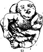


fetus



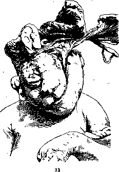
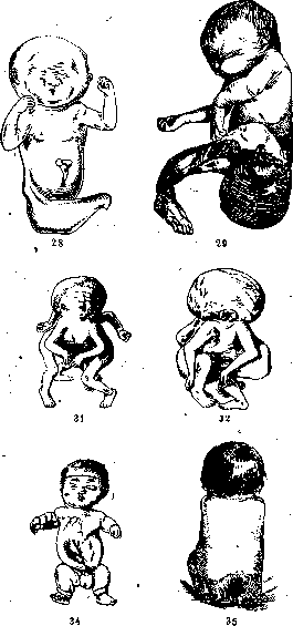
Figure 12. Spontaneous amputation of limbs (after Timofeev). Figure 13. Amelus (after Selitsky). Figure 14. Curvature of the fetal body due to insufficient dimensions of the amniotic cavity (after Timofeev). Figure 15. Preparation of partially dissected thoracopagus. Figure 16. Twins connected by the lower-anterior surfaces of the trunk. Figure 17. Cleavage of the abdominal wall with twisting of the lower extremities: 1—amnion; 2—hepar; 3—vesica urinaria; 4—placenta; 5—velamenta; 6—small intestines (after Krivsky). Figure 18. Cyclops (after Ilkevich). Figure 19. Epignathus with partial bifurcation of the trunk and four limbs. Figure 20. Hydrops fetus (after Sielart). Figure 21. Syncephalus monoprosopus (after Semyannikov). Figure 22. Acardiacus amorphus (after Kalmykov and Obraztsov). Figure 23. Two-headed monster (after Yurasovsky). Figure 24. Hernia cerebri through the canalis craniopharyngeus (after Lutokhin). Figure 25. Fissura abdominalis completa (after Bruno). Figure 26. Anencephalus. Figure 27. Craniorachischisis: 1—opening into the brain (after Blinnikov). Figure 28. Sireniform malformation (after Shubina). Figure 29. Sacrococcygeal teratoma (after Untilov). Figure 30. Hydrocephalus and orbital tumor (after Pisemsky). Figure 31. Cephalothoracopagus from the front. Figure 32. Cephalothoracopagus from behind. Figure 33. Rare malformation of the face and skull (after F. A. Solovyov). Figure 34. Micromelia. Figure 35. Hypertrichosis (after Krupenina). (Figures 15, 16, 19, 26, 31, 32, and 34 after Gruzdev.) 499 As for the causes leading to the occurrence of fetal malformations, they are usually divided into internal and external, and regarding many malformations it remains still unclear whether the causes provoking them belong to one or the other category. Causes of the first kind are rooted in the germ cells and cause abnormal development of the fertilized eggs, even if in the future the latter were under completely normal conditions of development; such causes include heredity, atavism, and so-called primary germinal variation; fetal malformations caused by these causes in most cases, if not always, are inherited. The external causes of malformations (called by some authors also intra-uterine) act only after the fertilization of the egg cell and can be either physical (thermal, mechanical, etc.) or chemical (various intoxications), and in some cases undoubtedly also psychological, and in modern times by such researchers as Dareste, Schwalbe, Winckel, a particularly important role in the genesis of fetal malformations is attributed to physical causes and predominantly to the discrepancy between the length of the fetus and the dimensions of the amniotic cavity (see Amniotic bands). In the very last years, extremely great attention not only of researchers but also of the governments of various countries has been attracted as a cause of the origin of fetal malformations by the influence of X-ray irradiation on both the ovaries and the pregnant uterus; the role of this factor is, however, still far from being fully elucidated. Anatomically, fetal malformations are extremely diverse, which naturally extremely complicates their classification. In modern science, there are several classifications of malformations—Saint-Hilaire, Marchand, Martius, and others. Based on the morphological principle, most authors first of all divide fetal malformations into two large groups: 1) single and 2) double, or better to say twin malformations. It is not devoid of interest that malformations of both kinds predominantly belong to different sexes: single malformations are more often observed in male fetuses (out of 158 cases of Marchand, in 103 they were observed in fetuses of precisely this sex and only in 55 in female fetuses), whereas double malformations are more characteristic of female fetuses, in which, according to Birnbaum, they are observed three times more often than in male fetuses. Among single malformations, one can distinguish, on the one hand, malformations of a qualitative and, on the other hand, quantitative character, and in the second category of single malformations, those are distinguished where there is an excessive development of either the entire organism of the fetus or its individual parts—so-called progressive malformations, and those which owe their origin, on the contrary, to underdevelopment of either the entire fetus or its individual organs—so-called regressive malformations. Further, depending on the localization of single malformations, one can, together with Martius, distinguish malformations of the head of the fetus, neck, trunk, and limbs (Figures 12–35 and separate table, cols. 359–360, Figures 4 and 5). As for double monsters, usually known by the name of "joined twins" (although it would be more correct to call them "not fully separated twins," since they are the result of incomplete division of monozygotic twins), Geoffroy Saint-Hilaire divides them into two large classes: autasites and parasites, referring to the first those of the "joined twins" who stand at the same stage of development, possess the same physical activity, and live each their own life, and to the second those where there is a sharp difference in the development of the twins, and the smaller one is nourished by the juices of the larger one and is as if an appendage of the latter. Marchand also divides double malformations first into two large groups: duplicitas symmetros, in which both twins are developed equally, and duplicitas asymmetros, corresponding to the second class of Saint-Hilaire's classification. Cases where there is duplicitas symmetros are then subdivided, according to Marchand, into two subgroups: duplicitas symmetros completa, in which the twins are to a large extent separated, and duplicitas symmetros incompleta, in which one end of the body is common to both twins, and the other is bifurcated. Cases of the first subgroup in turn are divided into three categories: twins connected by the lower ends, middle, and upper ends, and cases of the second subgroup into two: duplicitas symmetros incompleta inferior, in which the lower end is bifurcated, and duplicitas symmetros incompleta superior, in which the upper end of the monster's body is bifurcated. In duplicitas asymmetros, Marchand distinguishes a more developed autasite and a less developed parasite (for details on individual malformations, see the corresponding articles; on the influence of various malformations on the course of labor, see Labor). V. Gruzdev.
Related articles
Mentioned in
Cite this page
“Fetus.” Soviet Medical Encyclopedia. English translation of Bolshaya Meditsinskaya Entsiklopediya, 1st ed. (Moscow, 1928–1936), ed. N. A. Semashko. https://sovietmedicalencyclopedia.pages.dev/article/fetus/