Germ Layers
Historical document, translated for reference. It reflects medical knowledge of the 1920s–30s and is not medical advice.
Summary
Germ layers are the primary cellular layers in early embryonic development that give rise to all tissues and organs in the body. This article explains the three germ layers (ectoderm, endoderm, and mesoderm), their historical discovery, and their significance in comparative embryology.
Encyclopedia article (1928–1936)
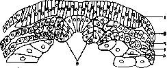
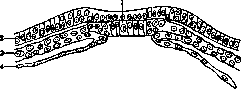
GERM LAYERS, layers or plates (German: Keimblätter, French: feuillets germinatifs, English: germinal layers), the basic embryological term denoting the layers of embryonic cells that form the body of the embryo in the early stages of development and in most cases have an epithelial character. Three germ layers are distinguished: 1) the outer (ectoderm, ectoblast, epiblast, sensory-skin layer), 2) the inner (endoderm, entoblast, hypoblast, intestinal-glandular layer) and 3) the middle (mesoderm, mesoblast) (fig. 1, 2, 3). Of these, the first two form earlier, and the third joins them later. The outer germ layer usually consists of tall, light-colored cells resembling cylindrical epithelium; the inner germ layer may consist of large cells filled with yolk plates and forming compact masses in places (amphibians), or conversely, of cells flattened like squamous epithelium (birds, mammals); the middle germ layer at the time of formation may consist of loosely arranged spindle-shaped or stellate cells which subsequently form an epithelial layer. Some authors consider the middle germ layer as two layers (parietal and visceral mesoblast, i.e., skin-muscular and intestinal-fiber layers), since over a large extent it is split. The doctrine of germ layers, their origin and further fate runs through the entire history of embryology; after Darwin it becomes closely connected with evolutionary theory and becomes the basis of comparative embryology; in the early 1880s the Hertwig brothers bring it into a systematic system, in which form it is usually presented in textbooks. But on the other hand it is subjected to strong criticism, and at present views on germ layers are far from being unified. Therefore, a proper understanding of germ layers without acquaintance with the history of the question is difficult to form.
c:=
Figure 1. Germ layers of a newt; 1-medullary plate; 2-ectoderm; 3-parietal layer of mesoderm; 4-visceral layer of mesoderm; 5-endoderm; 6-notochord. (After Hertwig.)
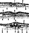
Figure 2. Germ layers of a chick. Sections of the blastoderm of three consecutive stages—A, B, C: 1-primary groove; 2-ectoderm; 3-endoderm; 4-mesoderm; 5-yolk; 6-primordium of the neural tube; 7-notochord; 8-body cavity; 9-body cavity mesoderm; 10-somite. (After Meisenheimer.)
Figure 3. Germ layers of a rabbit: 1-notochord; 2-ectoderm; 3-mesoderm; 4-endoderm. (After Beneden.)
Historical data. K. F. Wolff, who laid the foundation of modern embryology with his research on the development of the chick, described (1768) the development of the intestinal canal from a primordium having the appearance of a skin or layer, which then rolls into a tube, and suggested that in the same way the other systems of the embryo develop: nervous, muscular, vascular. 50 years later Pander (1817), studying the blastoderm of a 12-hour-old chick, described in it two thin layers: serous and mucous layers; between them later develops the third—vascular. Following in Pander's footsteps came K. E. Baer (1828-1837), who found that the two primary layers (animal and vegetative) subsequently each split into two: from the outer, animal, the skin and muscular layers are formed, from the vegetative—vascular and mucous. Subsequently they roll into tubes, forming primary organs. Further research on the germ layers of the chick belongs to Remak (1851), who distinguished three layers in total, calling them according to their physiological significance: the outer—sensory, the inner—trophic, and the middle—motor-generative. The middle layer splits into two only on the sides (lateral plates); it forms the skin-fiber and intestinal-fiber layers, bounding the body cavity. At the same time zoologists Huxley (1849) and Allman (1853) pointed out the homology between the first two germ layers and the body layers of lower invertebrates (coelenterates); Allman owns the terms "ectoderm" and "endoderm", which have become widespread and displaced the terms of previous embryologists. Extensive research on the development of different classes of invertebrates and the lancelet was carried out by the Russian scientist A. Kowalewsky; they provided the factual material for the theories of Ray Lankester (1873) and Haeckel (1874), which connected embryology with phylogeny. These scientists assumed that the simplest form, which gave rise in the process of evolution to all other invertebrates and vertebrates, consisted of two layers, which subsequently appear during the development of all animals in the form of two primary layers. Ray Lankester considered such a form to be the planula-blastula, in which a second layer is detached inward from the cellular layer; due to the rupture of the wall, the cavity of the planula communicates with the external environment and turns into the primary intestine. Haeckel saw the primary form in the gastrula, formed by invagination, and called it "gastraea" (Gastraea theory). The transition from a two-layered form to a three-layered one occurs by the detachment of cells from both layers. Haeckel's theory became widespread, and embryologists directed their efforts to prove the formation of the first two layers by the process of invagination. (In the first editions of O. Hertwig's "Lehrbuch der Entwicklimgsgeschichte" this method of formation is consistently carried out for all vertebrates.) Further work was directed to the study of the middle germ layer, which due to its heterogeneity presented difficulties for understanding; they were overcome by the work of Oscar and Richard Hertwigs (1881), who created the theory of the coelom (Coelomtheorie), analogous to the theory of gastraea. The Hertwig brothers first excluded mesenchyme (cell groups that detach from both layers and give rise to connective tissue and blood) from the composition of the middle germ layer, leaving the name mesoderm only for areas of an epithelial nature, and then connected the formation of mesoderm with the development of the body cavity (coelom). The development of the lancelet (Amphioxus), studied by Kowalewsky and Hatschek, was taken as a model, where this connection appears with full clarity (fig. 4). At a known stage, the primary endoderm of the gastrula gives a series of sac-like protrusions on both sides of the median axis—these are the primordia of the body cavity, lined with mesoderm. Subsequently they deepen between the ectoderm and endoderm and divide into areas: the proximal ones form somites (primary vertebrae), the distal ones merge with subsequent and preceding ones, forming the body cavity located between the mesoderm layers—parietal and visceral. These are the nearest derivatives of mesoderm. The same method of formation is observed in the newt (fig. 5); in others it is obscured, since the mesoderm grows in the form of solid masses, subsequently splitting. The matter is further complicated by the fact that in selachians, reptiles, and birds the mesoderm develops from two places (peripheral and axial), with the parietal layer of mesoderm growing from ectoderm (fig. 2), but if we consider the primary streak of birds as a blastopore and pay attention to the depression in Hensen's node, the formation of mesoderm can here also be connected by gradual transitions with the basic scheme.
Figure 4. Formation of mesoderm in the lancelet (A, B, C and D): 1-ectoderm; 2-medullary plate; 3-notochord; 4-mesoderm; 5-endoderm; 6-body cavity; 7-intestinal cavity; 8-neural tube; 9-somite; **-site of invagination of the body cavity. (After Hatschek.)
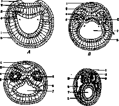
Figure 5. Formation of mesoderm in the newt: 1-blastopore; 2-parietal layer of mesoderm; 3-yolk plug; 4-visceral layer of mesoderm.
The doctrine of germ layers based on the theory of gastraea, coelom, and blastopore (Urmundtheorie) was expounded in a complete and systematic form in the aforementioned textbook by O. Hertwig, which represents the best monument
c
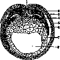
mesoderm layer; 5-ectoderm; 6-yolk cells; 7-entoderm; 8-intestinal cavity. (According to Hertwig.) ♦17 a figure in comparative embryology of vertebrates of that period when the ideas of evolution began to win recognition from the broad masses of naturalists, a figure who has not lost his significance even at the present time. Criticism of the doctrine of germ layers, which did not have particular success in the 19th century, currently attracts more attention in connection with the change in the course of embryology, which has moved from description and comparison to the elucidation of the causes of development with the help of experiment. The main objection against the doctrine of germ layers was given back by Reichert (1843), who, instead of layers, brought to the fore the rudiments of organs (primary organs), arising either directly as such or several together in a common rudiment. In contrast to germ layers, these primary organs are not strictly fixed concepts and vary in number, form, and position in different animals. In subsequent times, the main blows of criticism were directed at the middle germ layer (Kleinenberg, 1886; Bergh, 1896), which, both in vertebrates and especially in invertebrates, often represents a collection of completely heterogeneous rudiments and does not exist as a single layer. The differentiation of mesenchyme and mesoderm likewise cannot be carried out in the entire animal kingdom and encounters numerous contradictions. The main opponent of the doctrine of germ layers in recent times is the zoologist Meisenheimer, who fully shares Reichert's point of view. But, while acknowledging the full validity of the objections against the middle germ layer, it is hardly possible to agree with the deletion of the term germ layer itself, since the ectoderm and entoderm exist as quite definite morphological formations and are striking to everyone studying development. Their formation is a different matter: they can arise and indeed do arise in different animals in different ways depending on the amount of yolk and other causes; therefore, it is not possible to support the Hertwigs' theory in full. Fate of germ layers and their specificity. Already by the first researchers, it was clarified in general terms which organs or parts of them are given rise to by each germ layer, in other words, their "prospective significance." The outer germ layer produces the nervous system, the epidermis of the skin, the epithelium and smooth muscles of skin glands, the epithelium of the auditory organ, the nasal cavity, the anterior part of the oral cavity (including the glandular part of the pituitary gland and tooth enamel), the anal part of the rectum, the lens, and the epithelium of the amnion. The inner [layer produces] the epithelial lining of the intestinal canal and the glands formed in it, including the liver and pancreas. The middle [layer], the mesoderm proper, in the region of the somites gives rise to the musculature of the body (myotome) and connective tissue (sclerotome), in the region of the nephrotome—the excretory organs; the mesoderm lining the body cavity forms its endothelium (mesothelium) and the epithelial parts of the gonads. Primary germ cells in some cases can be located in the entoderm and from there move to the genital ridge. As for the mesenchyme, it forms the cellular elements of connective tissue and blood, although some authors derive the first rudiments of blood from the entoderm. There is no complete clarity in the distinction between mesoderm and mesenchyme. The doctrine of the fate of germ layers was subsequently supplemented by the proposition about their histological specificity, according to which the ectoderm, entoderm, mesoderm, and mesenchyme possess limited "prospective potency" and can
produce only
t
cells and tissues. For example, ectodermal epithelium can never give rise to connective tissue or the epithelium of entodermal glands—to leukocytes. Assertions contradicting this by Retterer regarding the transition of crypt epithelium into leukocytes or by Stöhr regarding the origin of lymphocytes [Fig. 6 longitudinal section of the embryo of Triton cristatus]

glands from the epithelial rudiment were met by histologists with distrust and forced one to assume errors in observation. On this same basis, in recent times, attempts have been made to draw a distinction between the endothelium of vessels and the peritoneum: the former, as a derivative of mesenchyme, can give rise to blood elements, whereas the mesodermal epithelium of the peritoneum (mesothelium) is not capable of this (Maximov). Although the proven origin of smooth muscles of glands from ectodermal and entodermal epithelium breached the doctrine of the strict specificity of germ layer derivatives, in general, it continues to prevail even today. The question of the fate of germ layers at early stages of development is being resolved in modern times by means of experiment. Spemann and Mangold, by transplanting various sections from embryos of pigmented newts (Triton taeniatus) to pigmentless ones (Triton cristatus) (which made it possible to trace their fate), found that in the blastula stage, sections of the animal and vegetative poles and the intermediate zone are determined, i.e., they give rise to specific layers, but in the gastrula stage, the formed layers do not possess specificity. Transplanted sections of ectoderm could become part of the intestine or, along with the mesoderm, give rise to somites (Figure 6). From this, the conclusion is drawn that germ layers, not possessing specificity, have significance only as topographical concepts. At the same time, in the late stages of gastrula, the emerging organ rudiments are already determined, and the section of the neural plate, for example, produces brain everywhere. Experimental study of histological specificity in vital tissue cultures generally leads to the same results.
Related articles
Mentioned in
Cite this page
“Germ Layers.” Soviet Medical Encyclopedia. English translation of Bolshaya Meditsinskaya Entsiklopediya, 1st ed. (Moscow, 1928–1936), ed. N. A. Semashko. https://sovietmedicalencyclopedia.pages.dev/article/germ-layers/