Egg
Historical document, translated for reference. It reflects medical knowledge of the 1920s–30s and is not medical advice.
Summary
The egg is the germ cell of multicellular organisms, varying widely in form and size. This article describes the structure, classification, and development of eggs across different species, including their protective layers and nutritional components.
Encyclopedia article (1928–1936)
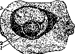
Egg, the germ cell of multicellular organisms. Depending on the stage of development, immature eggs (oogonia, oocytes of the 1st and 2nd order), mature eggs (suitable for fertilization), and finally, fertilized eggs—developing eggs of mammals—are distinguished. The form and size of eggs vary within extremely wide limits; however, it can generally be said that the eggs of many animals are the largest of cells. Many eggs are visible to the naked eye and sometimes reach very significant sizes (eggs of birds, reptiles, amphibians).-The first data on the structure of eggs appear in the works of the 17th and 18th centuries (Harvey, de Graaf, etc.), but the initial interpretation of the pictures observed here was largely incorrect; thus, de Graaf considered the egg of mammals to be the so-called Graafian follicle, which is a complex system of membranes covering the small egg contained within it. Only in the middle of the 18th century did Caspar Wolff describe the chicken egg in detail. The egg of mammals was studied much later (first by K. E. von Baer in 1827). The structure of the egg essentially depends on the amount of nutrient substance contained within it—the yolk (deutoplasm, para-plasm). Eggs are distinguished: 1) alecithal—practically devoid of yolk (mammals, lancelet), 2) mesolecithal—with a very even distribution of yolk (echinoderms), 3) telolecithal—with a large amount of yolk collected at one (lower) pole of the egg (many invertebrates, selachians, amphibians, reptiles, birds). In telolecithal eggs, the upper animal pole is distinguished, on which the cytoplasm of the egg is concentrated, and the vegetative pole, filled with heavier lumps of yolk. In some insects, 4) centrolecithal eggs occur—the protoplasm in the form of a sheath covers a significant mass of yolk located in the center of the egg. The yolk consists of grains, granules, or plates of various sizes and shapes. The arrangement of the yolk in the egg is also very different—in some cases it evenly fills all the protoplasm, in others it is located in the center or at the periphery of the egg. The nucleus of the egg undoubtedly participates in the formation of the yolk; in some cases (in Dytiscus, Proteus, etc.), it is possible to show the emergence of grains of chromatin from the nucleus and the deposition of the first granules of yolk around them (fig. 1). The formation of the yolk apparently also involves the rather mysterious organelle of the egg cell—the so-called yolk nucleus, or accessory nucleus, first described by Balbiani in the cells of a number of invertebrates. It arises from substances of the nucleus and represents a complex formation (fig. 2) with a lighter central part and darker areas on the periphery. The protoplasm free of inclusions (cytoplasm) of the egg is usually represented by small layers of foamy or homogeneous structure.-The nucleus of the egg cell during the longest stages of its development has the appearance of a vesicle (hence the name vesicula germinativa of old authors—Purkinje, 1824), extremely poor in chromatin. The shape of the egg nucleus is usually round or oval; in many cases, the appearance of pseudopod-like outgrowths directed toward the place of deposition of yolk plates in the egg can be noted; sometimes the nucleus moves entirely to the periphery of the egg into the area of penetration of nutrient substances. Inside the nucleus, nucleoli (one, two, or several) are clearly visible. One of them differs in special stainability (macula germinativa) (Wagner, 1834). Its significance in the biology of the egg is unclear.-The egg contains a number of organelles—the Golgi apparatus, chondriosomes, and centrosomes. In the eggs of amphibians and some echinoderms, there is a pigment localized in certain parts of the egg and marking the areas from which certain organ systems of the embryo will develop. The membranes of the egg can be divided into 5 groups. The primary membrane, the so-called vitelline membrane, oolemma, or zona pellucida, of the mammalian egg is the compacted outer edge of the protoplasm; in some cases it has a complex structure; provided with pores through which nutrient substances can pass into the egg. The time of appearance of the vitelline membrane varies greatly; sometimes (in mammalian eggs) it arises before fertilization, in which case it has a special opening for the penetration of spermatozoa—the micropyle; in other cases (echinoderms) the vitelline membrane arises only after fertilization and the penetration of the first spermatozoon into the egg.-In some animals and plants, the egg cell is surrounded by a group of poorly differentiated epithelial cells (follicular cells). The significance of these cells is very diverse; most often they transfer nutrient substances to the egg (nurse cells), in some cases the absorption of these cells is noted, just as of non-viable egg cells located in the same membrane with them, by the developing egg. Follicular cells secrete on the surface a dense membrane, the chorion, which in some cases reaches considerable thickness.-The third type of membranes is secreted outside the gonad during the passage of the egg through the genital ducts, uterus, etc. Very interesting examples of formations of this kind can serve as the cocoons of insects and the egg membranes of vertebrates (fish, amphibians, reptiles, birds). Here there is a deposition of additional reserves of nutrient substances (the white of the chicken egg, protein fluid in the cocoons of many invertebrates), which the long-developing embryo must feed on. The material of such formations is very different—chitin in invertebrates, dense substance in lower vertebrates, a hard shell impregnated with lime in birds and some reptiles. The chicken egg represents an extremely complex formation. Besides the embryonic disk itself, i.e., the area of pure protoplasm located on the animal pole, where the embryo develops, and the enormous amount of yolk, a number of additional parts are distinguished. These are the concentrically arranged areas of white and yellow albumen, differing from each other in the size and density of arrangement (yellow—smaller and denser) of the protein plates composing them; further, the chalazae—two threads of albumen stretching to the edges of the egg and supporting it in a suspended state inside the shell; further, two membranes, the so-called albumen membranes, diverging at the blunt end of the egg and forming there an air chamber; and finally the calcareous shell of the egg. However, naked eggs are not lacking, especially in a number of lower invertebrates, where in sponges and coelenterates, eggs capable of forming pseudopodia and amoeboid movement are described.-The presence of one or another type of membrane, as well as the total number of eggs, is essentially connected with the living conditions of the animal, the duration and circumstances of embryo development. In the presence of unfavorable development conditions (parasites, animals with external fertilization, etc.), the number of eggs increases significantly, and the number of protective adaptations of the egg ensuring its survival also increases; it is sufficient to point to the thick membranes of the eggs of parasites. Within the same species, in some cases, seasonal dimorphism of eggs is noted (winter and summer eggs), associated with the presence of fertilization and parthenogenesis.
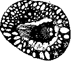
fig. 1. The nucleus of the egg of Pholcus phalangioideus during the formation of yolk; the longest pseudopodia are directed toward the dense accumulation of yolk. (After van Bambeke from Korschelt and Heider.)
The free protoplasm (cytoplasm) of the egg is usually represented by small layers of foamy or homogeneous structure.-The nucleus of the egg cell during the longest stages of its development has the appearance of a vesicle (hence the name vesicula germinativa of old authors—Purkinje, 1824), extremely poor in chromatin. The shape of the egg nucleus is usually round or oval; in many cases, the appearance of pseudopod-like outgrowths directed toward the place of deposition of yolk plates in the egg can be noted; sometimes the nucleus moves entirely to the periphery of the egg into the area of penetration of nutrient substances. Inside the nucleus, nucleoli (one, two, or several) are clearly visible. One of them differs in special stainability (macula germinativa) (Wagner, 1834). Its significance in the biology of the egg is unclear.-The egg contains a number of organelles—the Golgi apparatus, chondriosomes, and centrosomes. In the eggs of amphibians and some echinoderms, there is a pigment localized in certain parts of the egg and marking the areas from which certain organ systems of the embryo will develop. The membranes of the egg can be divided into 5 groups. The primary membrane, the so-called vitelline membrane, oolemma, or zona pellucida, of the mammalian egg is the compacted outer edge of the protoplasm; in some cases it has a complex structure; provided with pores through which nutrient substances can pass into the egg. The time of appearance of the vitelline membrane varies greatly; sometimes (in mammalian eggs) it arises before fertilization, in which case it has a special opening for the penetration of spermatozoa—the micropyle; in other cases (echinoderms) the vitelline membrane arises only after fertilization and the penetration of the first spermatozoon into the egg.-In some animals and plants, the egg cell is surrounded by a group of poorly differentiated epithelial cells (follicular cells). The significance of these cells is very diverse; most often they transfer nutrient substances to the egg (nurse cells), in some cases the absorption of these cells is noted, just as of non-viable egg cells located in the same membrane with them, by the developing egg. Follicular cells secrete on the surface a dense membrane, the chorion, which in some cases reaches considerable thickness.-The third type of membranes is secreted outside the gonad during the passage of the egg through the genital ducts, uterus, etc. Very interesting examples of formations of this kind can serve as the cocoons of insects and the egg membranes of vertebrates (fish, amphibians, reptiles, birds). Here there is a deposition of additional reserves of nutrient substances (the white of the chicken egg, protein fluid in the cocoons of many invertebrates), which the long-developing embryo must feed on. The material of such formations is very different—chitin in invertebrates, dense substance in lower vertebrates, a hard shell impregnated with lime in birds and some reptiles. The chicken egg represents an extremely complex formation. Besides the embryonic disk itself, i.e., the area of pure protoplasm located on the animal pole, where the embryo develops, and the enormous amount of yolk, a number of additional parts are distinguished. These are the concentrically arranged areas of white and yellow albumen, differing from each other in the size and density of arrangement (yellow—smaller and denser) of the protein plates composing them; further, the chalazae—two threads of albumen stretching to the edges of the egg and supporting it in a suspended state inside the shell; further, two membranes, the so-called albumen membranes, diverging at the blunt end of the egg and forming there an air chamber; and finally the calcareous shell of the egg. However, naked eggs are not lacking, especially in a number of lower invertebrates, where in sponges and coelenterates, eggs capable of forming pseudopodia and amoeboid movement are described.-The presence of one or another type of membrane, as well as the total number of eggs, is essentially connected with the living conditions of the animal, the duration and circumstances of embryo development. In the presence of unfavorable development conditions (parasites, animals with external fertilization, etc.), the number of eggs increases significantly, and the number of protective adaptations of the egg ensuring its survival also increases; it is sufficient to point to the thick membranes of the eggs of parasites. Within the same species, in some cases, seasonal dimorphism of eggs is noted (winter and summer eggs), associated with the presence of fertilization and parthenogenesis.
The free protoplasm (cytoplasm) of the egg is usually represented by small layers of foamy or homogeneous structure.-The nucleus of the egg cell during the longest stages of its development has the appearance of a vesicle (hence the name vesicula germinativa of old authors—Purkinje, 1824), extremely poor in chromatin. The shape of the egg nucleus is usually round or oval; in many cases, the appearance of pseudopod-like outgrowths directed toward the place of deposition of yolk plates in the egg can be noted; sometimes the nucleus moves entirely to the periphery of the egg into the area of penetration of nutrient substances. Inside the nucleus, nucleoli (one, two, or several) are clearly visible. One of them differs in special stainability (macula germinativa) (Wagner, 1834). Its significance in the biology of the egg is unclear.-The egg contains a number of organelles—the Golgi apparatus, chondriosomes, and centrosomes. In the eggs of amphibians and some echinoderms, there is a pigment localized in certain parts of the egg and marking the areas from which certain organ systems of the embryo will develop. The membranes of the egg can be divided into 5 groups. The primary membrane, the so-called vitelline membrane, oolemma, or zona pellucida, of the mammalian egg is the compacted outer edge of the protoplasm; in some cases it has a complex structure; provided with pores through which nutrient substances can pass into the egg. The time of appearance of the vitelline membrane varies greatly; sometimes (in mammalian eggs) it arises before fertilization, in which case it has a special opening for the penetration of spermatozoa—the micropyle; in other cases (echinoderms) the vitelline membrane arises only after fertilization and the penetration of the first spermatozoon into the egg.-In some animals and plants, the egg cell is surrounded by a group of poorly differentiated epithelial cells (follicular cells). The significance of these cells is very diverse; most often they transfer nutrient substances to the egg (nurse cells), in some cases the absorption of these cells is noted, just as of non-viable egg cells located in the same membrane with them, by the developing egg. Follicular cells secrete on the surface a dense membrane, the chorion, which in some cases reaches considerable thickness.-The third type of membranes is secreted outside the gonad during the passage of the egg through the genital ducts, uterus, etc. Very interesting examples of formations of this kind can serve as the cocoons of insects and the egg membranes of vertebrates (fish, amphibians, reptiles, birds). Here there is a deposition of additional reserves of nutrient substances (the white of the chicken egg, protein fluid in the cocoons of many invertebrates), which the long-developing embryo must feed on. The material of such formations is very different—chitin in invertebrates, dense substance in lower vertebrates, a hard shell impregnated with lime in birds and some reptiles. The chicken egg represents an extremely complex formation. Besides the embryonic disk itself, i.e., the area of pure protoplasm located on the animal pole, where the embryo develops, and the enormous amount of yolk, a number of additional parts are distinguished. These are the concentrically arranged areas of white and yellow albumen, differing from each other in the size and density of arrangement (yellow—smaller and denser) of the protein plates composing them; further, the chalazae—two threads of albumen stretching to the edges of the egg and supporting it in a suspended state inside the shell; further, two membranes, the so-called albumen membranes, diverging at the blunt end of the egg and forming there an air chamber; and finally the calcareous shell of the egg. However, naked eggs are not lacking, especially in a number of lower invertebrates, where in sponges and coelenterates, eggs capable of forming pseudopodia and amoeboid movement are described.-The presence of one or another type of membrane, as well as the total number of eggs, is essentially connected with the living conditions of the animal, the duration and circumstances of embryo development. In the presence of unfavorable development conditions (parasites, animals with external fertilization, etc.), the number of eggs increases significantly, and the number of protective adaptations of the egg ensuring its survival also increases; it is sufficient to point to the thick membranes of the eggs of parasites. Within the same species, in some cases, seasonal dimorphism of eggs is noted (winter and summer eggs), associated with the presence of fertilization and parthenogenesis.
The free protoplasm (cytoplasm) of the egg is usually represented by small layers of foamy or homogeneous structure.-The nucleus of the egg cell during the longest stages of its development has the appearance of a vesicle (hence the name vesicula germinativa of old authors—Purkinje, 1824), extremely poor in chromatin. The shape of the egg nucleus is usually round or oval; in many cases, the appearance of pseudopod-like outgrowths directed toward the place of deposition of yolk plates in the egg can be noted; sometimes the nucleus moves entirely to the periphery of the egg into the area of penetration of nutrient substances. Inside the nucleus, nucleoli (one, two, or several) are clearly visible. One of them differs in special stainability (macula germinativa) (Wagner, 1834). Its significance in the biology of the egg is unclear.-The egg contains a number of organelles—the Golgi apparatus, chondriosomes, and centrosomes. In the eggs of amphibians and some echinoderms, there is a pigment localized in certain parts of the egg and marking the areas from which certain organ systems of the embryo will develop. The membranes of the egg can be divided into 5 groups. The primary membrane, the so-called vitelline membrane, oolemma, or zona pellucida, of the mammalian egg is the compacted outer edge of the protoplasm; in some cases it has a complex structure; provided with pores through which nutrient substances can pass into the egg. The time of appearance of the vitelline membrane varies greatly; sometimes (in mammalian eggs) it arises before fertilization, in which case it has a special opening for the penetration of spermatozoa—the micropyle; in other cases (echinoderms) the vitelline membrane arises only after fertilization and the penetration of the first spermatozoon into the egg.-In some animals and plants, the egg cell is surrounded by a group of poorly differentiated epithelial cells (follicular cells). The significance of these cells is very diverse; most often they transfer nutrient substances to the egg (nurse cells), in some cases the absorption of these cells is noted, just as of non-viable egg cells located in the same membrane with them, by the developing egg. Follicular cells secrete on the surface a dense membrane, the chorion, which in some cases reaches considerable thickness.-The third type of membranes is secreted outside the gonad during the passage of the egg through the genital ducts, uterus, etc. Very interesting examples of formations of this kind can serve as the cocoons of insects and the egg membranes of vertebrates (fish, amphibians, reptiles, birds). Here there is a deposition of additional reserves of nutrient substances (the white of the chicken egg, protein fluid in the cocoons of many invertebrates), which the long-developing embryo must feed on. The material of such formations is very different—chitin in invertebrates, dense substance in lower vertebrates, a hard shell impregnated with lime in birds and some reptiles. The chicken egg represents an extremely complex formation. Besides the embryonic disk itself, i.e., the area of pure protoplasm located on the animal pole, where the embryo develops, and the enormous amount of yolk, a number of additional parts are distinguished. These are the concentrically arranged areas of white and yellow albumen, differing from each other in the size and density of arrangement (yellow—smaller and denser) of the protein plates composing them; further, the chalazae—two threads of albumen stretching to the edges of the egg and supporting it in a suspended state inside the shell; further, two membranes, the so-called albumen membranes, diverging at the blunt end of the egg and forming there an air chamber; and finally the calcareous shell of the egg. However, naked eggs are not lacking, especially in a number of lower invertebrates, where in sponges and coelenterates, eggs capable of forming pseudopodia and amoeboid movement are described.-The presence of one or another type of membrane, as well as the total number of eggs, is essentially connected with the living conditions of the animal, the duration and circumstances of embryo development. In the presence of unfavorable development conditions (parasites, animals with external fertilization, etc.), the number of eggs increases significantly, and the number of protective adaptations of the egg ensuring its survival also increases; it is sufficient to point to the thick membranes of the eggs of parasites. Within the same species, in some cases, seasonal dimorphism of eggs is noted (winter and summer eggs), associated with the presence of fertilization and parthenogenesis.
The free protoplasm (cytoplasm) of the egg is usually represented by small layers of foamy or homogeneous structure.-The nucleus of the egg cell during the longest stages of its development has the appearance of a vesicle (hence the name vesicula germinativa of old authors—Purkinje, 1824), extremely poor in chromatin. The shape of the egg nucleus is usually round or oval; in many cases, the appearance of pseudopod-like outgrowths directed toward the place of deposition of yolk plates in the egg can be noted; sometimes the nucleus moves entirely to the periphery of the egg into the area of penetration of nutrient substances. Inside the nucleus, nucleoli (one, two, or several) are clearly visible. One of them differs in special stainability (macula germinativa) (Wagner, 1834). Its significance in the biology of the egg is unclear.-The egg contains a number of organelles—the Golgi apparatus, chondriosomes, and centrosomes. In the eggs of amphibians and some echinoderms, there is a pigment localized in certain parts of the egg and marking the areas from which certain organ systems of the embryo will develop. The membranes of the egg can be divided into 5 groups. The primary membrane, the so-called vitelline membrane, oolemma, or zona pellucida, of the mammalian egg is the compacted outer edge of the protoplasm; in some cases it has a complex structure; provided with pores through which nutrient substances can pass into the egg. The time of appearance of the vitelline membrane varies greatly; sometimes (in mammalian eggs) it arises before fertilization, in which case it has a special opening for the penetration of spermatozoa—the micropyle; in other cases (echinoderms) the vitelline membrane arises only after fertilization and the penetration of the first spermatozoon into the egg.-In some animals and plants, the egg cell is surrounded by a group of poorly differentiated epithelial cells (follicular cells). The significance of these cells is very diverse; most often they transfer nutrient substances to the egg (nurse cells), in some cases the absorption of these cells is noted, just as of non-viable egg cells located in the same membrane with them, by the developing egg. Follicular cells secrete on the surface a dense membrane, the chorion, which in some cases reaches considerable thickness.-The third type of membranes is secreted outside the gonad during the passage of the egg through the genital ducts, uterus, etc. Very interesting examples of formations of this kind can serve as the cocoons of insects and the egg membranes of vertebrates (fish, amphibians, reptiles, birds). Here there is a deposition of additional reserves of nutrient substances (the white of the chicken egg, protein fluid in the cocoons of many invertebrates), which the long-developing embryo must feed on. The material of such formations is very different—chitin in invertebrates, dense substance in lower vertebrates, a hard shell impregnated with lime in birds and some reptiles. The chicken egg represents an extremely complex formation. Besides the embryonic disk itself, i.e., the area of pure protoplasm located on the animal pole, where the embryo develops, and the enormous amount of yolk, a number of additional parts are distinguished. These are the concentrically arranged areas of white and yellow albumen, differing from each other in the size and density of arrangement (yellow—smaller and denser) of the protein plates composing them; further, the chalazae—two threads of albumen stretching to the edges of the egg and supporting it in a suspended state inside the shell; further, two membranes, the so-called albumen membranes, diverging at the blunt end of the egg and forming there an air chamber; and finally the calcareous shell of the egg. However, naked eggs are not lacking, especially in a number of lower invertebrates, where in sponges and coelenterates, eggs capable of forming pseudopodia and amoeboid movement are described.-The presence of one or another type of membrane, as well as the total number of eggs, is essentially connected with the living conditions of the animal, the duration and circumstances of embryo development. In the presence of unfavorable development conditions (parasites, animals with external fertilization, etc.), the number of eggs increases significantly, and the number of protective adaptations of the egg ensuring its survival also increases; it is sufficient to point to the thick membranes of the eggs of parasites. Within the same species, in some cases, seasonal dimorphism of eggs is noted (winter and summer eggs), associated with the presence of fertilization and parthenogenesis.
S. Zalkind. The woman's egg. The question of the embryonic pathway (see Oogenesis) in humans cannot be considered resolved, although the data of G. Politzer on the very early stages of development of human embryos (starting from 0.3 mm in length), in which he discovered primary germ cells (Urkeimzellen), speak in favor of its existence. The oogonia of the embryo first become morphologically different at the moment of sex differentiation, i.e., at about 2 months of age. They multiply intensively throughout the first half of intrauterine life; their multiplication ceases by 6-7 months. Even during the period of multiplication, other processes are observed in a number of oogonia. These latter proceed in three directions. Part of the oogonia transforms into oocytes of the first order, which is morphologically characterized by typical changes in the nucleus. In the nucleus of the oogonium, which has the appearance of a lightly colored and almost structureless vesicle with 1-3 nucleoli, chromatin accumulates in the form of lumps, clots, and short threads, as a result of which the nucleus begins to stain darker. Such nuclei, according to Vinivarter, already belong to oocytes of the first order. Further, the entire nucleus is filled with a ball of thin, twisted threads, which fold in pairs. The 'bouquet' considered by some authors characteristic of this stage, in the form of a dense ball of threads pushed to one pole, is in fact a vital artifact that easily arises due to the particularly high lability of nuclear structures at this stage. Another direction of changes in germ cells is the degeneration of oogonia and especially oocytes. The vast majority of the latter perish in the embryonic ovary with phenomena of chromatopyknosis: the chromatin components of the nucleus shorten, showing at the same time a tendency to pairwise arrangement. The karyoplasm swells, becomes more transparent and lighter. The general appearance of the nucleus of such degenerating cells resembles its structure during reduction division, which gave rise to erroneous interpretations of such pictures in this sense, but the similarity here is purely external. Maturation divisions do not occur in the embryonic ovary. The process of degeneration of germ elements is most pronounced in the ovary of fetuses 6-7 months. Finally, a certain part of the first-order oocytes, surrounded by a layer of follicular cells, forms a primordial follicle; by 7-8 months, all remaining germ cells are enclosed in them. If not one but several oocytes enter the composition of the primordial follicle, then a polyovular primordial follicle arises. The further development the egg cell undergoes already inside the follicle. It is studied still quite insufficiently, especially little is known about the nuclear changes during the process of maturation itself. One should distinguish between the processes of egg cell maturation, for which both maturation divisions are decisive, apparently occurring at the moment of ovulation or during the passage of the egg through the tubes (Allen, Bland, Newell and Pratt), and the maturation of the primordial follicle, which transforms into a Graafian vesicle. The latter also as a rule takes place during the period of sexual maturity, however, as an exception, it may occur in the embryonic ovary, and in such a case the egg cell still remains an oocyte of the first order. The mature egg see Ovulation, Embryo.

Related articles
Cite this page
“Egg.” Soviet Medical Encyclopedia. English translation of Bolshaya Meditsinskaya Entsiklopediya, 1st ed. (Moscow, 1928–1936), ed. N. A. Semashko. https://sovietmedicalencyclopedia.pages.dev/article/egg/