Obstetric Manual Maneuvers
Historical document, translated for reference. It reflects medical knowledge of the 1920s–30s and is not medical advice.
Summary
This article describes various manual techniques used in obstetrics, including Leopold's maneuvers for determining fetal position, Müller and Hofmeyer's techniques for assessing fetal head compatibility with the pelvis, and methods for assisting in breech and footling births.
Encyclopedia article (1928–1936)
OBSTETRIC MANUAL MANEUVERS. These include: 1) Leopold's maneuver for determining the position of the fetus in the uterus at the end of pregnancy or at the beginning of labor; 2) Müller and Hofmeyer's maneuver for determining the correspondence between the size of the fetal head and the size of the pelvic inlet; 3) maneuvers of Mauriceau, Smellie and others as an aid in delivering the infant in breech or footling presentations; 4) expression of the placenta according to Credé; 5) manual separation and removal of the afterbirth; 6) massage of the uterus by introducing a hand into its cavity to combat atonic bleeding; 7) pressing out of the fetus according to Kristeller; 8) manual dilation of the cervix and 9) separation and removal with fingers of the ovum and its parts. - Methods for determining the size and position of the fetus in the uterus at the end of pregnancy were proposed by Leopold and consist of four maneuvers. In the first maneuver (see table 11, fig. 5), the physician stands to the side of the pregnant woman facing her head and places the medial borders of the palms of both hands on the fundus of the uterus. By pressing them slightly inward, he determines the fundus and simultaneously determines the month of pregnancy and the parts of the fetus located near the fundus. - In the second maneuver (see table II, fig. 6), the physician remains standing in the same position as in the first, but lowers the palmar surfaces of both hands from the fundus to the lateral parts of the uterus and feels for the position of the fetus's back and small parts. When pressing with the palms and fingertips, a dense surface of the back is palpated on one side of the uterus, and on the other - the small parts of the fetus, which sometimes produce "quick, jerky movements, easily felt through the uterine wall and abdominal coverings." If the fetus's back is turned backward, then many small parts of the fetus are encountered under the fingers at the anterior uterine wall, while the back cannot be palpated. In transverse or oblique positions of the fetus, the head or buttocks are palpated through the lateral surfaces of the uterus, while the back of the fetus is determined either closer to the fundus or closer to the pelvic inlet. In hydramnios, the obtained data are less pronounced, since the parts of the fetus are removed from the uterine wall by an increased amount of amniotic fluid, and here one can obtain a clear fluctuation (shaking) of the amniotic fluid. For this, one hand is firmly applied to the uterus and the fingertips of the other hand are used to create wave-like movements in the fluid, which is felt by the first hand. If the head is presenting above the pelvic inlet, then an unevenly dense, elastic, large part of the fetus - the buttocks - is palpated at the fundus of the uterus, and conversely, if the buttocks are presenting above the pelvic inlet, then a round, evenly dense and easily ballotable head is palpated at the fundus. - In the third Leopold maneuver (see table II, fig. 8), it is determined which large part of the fetus is presenting and where it is located. For this, the physician remains in the same position relative to the pregnant woman as in the first two maneuvers, using only one hand for the examination, the palm of which is pressed against the lower part of the uterus above the pubis. With the fingertips of this hand - the thumb on one side, and the other four on the other - an attempt is made to grasp the presenting part. On one side, the anterior part of the head comes under the fingers, and on the other - the posterior part. The aforementioned fingers can be advanced inward and with a quick upward push, the presenting part is pushed upward. If the head is presenting, it gives a sensation of a counter-blow against the fingers, called ballotment in obstetrics. If the presenting part does not ballot clearly or does not ballot at all, and on palpation it appears rounded but unevenly dense, then these are the buttocks. All these maneuvers should be performed without sudden movements to avoid causing pain and uterine contractions. - In the fourth maneuver (see table II, fig. 7), the physician stands with his back to the face of the pregnant woman or parturient and applies the palms of both hands with the fingertips pointing downward to her groin areas above the Poupart's ligaments. The fingertips are slowly and carefully advanced into the pelvis to avoid contractions, with the aim of determining, in the case of a presenting head, where the occiput (the flatter surface) is located and where the forehead (the more convex surface) is, and how much the entire head has descended into the pelvic cavity. If the buttocks are presenting, then it is determined where the sacrum and the fetus's legs are located. In a transverse position of the fetus, no presenting part is determined above the inlet, and the fingertips can be deeply advanced into the pelvis. In order to determine whether the fetal head at the end of pregnancy corresponds in size to the pelvic inlet and whether it can pass through it, P. Müller proposed the following manual maneuver: the obstetrician stands to the left of the pregnant woman or parturient, facing her feet, introduces previously washed index and middle fingers of one hand (usually the left) into the vagina, and with the palm and all fingers of the other hand tries to press the head into the pelvis (the work of this hand can be done by the assisting assistant's hands), judging by the degree of descent of the head into the pelvis regarding its size and its relation to the pelvis. The narrower the pelvis and the larger the head, the more it protrudes above the upper edge of the pubic arch; the farther the sagittal suture is from the pelvic conjugate line and the less both parietal bones can be encircled by a finger from the vaginal side (see table III, fig. 10), the greater the discrepancy between the pelvis and the head. In occiput posterior position, the head protrudes significantly above the edge of the symphysis, and the discrepancy is great. Conversely, the more both parietal bones can be encircled by a finger (see table III, fig. 9) and the closer the sagittal suture approaches the pelvic conjugate line, the greater the correspondence and the sooner the head will pass through the given pelvic ring without delay. Müller's maneuver was proposed mainly for cases of narrow pelvis in order not to miss the moment for inducing premature labor. - For the same purpose and to determine whether the head can pass through the pelvis with natural forces during labor, Hofmeyer proposed the following maneuver: standing to the left of the chloroformed parturient, with his back to her face, the obstetrician, grasping the head through the lower uterine segment (through the abdominal walls) with one hand so that the thumb is on the occiput and the others are on the lower jaw above the chin (or this grasping is done with two hands so that four fingers of each hand are on the occiput and near the chin, while the thumbs remain free) - exerts strong downward pressure, trying to press the head into the pelvis. If this cannot be done, the same maneuver is attempted in the Walcher position, in which the true conjugate increases by 0.5 cm. Depending on whether the maneuver succeeds or not, labor is left to natural forces or resort to one or another operative intervention to artificially terminate it. A. r. p. as an aid in delivering the infant in breech presentation. These maneuvers are used only from the moment the child is born to the navel, and before that, labor is left to natural forces so that the fetus's hands maintain their normal position, i.e., remain pressed against the chest, entering the pelvis together with the upper part of the trunk, while the head remains in a flexed position. From the moment the navel appears externally, the obstetrician assisting in the delivery with breech presentation performs the following three manual maneuvers, in which one should never forget that the midwife presses on the head from above through the lower uterine segment. If the umbilical cord is strongly stretched, it is necessary to relax this tension and remove the loop from the leg if it is wrapped around it and the infant is sitting astride it. If this cannot be done, then in order to avoid rupture of the umbilical cord or premature separation of the placenta during further delivery of the fetus, it must be cut, clamping both cut ends with two Kocher clamps. Only after this is the first maneuver undertaken, which consists of grasping the infant's buttocks with both hands so that the thumbs of both hands lie on the sacrum and along the spine, while the others are on the infant's thighs and pelvic bones. It is forbidden to grasp the infant's abdomen, so as not to rupture the liver and other abdominal organs. After this, the extraction of the infant is performed so that its shoulder dimension passes through the anteroposterior dimension of the pelvic outlet, until the inferior angles of the scapulae appear externally. - The second manual maneuver consists in freeing the fetus's hands. The posterior hand is freed first; the obstetrician grasps it with two fingers of the corresponding hand, passed into the sleeve of the parturient along the dorsal surface of the fetus (see table IV, fig. 12). Therefore, if such a hand is the right one, then the obstetrician inserts his right hand into the sleeve, and with his left hand strongly displaces the fetus's legs to the right groin of the parturient, grasping them in the area of the condyles. The insertion of two fingers into the vagina is performed along the infant's back from the perineal side of the parturient; Reaching the infant's right shoulder with them, they then descend along the shoulder to the elbow flexure; Then, by pressing on the latter, they cause the forearm to slide downward, closely pressed against the anterolateral surface of the chest.
Once this is accomplished, the obstetrician grasps the fetus's legs with their right hand, diverting them to the opposite side of the mother's pubis, and with two fingers of the left hand, similarly frees the infant's left arm. If this arm is not bent back over the head, it is freed just as easily as the right one. If the arm is bent back over the head or extended upward, it is necessary to turn the fetus's shoulders so that the left shoulder, located at the pubis, turns toward the sacrum. For this, the physician lowers the fetus's legs to hang down, and with the palms of both hands, encircles the infant's shoulder girdle and turns it from front to back so that the left shoulder is directed toward the mother's sacrum (see table V, fig. 15). As soon as this is done, the legs are again grasped and diverted to the left inguinal fold of the mother, and with two fingers of the other hand (the left hand in this case), the left arm is freed. -Müller, when extracting the fetus by the breech, recommended his manual technique for freeing the arms. The first act of extraction, according to Müller, consists of pulling the child's body downward until the anterior shoulder appears from under the pubis. After this, the body is lifted upward to deliver the posterior shoulder from under the perineum (second act) and, thus, to allow the posterior arm to be born freely. With a normal pelvis, Müller's technique works well. Once the arms are freed, it is necessary to quickly free the head. To accomplish this, one must never forget the technique mentioned above, which consists of having an assistant press on the head through the lower part of the uterus over the pubis, not only from top to bottom, but also from front to back toward the sacral promontory. This was already recommended by Celsus. Neither obesity of the abdominal walls nor their tension should serve as an obstacle to this. The physician conducting the delivery should ensure that the mechanism of birth of the subsequent head occurs correctly. For this, the head, while in the flexion phase, should pass through the pelvic inlet with the sagittal suture in an oblique or transverse diameter, and then, gradually straightening out toward the outlet, should be positioned with the same suture in the anteroposterior diameter of the outlet and be born in the same position. The suboccipital fossa presses against the lower edge of the pubic arch, and from the perineal side, the chin, forehead, and vertex gradually emerge; and only at the very end does the occipital exit from under the pubis. -If the head does not deliver by natural forces, it becomes necessary to apply the third manual technique for the birth of the subsequent head. There are many variations of this technique, but the task of all is the correct positioning of the head as it passes through the pelvis, first, and its extraction, second. The English method (Smellie's method) consists of introducing two or four fingers of the left hand into the vagina from the perineal side and positioning two of these fingers, the index and middle, on the infant's face on either side of the nose (on the fossa canina) with the aim of flexing the head. At the same time, the index and middle fingers of the right hand, inserted into the vagina from the side of the pubic arch, press on the occiput and thus enhance the flexion of the head. While holding the infant, who is sitting astride the left forearm, with squeezing movements of both forearms and hands, the obstetrician, after some extraction of the head in a horizontal direction, lifts the infant's body and chest upward to form a right angle with the horizontal and, imitating the natural mechanism, delivers the head from the vulva. If this is not successful, then after performing the first act of the third technique (flexion of the head), one proceeds to the artificial extraction of the head by the French method of Maurice (1668) and Levret (1747). For this (see table IV, fig. 14), the fingers of the left hand, which were on the face, are moved into the infant's mouth, positioning them on the dental processes of the lower jaw and pressing the chin to the chest, while the fingers of the right hand, which were pressing on the occiput, are placed in a V-shape on the fetus's shoulders on both sides of the neck and traction is applied to the lower jaw and shoulders so that the head turns with the face toward the perineum and the sagittal suture aligns with the anteroposterior diameter of the outlet. When the suboccipital fossa is brought under the lower edge of the pubic arch, the infant's body is lifted upward (see table IV, fig. 13), and the head emerges through the vulva with the chin, face, and forehead from the perineal side. During such delivery of the head, care must be taken to preserve the integrity of the perineum according to general rules. -Some modifications to the techniques are made when the head enters the pelvic cavity not only without rotating the chin backward, but is directed more toward the symphysis. Then, two fingers of each hand are inserted into the vagina and pressure is applied from opposite sides to the head - with one hand on the cheek on one side, and with the other on the occiput on the opposite side, and thus the head is forced to rotate around a vertical axis with the chin backward. If this is not successful and the chin remains at the symphysis, the head is delivered with the chin directed forward. If the head is flexed with the chin to the chest, then by simultaneous downward traction on the shoulders with the index and middle fingers of the left hand, on whose forearm the infant is lying prone, and traction on the lower jaw with the index finger of the right hand, inserted into the vagina from the side of the pubic bones (see table V, fig. 16), the head is made to deliver until the anterior edge of the large fontanel emerges from under the pubis, and then, by rotating the head around this point around the lower edge of the pubic arch forward, the occiput is made to emerge from behind the perineum. If the head is extended and the chin has receded far from the chest, then with the index finger of the hand inserted under the symphysis, strong pressure is applied to the lower edge of the lower jaw, forcing the chin to go over the upper edge of the pubic bones, and then by traction on the shoulders with the posterior hand, on whose forearm the infant is lying prone, with simultaneous lifting of the entire infant (see table V, fig. 17) upward and forward, the head is made to emerge externally, pressing with the anterior edge of its neck against the lower edge of the pubic arch. Expression of the placenta according to Credé was proposed in the middle of the last century (1853) as a replacement for manual separation of it immediately after delivery to combat puerperal fever. At present, no one uses this method for this purpose, but it is applied in cases of bleeding after the birth of the fetus, and only when, despite the placenta having already separated and lying in the lower part of the uterus, bleeding continues due to weak uterine contractions. This more often occurs in women with an infantile uterus and usually happens 30-40 minutes after the birth of the fetus, when the uterine fundus appears pointed and raised above the navel. -Expression of the placenta according to Credé is performed as follows. The well-contracted uterine fundus, from massage, is grasped with the entire right hand so that the thumb is placed on the anterior surface and the other four fingers on the posterior surface. The palm should be placed on the uterine fundus. The palm and fingers act in such a way that, in addition to compressing the uterus from front to back, pressure is applied to the uterine fundus toward the sacral cavity. From these movements of the hand, the placenta immediately exits externally. Before the separation of the placenta from the inner surface of the uterus, its expression according to Credé should not be performed, as this leads to increased bleeding due to bruising of placental tissue and to the tearing off of individual lobes from it and the membranes. In women who suffered from anteversion of the uterus, before applying Credé's method, it is necessary to lift the uterus and correct the collapse of its walls in the cervical region. In cases where the placenta has separated, but has become trapped in the tubal angle or at the internal os of the uterus, the latter has an hourglass shape. In these cases, it is necessary to first administer anesthesia, and in case of entrapment of the placenta in the tubal angle, to express it out of the angle first, and then out of the uterus. Manual separation of the placenta is performed as follows: the hands and vagina are washed according to general rules, treated with 5% tincture of iodine and sterilized vaseline oil. The abdomen is treated with 10% tincture of iodine and covered with a sterile diaper. Ether anesthesia is administered. The operator, forming a cone with the fingers of the right hand (see table V, fig. 18), inserts them through the vagina (while holding the umbilical cord) into the uterus, the fundus of which is fixed with the left hand through the abdominal wall. After finding the edge of the placenta and proceeding along the outer surface of the membranes, the operator slides the edge of their elbow between the placenta and the uterine wall and separates the uterus from the placenta with more or less effort. If the placenta is in a location that is difficult to reach when the woman is lying on her back, the woman is turned to one side or the other, or even onto her abdomen. The placenta is removed from the uterus by causing it to slide along the anterior surface of the forearm inserted into the uterus - by constantly clenching the fingers of this hand into a fist without withdrawing them from the uterus. The emergence of the placenta externally is facilitated by gentle traction on the umbilical cord with the left hand. Then, having verified with the internal hand that the uterus is empty and its walls are well contracted (the uterus has become flat, firm, and the hand cannot be clenched into a fist inside it), the operator withdraws their hand externally. Massage of the uterus.
If after the expulsion of the placenta from the uterus the uterus continues to contract poorly and at the same time threatening hemorrhage appears, the operator, without removing the hand from the uterus, compresses it into a fist and with the outer hand performs a massage of the uterus, i.e., rubs it through the abdominal coverings until it contracts completely; under the skin is usually injected 1-2 cubic cm of a 5% solution of ergotin. Washing the uterus after separation of the placenta is not necessary. One can smear its inner surface with tincture of iodine by means of a strip of gauze on a forceps. Expression of the fetus according to Kristeller. When the activity of the abdominal press in the second stage of labor is weak, it can be strengthened and supplemented by squeezing the fetus out of the uterus through the abdominal coverings. Manual maneuvers suitable for this were known to obstetricians at the dawn of obstetric science and were widely used before the invention of forceps. Ambroise Pare, who died in 1501, widely used these maneuvers. In 1867, Kristeller described these maneuvers and established the indications for them, which is why they bear his name. They are used in cases of weakened cardiac activity of the fetus, after full dilation has occurred. After, in a normal pelvis, the head has descended into its outlet and begun to crown, the operator, standing with his back to the face of the parturient woman on her left side, performs a massage of the uterus. After the onset of a contraction, he grasps the uterus through the abdominal coverings with both hands so that the palms rest on the fundus of the uterus, the thumbs on the anterior surface and the remaining fingers on the posterior. By squeezing the fingers of the hands from front to back and pressing them on the fundus of the uterus toward the sacrum, pressure is exerted on the fetus along its axis and this forces the head to crown. Each pressure lasts 5-8 seconds and is repeated with the renewal of contractions. No more than 10-15 pressures are made. Rough application of these maneuvers is not recommended, since atonic hemorrhages are observed in the placental and postpartum periods. Manual dilation of the cervix is performed in cases when the cervical canal is effaced but little dilated, and labor or miscarriage requires termination for indications from the mother or fetus. If this is done in the case of an abortion, then after washing the hands and vagina according to general rules, four fingers of the hand (except the thumb) are introduced into the vagina. Of these, one index finger is inserted into the cervix and bent strongly in it. By pulling the finger downward, the cervix dilates to such an extent that in a short time two fingers can be inserted into it, and if it concerns an indication for termination of labor, then two fingers are bent in half, then three fingers are inserted, they are handled in the same way, and finally the entire hand is inserted to perform, for example, a rotation, if such is indicated. Sometimes the same is achieved by the so-called method of Bonnaire, which consists in inserting the index fingers of both hands into the cervix and stretching the cervix to the sides, then inserting 2-3 fingers, stretching the cervix to the required size and performing the indicated operation. Removal of the ovum by fingers. If a woman has bleeding due to miscarriage, then under anesthesia various manual maneuvers are performed [expression of the ovum according to Budin, digital removal of it according to Bumm and Honing], which aim to separate and remove the entire ovum or its remnants. The operator, having washed his hands according to general rules, inserts one or two fingers of one hand into the uterus, and with the palm of the other hand pushes the grasped uterus toward the finger inserted into the cavity, which, upon reaching the site of attachment of the ovum, detaches it and removes it outward with scooping motions. Then the uterine cavity is smeared with tincture of iodine. Finally, some manual maneuvers used in obstetrics should be mentioned. - The manual maneuver of the bowling ball (Kegelkugelhandgriff) of Liepmann - a combined manual maneuver for the extraction of small fetuses and premature infants. To perform it, the left hand is inserted into the vagina, grasping the child's head like a bowling ball, while the right hand, strongly pressing on the fundus of the uterus (on the pelvic end), produces the same pressure as in the Kristeller method. Of course, the pressure by the outer hand should be exerted during uterine contraction. - Combination of maneuvers [G. A. Solovyev, for converting brow presentation to face presentation, Thorn for correcting face presentation to occipital presentation, internal maneuver of Bodelok and external Schatz], - in fact, represents the Thorn method. Recently, Zangemeister proposed his modification of the manual correction of face presentation. - The Ritgen-Olshausen maneuver in extracting the head with forceps. The Olshausen maneuver consists in that, after the small fontanelle (or other guiding point) has been extracted with forceps from under the pubis, two fingers in a glove are inserted into the anus, which fix the head, holding the chin. According to Ritgen, fingers are not inserted into the rectum, but the same fixation of the head is done with the thumb and index finger of one of the hands, exerting pressure on the soft parts between the tip of the coccyx and the anus. Double manual maneuver according to Smellie-Siegemundin - see Obstetric version.
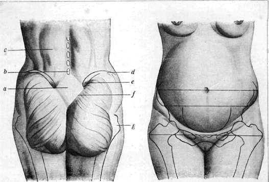
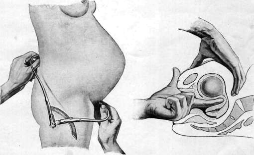
1, Michaelis' rhombus in a well-built woman: a - rhombus; b - spinous process of the 5th lumbar vertebra; c - erector trunci muscle; d - crest of the ilium; e - posterior superior iliac spine; f - line of attachment of the gluteus maximus; g - greater trochanter. 2. External dimensions of the pelvis: intertrochanteric distance (32 cm), distance between spines (26 cm); distance between crests (29 cm). 3. Measurement of the external conjugate (conjugata externa - 20 cm). A. Determination of pregnancy by internal obstetric examination: first sign of Hegar.
B. I. E, Obstetric Manual Maneuvers and Examination II
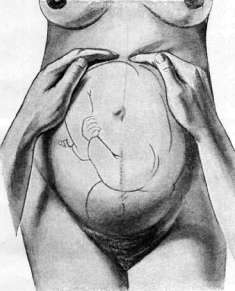
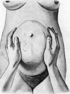
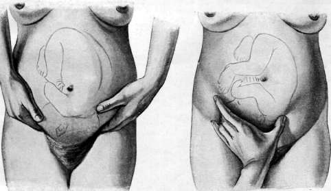
5. Determination of the position of the fundus of the uterus. G. Determination of the fetal spine (schoenzeit). 7. Determination of the presenting part*. 8. Grasping the presenting part (the third manual maneuver of Leopold).
Obstetric Manual Maneuvers and Examination III
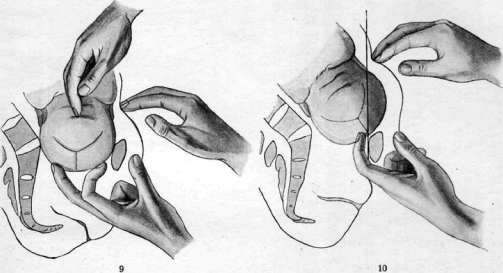
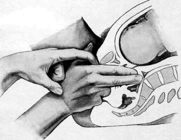
and 9. Depression of the head into the narrowed pelvic inlet according to R. Muller: the head with the help of the assistant's hands is depressed into the inlet, the discrepancy is small. 10. Depression of the head into the narrowed pelvic inlet according to R. Mй11et*: the head4 with significant protrusion beyond the edge of the symphysis, the discrepancy is large. 11. Measurement of the diagonal conjugate. Obstetric Manual Maneuvers and Examination IV 12. Delivery of the fetal posterior hand. 13. Lifting the body of the fetus upward in the method of Mor and co. U. Manual maneuver of Morriso-Levres for introducing the head. Obstetric Manual Maneuvers and Examination V
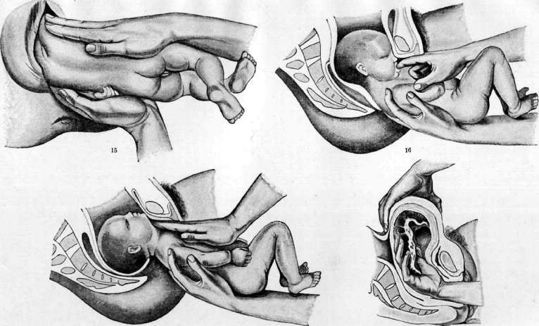
13 15. Turning the anterior sac of the fetus backward. 16. Delivery of the subsequent head with the chin pressed to the chest and turned toward the pubic arch. 17. Delivery of the subsequent head with the chin strongly abducted from the chest. 18. Manual separation of the afterbirth.
v. Ilyuvich.
Related articles
Mentioned in
Cite this page
“Obstetric Manual Maneuvers.” Soviet Medical Encyclopedia. English translation of Bolshaya Meditsinskaya Entsiklopediya, 1st ed. (Moscow, 1928–1936), ed. N. A. Semashko. https://sovietmedicalencyclopedia.pages.dev/article/obstetric-manual-maneuvers/