Seminal Vesicles
Historical document, translated for reference. It reflects medical knowledge of the 1920s–30s and is not medical advice.
Summary
This article from the 1928–1936 Soviet medical encyclopedia provides a detailed anatomical description of the seminal vesicles, including their structure, blood supply, innervation, and secretions. It also discusses their physiological role, pathological conditions, and the methods used to diagnose and treat related disorders.
Encyclopedia article (1928–1936)
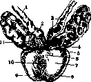
SEMINAL VESICLES (vesiculae seminales, glandulae seminales) represent a paired organ developing from the Wolffian ducts, consisting of two bag-like formations located between the posterior inferior wall of the bladder and the ampulla of the rectum, above the upper edge of the prostate gland; directed by their longitudinal axis from above and outside downwards and inwards, with their apex extending 4-5 cm above the Littre's triangle and the ureteral orifices. The lower end of the seminal vesicles approaches the upper edge of the prostate gland, so that the latter rests upon them. The necks of the seminal vesicles form an almost closed angle, which in newborns and in persons over 60 years of age is obtuse, while in maturity and old age it equals 60-80°. The peritoneum, passing from the bladder to the upper surface of the seminal vesicles, covers them for 1-2 cm, then ascends along the rectum. The seminal vesicles of an adult are usually 4-5 cm long, 1.5-2 cm wide, and 1-1.5 cm thick, presenting a convoluted tube, prostatic and ductus deferens: 1- ductus deferens; 2-ampulla of ductus deferens; 3-diverticula of ampulla; 4-excretory duct of seminal vesicle; 5-ejaculatory duct; 6-isthmus of prostate; 7-basis of prostate; 8-lobus sinister; 9-lobus dexter; 10-urethra; 11-excretory duct of seminal vesicle (see figure), corpus vesiculae seminalis. Which together with the ampulla duct forms the ejaculatory duct (d. ejaculatorius). The seminal vesicles are situated in a sac consisting mainly of connective and muscular tissue; their surface is uneven, the shape is elliptical, slightly flattened in the anteroposterior direction. The abundant blood supply originates from the hypogastric artery by vessels branching from it: inferior vesical (a. vesicalis inf.), middle hemorrhoidal (a. haemorrhoidal med.). The deferential artery (a. deferentialis) is located on the anterior wall of the duct and gives a branch anastomosing with the middle hemorrhoidal artery and with the lower branch of the testicular artery (a. spermatica int.). The veins generally follow the course of the arteries; the duct deferens vein, coming from the tail of the epididymis, joins the external spermatic vein. These veins form a plexus surrounding the duct deferens, ampulla, and passing into the seminal vesicle plexus (pi. venosus seminalis) and further into the vesico-prostatic venous plexus (pi. venos. vesico-prostaticus).-Lymphatic vessels. According to Sappey, are numerous and densely anastomose with each other; collecting then into 2-3 trunks, they empty into the hypogastric lymphatic vessels. The nerves of the seminal vesicles originate from fibers of the hypogastric sympathetic plexus, partly from the plexus pelvis, forming together with spinal nerves plexuses enveloping the seminal vesicles. The walls of the seminal vesicles consist of three layers - connective tissue, muscular, and mucous membrane. The medial wall, i.e., facing the duct deferens, is thicker than the lateral. The mucous membrane has toothed elevations of the most diverse forms, from small individual projections to a reticular structure, sometimes filling the entire lumen of the seminal vesicle. The epithelium is single-layered cylindrical, partly multi-layered cylindrical, sometimes cubic (Zaigraev).-Physiological significance of the seminal vesicles is still not sufficiently elucidated. The secretion of the seminal vesicles is transparent glass-like, sometimes yellowish, odorless, alkaline reaction (pH 7.6 according to Armistead). It contains fat-like droplets of various sizes, some 5-10 times larger than leukocytes, viscous, gelatinous granules called "sago grains." Upon staining, the latter give a reaction for mucus and a weakly positive reaction for fat, easily dissolve in acetic acid and were named by Robin simpexions (simpexions). In the secretion of the seminal vesicles, according to the research of Con, crystals of three types are contained: Reichmann's, Lubarsch's, and Béth's. The purpose of the secretion of the seminal vesicles lies in thinning the medium, which ensures greater motility of spermatozoa, in nourishing the spermatozoa not only inside the seminal vesicles, but also in the semen ejected into the vagina. According to recent research, the seminal vesicles apparently participate in internal secretion. Method of research of the seminal vesicles - see Vesiculitis and Vesiculography. Malformations of the seminal vesicles occur generally rarely. The most frequent is underdevelopment or absence of one seminal vesicle. In these cases, there is usually no corresponding testicle or kidney. Cases have been observed of the fusion of both seminal vesicles into one located in the middle, as well as cases of absence of both seminal vesicles. The method of vesiculography revealed other, smaller malformations, for example, connection of the two seminal vesicles to each other by a tube. The practical significance of these anomalies is insignificant.--Inflammation of the seminal vesicles is observed extremely rarely due to their deep position and easy mobility.--Cysts, ectasias, hydrocele, and empyemata of the seminal vesicles are observed most often due to obstruction or obliteration of the ejaculatory duct as a result of a chronic inflammatory process or in prostate hypertrophy. Their contents are either pure serous fluid, or turbid with admixture of mucus, or pus. The size of these formations is extremely diverse - from a small nut to a tumor protruding into the pelvic cavity. Thus, Frönshtein described an empyema in which the amount of pus in the seminal vesicles exceeded 700 cm3. The diagnosis of these formations does not present difficulties: upon palpation of the seminal vesicles through the rectum, an elastic tumor of corresponding size is felt. In doubtful cases, vesiculography can be performed, and in extreme cases - puncture of the seminal vesicles. Stones of the seminal vesicles are a relatively rare disease; they are formed due to stagnation of secretion as a result of narrowing of the ejaculatory duct, more often in old age. They consist mainly of calcium phosphate. The diagnosis of them is based on palpation of the seminal vesicles through the rectum, better on a curved metal bougie introduced through the urethra, and on roentgenography. Subjective symptoms are expressed in the form of pain during ejaculation, which can reach spermatic colic (colique spermatique). Other symptoms differ little from the subjective disorders caused by prostatitis, vesiculitis, and posterior urethritis. Treatment consists in striving to crush and remove the stone through the ejaculatory duct by pressing on the seminal vesicles through the rectum. For pain, hot sitz baths and even narcotics are prescribed. In severe cases, surgical intervention should be resorted to.- Tumors of the seminal vesicles, both benign and malignant, are extremely rare. Cancer of the seminal vesicles is most frequently encountered as primary, as well as in the form of metastases or as invaginated from neighboring organs. Their recognition is quite difficult, but pain in the inguinal region, large intestine, and perineum, difficulty in defecation and urination should cause suspicion of this disease. Cystoscopically, bullous edema is present in the area of the corresponding seminal vesicle. Upon palpation through the rectum, a roughened, enlarged seminal vesicle can be felt. - About syphilitic disease of the seminal vesicles is known little. Individual cases of this disease have been described.
Related articles
Mentioned in
Cite this page
“Seminal Vesicles.” Soviet Medical Encyclopedia. English translation of Bolshaya Meditsinskaya Entsiklopediya, 1st ed. (Moscow, 1928–1936), ed. N. A. Semashko. https://sovietmedicalencyclopedia.pages.dev/article/seminal-vesicles/