Sexual Sterilization
Historical document, translated for reference. It reflects medical knowledge of the 1920s–30s and is not medical advice.
Summary
This article from the 1928-1936 Soviet Medical Encyclopedia discusses sexual sterilization as a method of negative eugenics aimed at preventing the transmission of hereditary pathological traits. It describes the surgical procedures for men and women, differentiates sterilization from castration, and reviews the history of its legal implementation in the United States and Europe up to 1933.
Encyclopedia article (1928–1936)
SEXUAL STERILIZATION. Sterilization is one of the methods of negative eugenics (see), pursuing the goal of preventing the transmission of hereditary pathological traits to offspring. Sexual sterilization is achieved by means of an operation that leads to the cessation of patency in the excretory ducts of the gonads. Surgical intervention is reduced to cutting or excision of a certain part, or ligation of the spermatic cords in men and the Fallopian tubes in women. The operation of sterilization should not be confused with castration. In practice, there is a fundamental difference between the two, because in sterilization, as a rule, no harm is caused to the subject, whereas castration leads to a whole series of more or less serious disturbances in the proper functioning of the organism. Sterilization as a eugenic method was first introduced in the state of Indiana (USA) in 1909; therefore, this method in the literature is often called the Indiana system of practical eugenics. Subsequently, sterilization was also introduced into the practice of other states of North America and partially Europe (Switzerland, in 1922).

In total, in all countries of the world up to 1933, about 15-17 thousand operations had been performed.
Proponents of sterilization support their positions with two considerations. First, that a hereditarily defective subject, being frequently a helpless and sometimes a dangerous member of society, costs the state and society very dearly and tends, due to the absence of inhibiting factors, to produce numerous offspring, and thus the hereditary pathological predisposition with each generation acquires ever greater and greater distribution, and this fact already constitutes a threatening danger to the "well-being of the race." Second, surgical intervention in sterilization is not dangerous in itself, especially for men, and exerts a rather favorable than negative effect on the general (physical and mental) condition of the operated person. Among the ardent champions of sterilization, mention should be made of the American doctors Sharp, Oксner [Ochsner], the English doctor Rentoul, the German Beters, Neckke, and from the latter, fascist period—Lenz, Luxemburger, Rüdin, etc., the Italian doctor Zuccarelli, the Romanian Benedec. In the Soviet Union, the biologist Volotskoy spoke out in favor of sterilization. From the very beginning, the initiative of the state of Indiana, and following it other states of North America, provoked fierce criticism both from jurists and representatives of medicine. The former protested against compulsory sterilization as an attempt to violate personal rights, especially when persons who committed crimes against the person and vagrants were included among the persons subject to sterilization. Medical circles objected to sterilization mainly from the consideration that our current knowledge of the transmission of hereditary ailments from generation to generation is extremely insufficient, as is our knowledge regarding the identification of carriers of the morbid predisposition, in order to intervene in very intimate matters by such a radical method. In particular, very many German doctors spoke out against sterilization, who now, with the transfer of power to the fascists, have become ardent proponents of sterilization; a negative position is held by Ulbricht, Verweck, Hacking, Schmidt, and others. Among Russian authors, Bekhterev, Filipchenko, Koltsov, Yudin, and Davidenkov spoke out against sterilization. By and large, the method of sterilization has not yet received due recognition either in America or in Europe. Some of the proponents of sterilization (such as, for example, Sano, Cahane) have recently referred to the supposedly favorable results of the American experience. These references cannot be considered at all convincing, because from a eugenic point of view, making conclusions before a period of several generations has passed, subject to strict accounting of social and other factors, is premature to say the least. The question of sterilization acquires particular acuteness in our time, in connection with the fascist German law on the compulsory sterilization of the hereditarily defective, of July 1933. From January 1934, the law entered into force. According to the first paragraph of this law, the following categories of patients are subject to compulsory sterilization: 1) congenitally feeble-minded, 2) schizophrenics, 3) persons suffering from manic-depressive psychosis, 4) epileptics, 5) patients with Huntington's chorea, 6) hereditarily blind, 7) hereditarily deaf, 8) sufferers from severe hereditary physical deformities, and in addition it is stated that severe alcoholics may also be sterilized. According to indirect indications in the following paragraph of the law, as well as in the psychiatric commentary article on the law, repeat-offender thieves are also subject to compulsory sterilization. At first glance, modern data of psychiatric genetics may speak in favor of sterilization. According to the research data of Rüdin's school (Luxemburger, Schulz, Brugger, and others), it can be considered established that the incidence of psychoses among children and immediate relatives of the mentally ill is significantly greater than in the population in general. Thus, for example, the frequency of psychoses in the children of schizophrenics is more than 10 times greater compared to the prevalence of this disease in the population (9–10% : 0.6–0.85%), in brothers and sisters—8 times (6.5% : 0.6–0.85%), in uncles and aunts—3 times. Thus, the sterilization of the mentally ill would seem to acquire great meaning and significance. However, closer acquaintance with the issue forces one to evaluate this method differently. Most hereditary diseases are of a recessive nature; dominant diseases either occur very rarely (such as, for example, Huntington's chorea) or raise great doubts on the question of the undesirability of having offspring from them (manic-depressive psychosis). In recessive forms, as should be expected theoretically, the hereditary disease is obtained not as a result of the marriage of a sick person (homozygote) with a healthy one or with a sick one, but from so-called healthy people, heterozygotes. Clinical observations confirm these theoretical expectations. Thus, for example, it is rare when the parents of schizophrenics are mentally ill; the parents of epileptics usually do not suffer from epilepsy themselves. The data of Rüdin, Zoller, M. Bleuler, and others show an insignificant percentage of mentally ill parents of probands. When attempting to eradicate more or less widespread hereditary diseases through sterilization, according to Hardy's formula, more than a dozen centuries would be required, and that on the condition of ideal coverage of all homozygous cases by sterilization. And this is practically unfeasible, because, first, homozygosity for one or another pathological gene can be established only by the clinical picture of the affliction, but we know that a number of hereditary diseases are detected clinically late, after the carrier of the predisposition has produced significant offspring; second, a part of the homozygotes remains inhibited, with an unrealized disease, in some cases due to paratypic, in others—genotypic factors, and thus remains unrecognized and freely transmits the predisposition to offspring. Furthermore, in most forms of hereditary diseases, it is sometimes extremely difficult to distinguish the genotypic form from paratypic formations that are outwardly similar to it. The phenotypic range of a hereditary disease is very large, from massive ones, causing no doubts in anyone, to controversial, mild, poorly expressed cases. Applying sterilization only to severe cases, which is what the practice of America and Europe reduces to and as some authors still propose (Hübner), is incorrect, because in eugenic terms, mildly expressed cases are more dangerous, since they usually remain in life and can freely reproduce, while the former mostly, due to the severity of the disease, are torn away from life. Thus, sterilization in relation to homozygotes can hardly yield significant results while fully capable of serving as a source of all sorts of errors and abuses. The main distributors of hereditary diseases are recessive heterozygotes, and the main blow of eugenic measures should be directed in this direction. The use of sterilization in relation to this category of burdened people is completely unthinkable, because, first, in that case it would be necessary to subject every 15th to 20th person for the first disease and every 7th to 10th for the second to sterilization in relation to only such diseases as epilepsy and schizophrenia; second, at the present time we do not possess sufficient data that could make it possible to correctly recognize these heterozygous carriers of the morbid predisposition, despite the fact that it is known that a significant number of psychopathic personalities are often noted in the families of the mentally ill. The possibility of abuses acquires particular significance when the question of the sterilization of criminals is raised, proceeding from the unsubstantiated premise that a criminal act, anti-social behavior, is the phenotypic expression of the hereditary disadvantage of the subject, and not the result of unfavorable social and living conditions. These Lombrosian assertions have recently been tried to be revived again (Lange). If in an insignificant part of cases a criminal act can be linked to a psychopathic soil, on which these peculiar reactions to corresponding environmental irritations arise, then in the overwhelming majority of cases, the phenomena of criminality cannot be viewed within the sphere and by the methods of biological sciences. Meanwhile, starting with the experiment of the state of Indiana, the center of gravity of sterilization, either openly or veiledly, as in the German law, falls precisely on the fight against "criminal persons." And in this point lies the main danger of sterilization as a eugenic method—serving as a source of arbitrariness and abuses. In relation to criminals with sexual perversions, castration was used, and recently there have been tendencies to draw a distinction in "indications" in some cases for castration, in others for sterilization (Weatherill). Sterilization as a practical eugenic measure does not withstand serious criticism. In the dimensions in which it is possible to implement it in life, it cannot yield significant results, and in the dimensions necessary for sterilization to justify itself, it is completely inapplicable. According to Lenz's calculations, in Germany alone it is now necessary to subject at least 60,000–70,000 people to sterilization and, over the course of many decades, about 10% of each newborn generation. Compulsory sterilization can only cause irreparable harm to the cause of eugenic education of the population.
The roots of the modern "campaign" for sexual sterilization should be seen solely in the peculiar conditions of the fascistization of Western Europe, in the attempt of capitalist states to shift the center of gravity of growing class contradictions, crisis, and unemployment onto chauvinistic racial problems, clouding1 people's minds with the necessity of preserving the "purity of the race" and purging it of "pathological elements".
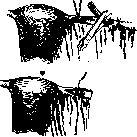
A- Hapachyan. Sterilization of the man (surgical methods)-see Castration-castration in the man, Rejuvenation. Sterilization of the woman. Modern surgical methods of sterilization of the woman. The idea of surgical sterilization of the woman was proposed by Blundell in 1819; initially, it pursued specific goals, namely permanent sterilization, and only gradually in the subsequent period, in connection with the development of obstetrics, the elaboration of the technique of certain obstetric procedures and gynecological operations, and changes in views on the indications regarding the admissibility of pregnancy under certain conditions of the woman, did methods of temporary sterilization begin to emerge. The multiplicity of surgical operations is the result of dissatisfaction with existing forms in terms of their reliability for the intended purpose.-According to the site of application, sterilization methods are divided into methods applied to: a) the vagina, b) the uterus, c) the ovary, d) the tubes. To approach the tubes and ovaries, various authors suggest using the abdominal, vaginal, or inguinal route. Operations on the vagina. In 1925, Haendly proposed achieving permanent or temporary sterilization by forming a double vagina. Surgical technique: a longitudinal incision along the vaginal mucosa on each side of the portio vaginalis, starting from the external os and ending at the entrance to the vagina 2 cm to the side of the urethra. Separation of the vaginal mucosa at a certain distance and approximation of the freshened surfaces to form a transverse septum. Freshening of the posterior surface of the cervix with the removal of the mucosa. Closure of the transverse septum from back to front produces a double vagina: a small anterior one for the drainage of uterine secretion and a posterior one for sexual intercourse (Fig. 1). Pomakion in 1924 reported two cases of restoration of a vaginal septum that had existed before childbirth and had been divided in one case and ruptured during childbirth in the other. The septa were located in such a way that during restoration it was possible to form a double vagina, one of which ended in a blind pouch, while the other looked toward the vaginal part of the uterus. Uterine methods. Among the methods of sterilization used on the uterus, mention must be made of the methods of Cox and Ursen, as well as the method of I. A. Golyanytsky and V. M. Kasogledov. Cox proposed an operation consisting in the artificial creation of mucous polyps of the cervix in the region of the external os. He was prompted to this idea by a case where a woman suffering from infertility and having a polyp in the region of the external opening of the cervix became pregnant shortly after its removal. The operation is performed as follows: above the external os, by means of a small incision parallel to the longitudinal axis of the uterus, the mucosa is detached, behind...

Figure 1. Haendly's method.
which three small stitches parallel to the direction of the incision are applied. The same is done on the posterior lip. Two small folds of mucosa are obtained, covering the vaginal part of the uterus (Fig. 2).-In 1899, experimenting on the question of the fixation of the ovum, Ursen made an incision in the uterus along the anterior wall and excised the mucosa. Golyanytsky, attaching importance to the chemism of the contents of the cervical canal, proposed the following operation technique. The operation is performed as follows: with a scalpel inserted parallel to the cervical canal, a cylinder of cervical mucosa 1--11/2 cm high is separated and completely excised. From the mucosa of the anterior fornix through its entire thickness together with the submucosal layer, a flap is formed - 21/2 cm wide and 3--4 cm high with its base toward the cervix, turned downwards and sewn with 3 interrupted catgut sutures to the freshened surface of the cervical canal. Next, a similar flap is excised from the mucosa of the posterior vaginal fornix and sewn in the same way as the first flap. Then, from the mucosa of the right fornix in the same manner, a longer flap (5--6 cm) is excised, 3 cm wide, placed over the sewn flaps of the blood-stained surface, for which purpose it is rotated, and it is sewn both to the denuded cervix and to the sewn flaps (Fig. 3). Tissue defects at the site of the excised flaps are closed by pulling together the surrounding tissues. Of 14 operated women under observation from 6 months to 3 years, only two became pregnant in the 8th and 9th month after the operation. In both women, the operation was accompanied by failure—non-fusion of the flaps with the freshened surface of the cervical canal. Kasogledov performed 28 operations according to Golyanytsky's method with certain modifications, consisting...

Figure 2. Cox's method.
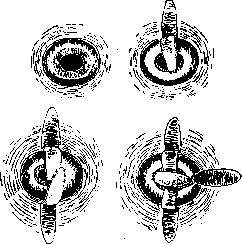
Figure 3. Golyanytsky's method.
in that he abandoned the excision of such a large amount of vaginal mucosa and took only 2 flaps. The first act of the operation was performed by Kasogledov in the same way as by Golyanytsky (Fig. 4). The first flap-pelot was taken from the posterior fornix and sewn to the anterior lip. The bridge-like flap, with a width in the middle of 31/3 cm, was lowered in the middle over the cervix and sewn to the upper and lower edge of the incisions in the fornices, with the corners at the base of the bridge left unstitched for 1 cm for drainage. Complete engraftment of the flaps takes about 2--3 weeks. 5 operations have been performed according to this modification. The result has not been ascertained. Out of 6 operations with one flap, as the author did initially, there were 3 failures, two patients were lost to follow-up, and one did not become pregnant (6 months of observation) (Fig. 5). Ovarian methods. These methods can be divided into three groups, namely: 1) methods of placing the ovary into the inguinal canal, 2) methods of placing the ovary into the broad ligament, and 3) methods of placing the ovary inside the peritoneum. Methods of placing the ovary in the inguinal canal. In 1905, van de Velde used the route previously used in the Alexander-Adams-Goldspohn operation, with opening of the peritoneal funnel to bring out the ovary and secure it there for the purpose of temporary sterilization. Upon opening the peritoneal funnel, finding the ovaries, and luxating them, a purse-string suture is applied to the peritoneum capturing the mesovarium to prevent the ovary from retreating into the abdominal cavity. The ovary was placed in front of the inguinal ring in the canal. After the operation, pain was observed in the area of the displaced ovary. Gutbrodt and Wessel performed this operation 6 times (Fig. 6). In one of these cases, the extraperitoneal location of the ovaries proved insufficient, since after 4 months the woman became pregnant again. No pain was observed. Menstruations were normal.- Methods of intra-ligamentous placement of the ovaries. The same van de Velde in 1907 reported a new operation of placing the ovaries between the leaflets of the broad ligament. The first step is the ligation of the ovarian fimbria. After lifting the tube and ovary, an incision is made in the posterior leaflet of the broad ligament, the peritoneal leaflets are separated, and the ovary is placed in this pocket. The edges of the incisions are joined with the capture of the mesosalpinx as well, to prevent the ovary from slipping out of the pocket. In 1909--10, Papan, Bucura, and Taddei performed this operation. Bucura proposed using it for temporary sterilization (Fig. 7). Van de Velde subsequently abandoned this method, stating that it creates the danger of altering the germinative epithelium when it is enclosed in connective tissue. Gottschalk, Mülberger
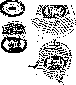
Figure 4. Kasogledov's method. Modification I.


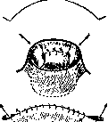
Figure 5. Kasogledov's method. Modification II.
Bucura, and Pestalozza consider that with this operation, in the final result, cystic degeneration of the follicles occurs with corresponding changes in menstruations. Method of placing the ovary inside the peritoneum. Blumberg in 1912 reported at the International Congress in Berlin on the operation he performed of placing the ovary in a peritoneal fold. He wrapped the broad ligament in the form of a cloak onto the posterior surface of the uterus and tightly sutured its free edges with silk sutures to the posterior surface of the uterus; the ovary is placed in the pocket thus formed and remains free there, since only the free edges of the broad ligament are sutured (Fig. 8). In six women operated on by Blumberg, no painful phenomena were observed, and menstruations proceeded normally. In 1912, van de Velde published a method for isolating the ovary in a hermetic cavity made of peritoneum: elevation of the uterus and transection of the ovarian fimbria, thanks to which the tubes approach the round ligament; a transverse incision is made in the broad ligament anteriorly and posteriorly, with a length corresponding to the length of the ovary, sparing the underlying tissues and vessels as much as possible. The connective tissue between the leaflets is also carefully separated with some blunt instrument. The ovary is guided through this opening onto the anterior surface of the broad ligament. With separate sutures...



Figure 8. Blumberg's method.
Figure 6. Gutbrodt and Wessel's method.
Figure 7. Papan and Bucura's method.
...

Figure 9. Van de Velde's method.
cover the edges of the posterior incision with the posterior leaflet of the mesovarium (take care of the vessels!). One should try to arrange the sutures so that the ovary does not move back completely or partially. Sutures on the anterior surface of the broad ligament, which can also be fixed to the anterior leaflet of the mesovarium (Fig. 9). The same is done on the other side. Then the ovaries lying on the anterior leaflets of the broad ligament are covered with the transitional peritoneal fold...
Figure 10. Flatau's method.
ligaments. In 1923, Lindig proposed placing the ovaries into a pouch made of appendices epiploicae. Grünwald experimented on animals by isolating the ovaries with the omentum, which easily adheres without being resorbed for 6 months. The ovaries do not change their histological structure. Liberation of the ovary presents no difficulties. Methods of tubal sterilization. These methods, the most numerous in quantity, originate from Blundell's proposal in 1819 to remove parts of the tubes on both sides at the end of a cesarean section so as not to resort to the latter a second time. In 1880, Lungren performed the first ligature of both tubes. This method was used for a long time by many gynecologists and not without success. There were reports from a number of authors about failures as well. Subsequently
Figure 12. Hans's method.
Figure 11. Madlener's method. the number of reports of failures increased, and the operation was finally discredited after Fränkel's experiment. A loop from the tube itself was made by Flatau by preliminarily separating the tubes from the broad ligament (Fig. 10). The broad ligament was then sutured. Flatau (1926) performed this operation six times, but it did not become widespread. Stolz proposed forming a fold from the round ligament via anterior colpotomy, which simultaneously leads to the formation of a tubal fold. By applying a silk ligature to the tubal fold, he simultaneously captures the fold itself. If the ligature were to cut through the tube, the fold of the round ligament would serve as a protection against the penetration of spermatozoa. Crushing of the tubes. Friedemann in 1906 proposed replacing the usual tubal ligation with ligation preceded by
through the vagina,
Figure 14. Markov's method.
crushing of the tube along with its mucosa. In 1910, Madlener used this proposal and in turn described the method of applying a ligature with preliminary crushing of the tubes. The tube is grasped in the middle and raised so high that a right angle is formed between the separate parts of the tube. The second hand, armed with Doyen's enterotribe, applies it under the forceps to both parts of the tube below the mesosalpinx and crushes the tube; a tight ligature is applied to the crushed site (Fig. 11). The operation can be performed both through the vagina and by laparotomy. Madlener reported 89 cases of this operation with remarkable success. The best success in terms of forming a dense scar was obtained if a small part of the mesosalpinx was captured during the crushing of the tube. In 1921, Hans described a method of crushing the tube in two places using Blunk's forceps with the application of ligatures, tightening them and forming a loop (Fig. 12). In 1925, Waser reported 225 cases of sterilization by a modified Madlener method; he applied the crushing instrument so that it captured a significant part of the mesosalpinx (Fig. 13). In 1919, Cupler ligated the vessels that nourish a part of the tube, excised this part, and freed the uterine part of the tube from the peritoneum surrounding it, crushed this part of the tube with an angiotribe, ligated it, and peritonealized it. Markov proposed modifying Madlener's operation and performing tubal ligation internal and external to the crushed areas. Baksht experimented on animals and confirmed the expediency of this modification (Fig. 14). Transection of the tubes between two ligatures. Simon Thoma in 1885 recommended double ligation and transection using a Paquelin cautery. Kehrer in 1897 proposed performing the operation via the vaginal route, using the principle of ligation and transection of the tube. Fabritius and Köhler modified Kehrer's method in the direction of transecting the tubes after preliminarily applying a ligature to the loop. The two stumps formed after transection are ligated to ensure better impassability (Fig. 15). Double ligature with transection. In 1898, Rühl proposed the following method of tubal transection with peritonealization of the stumps: via anterior colpotomy, the uterus is brought out, double ligatures are applied,
Figure 15. Fabritius's method. and the tube is transected at a distance of 5 cm from the uterine angle. The uterine stump of the tube is separated from the mesosalpinx, then pulled down and fixed in the vaginal incision. In view of the difficulty of placing the stump with a median incision, the author later proposed making small linear incisions on each side of the median incision. In 1897, Beuttner published a method of this kind for celiotomies. The tubes were transected at their distal end closer to the ampullary part, then the muscular layer of the tubes was sutured and above it the middle layer, and finally a suture was applied connecting both segments after their peritonealization (Fig. 16). In 1901, Kehrer proposed two methods of sterilization—one as an independent method, the other as a method accompanying some other surgical intervention. To perform sterilization alone, he proposed using colpotomy with a 1 cm incision of the tubal peritoneum at the uterine angle, isolating a 2 cm loop of the tube through the incision and its resection; the stumps are ligated and again buried between the leaflets of the peritoneum, which is sutured, separating the stumps. For cases of surgical interventions with concomitant sterilization, Kehrer proposed making a wedge-shaped excision of the uterine angle. Hofbauer in 1911 proposed two technical techniques that were supposed to give good results. The first modification consists in the fact that the tubes are transected approximately at the border of the middle and lower thirds; the distal end is ligated, and the proximal one is placed between the proper ligament of the ovary and the round ligament and attached here by suturing the round and ovarian ligaments. This method was used by the author in abdominal celiotomy. For vaginal operations, the author proposed: transection of the tube, ligation of the distal end, and implantation of the proximal end into the loop of the round ligament (Fig. 17). In 1905, Asch reported the following method: the tube is transected at the transition site of the middle part into the uterine part. A ligature is applied
Figure 18. Asch's method.
Figure 19. Rabinovich's method.
to the uterine stump, under the stump of the distal part a ligature is applied to the vessel running under the tube. The peritoneal leaflets are separated, and the ligated uterine stump is buried into the pocket formed between the leaflets. The distal end of the tube remains unligated (Fig. 18). In 1911, experiencing certain difficulties with this method in terms of using the separated thin leaflets of the peritoneum of the broad ligaments, Labhardt proposed his method: after transecting the tube near the uterus and ligating the distal end, the tube is isolated from the peritoneal covering, a part of it is excised, the tube is ligated at the uterine angle beyond the site of tubal excision, the stump is peritonealized, and the peritoneal cuff formed after pulling out the tube is sutured. Rabinovich applied two ligatures to the tubes and then isolated a segment of the tube with a linear incision of the peritoneum, trying not to injure the vessels of the mesosalpinx (Fig. 19). Suturing the leaflets of the broad ligament; the uterine stump of the tube is sutured to the uterus in such a way that it points toward the midline of the body of the uterus. Skrobansky uses a double ligature of the tube with its transection and burying of the uterine end; the uterine end is folded in half, ligated with the same ligature, and buried in the broad ligament behind the uterus (Fig. 20). Freund, after applying two ligatures at the uterine end and transecting the tube, fixed the uterine stump to the abdominal wall (Fig. 21). Pfeilsticker in 1925, via anterior colpotomy after bringing out the fundus of the uterus, severed the tube at a distance of 1 or 1.5 cm; after suturing the angle, he made a small 1 cm incision of the peritoneum here, into which he transplanted the tubal stump. In 1928, Matveev reported a method that he had been using for 15 years and which consists in the fact that, as in Pfeilsticker's method, the proximal end of the tube is buried and attached to the bottom of a pocket 1 cm deep, obtained by an incision on the anterior wall of the uterus, at the level of the lower edge of the round ligament and 1 cm from the edge of the uterus (Fig. 22). Irving proposed the following method of burying the transected tube: via colpotomy, the uterine angle is brought out, the tube
Figure 20. Skrobansky's method.



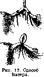




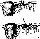
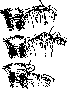

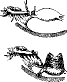
Figure 21. Freund's method. The tube is ligated with a double ligature at a distance of 3-5 cm from the uterine end; the proximal end of the tube is separated from the mesosalpinx (Fig. 23). With a straight needle threaded with a double thread, the proximal end is sutured in such a way that a loop is formed around it, then a small incision of the peritoneum near the corner of the tube and spreading of the musculature forms a pocket. The needle carrying the two threads fixed to the tube is passed through this pocket and brought out a few centimeters from the edge of the incision. Then the incision is closed
Figure 22. Matveyev's method.
with a suture, and the distal end is also buried between the leaves of the broad ligament. Gruzdev reported in 1922 on the method applied by him of sectioning the tubes with subsequent placement of the ends of the transected tubes one upon the other and tying them with peritonization (Fig. 24). Resection of the fallopian tube with processing of the stumps. An operation of this kind was first performed by Fritsch in 1897. After applying two ligatures to the tube at a distance of 2 cm from one another, he excised the part of the tube lying between these ligatures. In 1912, Liepmann proposed his method, which is basically similar to Kirchhoff's method (Fig. 25). Celiotomy: incision of the serous tube from the uterine angle by 5 cm, isolation of the tube from the peritoneum and excision of 1 cm. Suturing of the peritoneal edges with a Lembert suture. Hauch excises 4-5 cm
Figure 23. Irving's method.
of the tube after a preliminary longitudinal incision of the serosa and isolation of the tube, with the application of ligatures to the distal end through the serosa and burial of the proximal end between the leaves of the broad ligament, and for the burial of this stump, the ends of the ligature tying the tube stumps in the uterine part are used (Fig. 26). In 1928, Sergeyev, with the aim of subsequent use of the tube segment if it is necessary to eliminate female sterility, left the distal end of the tube open. In addition, he preserved the vascular arcade for normal nutrition of the remaining part of the tube; the uterine part of the tube was resected together with the serous cover; connection of the peritoneal leaves with a interrupted suture, and the last suture toward the distal end of the tube is located so that the knot lies on the anterior or posterior leaf of the broad ligament, so as not to irritate the tube segment (Fig. 27). Wedge excision of the tube and excision of its interstitial part. In 1898, Neumann and Rose simultaneously proposed a method similar in idea: excision of a wedge from the region of the uterine angle with the expectation of excising the interstitial part of the tube. The wound was sutured with catgut. The idea of such removal was used even earlier by Schauta in salpingitis isthmica nodosa (Fig. 28). Taussig proposed
Figure 26. Hauch's method.
Figure 24. Gruzdev's method. Figure 25. Liepmann's method. wedge excision without resection. The round ligament is pulled to the uterine angle with a suture; the excised tube is buried between the leaves of the peritoneum formed by an incision along the inner edge of the round ligament, where it is sutured; the peritoneal incision is restored (Fig. 29). Serdyukov in 1929 reported on the method used by him and consisting in wedge-shaped excision of the tube with ligation of the distal end and its burial, after suturing the uterine angle, between the muscles in the lower corner of the incision using the ends of that ligature which was applied to the tube. The author attributes to the merits of his method fixation in muscle tissue, the absence of trauma to the mesentery, better blood supply to the tube, and the possibility of its transplantation. Blagovolin as early as 1921 proposed a similar method, but with the amputation of a part of the tube (Fig. 30). Solovyov performs a shallow wedge-shaped excision of the tube during abdominal celiotomy, ligates the tube stump and with the ends of this same thread buries it at the lower corner of the wound between the muscles. The upper part of the incision is sutured with a continuous suture and covered over it again with the round ligament with its mesentery, having freshened the surface of the uterine angle (Fig. 31). Kiparsky reported in 1927 on a method of wedge excision with resection of the uterine part and leaving the distal end of the tube open, with the mucosa of the tube being sutured to the serosa (Fig. 32). Genter ligates the tube at a distance of 1-1.5 cm from the uterine angle; a piece of tube from the ligature to the uterus is excised with scissors. The ends of the ligature are used to bury the stump between the leaves. The uterine angle is sutured (Fig. 33). Amputation of the tube and displacement of the ends from the angles with their burial. In 1913, Tarnovsky proposed an operation which he called "tubal reimplantation" (Fig. 34). It consisted of the following: the tubes were amputated at the uterine angles. The uterine parts were buried and peritonized, the ends of the tube segments were moved into a through channel 6 cm long formed on the posterior surface of the uterus in its musculature, and the tube of each side was pulled to the edge of the channel of the opposite side and
Figure 27. Sergeyev's method.
SEXUAL STERILIZATION over them through the thickness of the channel a suture was applied. The author had in mind the possibility of reverse transplantation of the tubes into the uterus. Resection of the ampullary part of the tubes. Menge, during the operation for prolapse of the uterus and vagina during the performance of ligament shortening according to Alexander-Adams, performed amputation of the ampullary end of the tube, opening the peritoneum and suturing the tube stumps to the aponeurosis
Figure 28. Peay's method
Figure 29. Taussig's
method.
of the large oblique muscle. Krönig also considered resection of the abdominal end to be a simple and reliable operation, but he proposed doing this via colpotomy. Spinelli proposed performing the same operation via posterior colpotomy. Lvov as early as 1897 proposed this operation. Mironov proposed using anterior colpotomy as the route and, having brought out the uterus, separating the tubes from the broad ligament, resecting the distal end of the tube and placing the stump of the amputated tube into the vaginal incision. Mermann moved the tubes, having previously amputated the abdominal ends, to the anterior surface of the uterus, attaching them here and suturing both lumens of the tubes. Such a position of the tubes, giving a kink in their uterine part, further protected the uterine cavity from the lumen in the tube and from the abdominal cavity (Fig. 35). Faynberg recently proposed
Figure 30. Blagovolin-Serdyukov's method.
are placed on the ampullary end of the tube and tied in such a way that the knot falls on the lateral side of the tube opposite the round ligament; the ampullary end is amputated, and the stump is cauterized (Fig. 36). "Here I must point out," says the author, "that one should not amputate a large section of the ampullary end and one must try to ensure that the resulting suture around the tube is in a strictly frontal plane, because otherwise the tube can slip out of the thread embracing it and this will force us to apply new sutures. Then a needle is put on the free end of the threads in turn and the first (a) is injected into the outer edge of the mesosalpinx and under the round ligament, and the second (b) earlier under the round ligament, stepping back from the previous injection at a distance of 1 cm outward from a, and into the posterior leaf of the mesosalpinx, trying to catch a sufficient fold with the needle (if the anterior leaf of the mesosalpinx is also punctured during this, it does not matter). With such a typical management of the threads, the latter cross and when they are tied, the stump is inverted and very well peritonized, and the stump rests tightly against the posterior wall of the broad ligament in the region of the alae vespertilionis. It is necessary to make the injections under the round
Figure 31. Schauta-Solovyov's method.
Figure 32. Kiparsky's method.
ligament at a distance from the uterine angle equal to the remaining part of the tube, then upon final tying of the suture the remaining part of the tube will lie parallel to the round ligament without forming an arc." Displacement and burial of the abdominal end of the tube. Rühl, Selheim, Nürnberger, and Mironov reported on the method of displacing the ampullary end of the tube into the vagina during colpotomy, and they used various approaches to the tubes:
Fig. 33. Genter's method.
Figure 34. Tarnovsky's method.

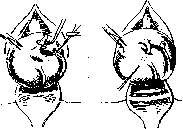



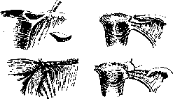
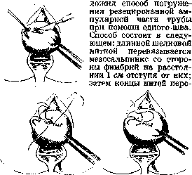


of the same. Selheim, Nürnberger-posterior, Mironov, and Pfeilsticker—via anterior colpotomy. Selheim fixed the exteriorized ampullae of the tubes in the vaginal scar. Nürnberger placed the tube ampullae between the peritoneum and the vaginal mucosa, suturing them to the peritoneum and carefully stitching the peritoneum around them. Pfeilsticker proposed a modification of his operation, consisting in the fact that after opening the peritoneum during colpotomy, the tubes are withdrawn with long forceps, their fimbriated ends are placed on the anterior surface of the uterus, where they are grasped with a fine suture to the uterine wall and with another suture are sewn to the plica; thus, upon closing the wound, they are located extraperitoneally.-Transplantation into the inguinal canal. Menge proposed in 1900, to ensure permanent results in operations for uterine prolapse and descensus, to combine perineal surgery and the Alexander-Adams operation with sterilization, in which the tube was pulled out through the opened peritoneal cone, separated from the broad ligament, and a large part of it was resected. The uterine end of the tube was sutured in the inguinal canal beneath the aponeurosis of the external oblique muscle. Stoeckel in 1915, with the aim of restoring the possibility of pregnancy, proposed suturing the tube at the abdominal ostium so that the ampulla was exteriorized and a significant part of the tube was extraperitoneal, placed in front of the fascia and muscles so that the ampullary end of the tube was located as laterally as possible. The round ligament is secured in the usual manner (Fig. 37). Küstner as early as 1911 spoke of positioning the fimbriated end in the inguinal canal. Elos sutured the fimbriated end into the inguinal canal.-Transplantation into the broad ligaments. In 1909, Selheim proposed embedding the abdominal end of the tube into the broad ligaments: liberation of the ampullary end by cutting the ligament, incision of the anterior leaf of the broad ligament, and securing the tube here. Turenne reported in 1911 on a method he had been using since 1911, which consisted in making an incision on the stretched broad ligament at a distance of 15-20 mm from the posterior edge of the tube near the ampulla, corresponding to the widened end of the tube; the peritoneal leaves were separated and the ampulla of the tube was placed here; fixation was achieved with a circumferential suture and a single suture of the free part of the surrounding peritoneum (Fig. 38). Merrmann's method.
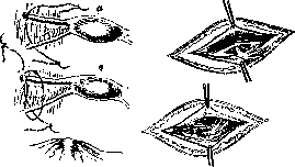
Fig. 36. Feinberg's method. Figure 37. Stoeckel's method. Intraperitoneal embedding of the ampulla. Littauer in 1927 proposed a method resembling Pfeilsticker's method, but performed via laparotomy. Liberation of the tube and passing it under the round ligament with embedding beneath the uterovesical fold (Fig. 39). Schweizer in 1921 proposed exteriorizing the fimbriated end of the tube onto the anterior leaf of the broad ligament, passing it under the round ligament and positioning it in the peritoneal depression located between the urinary bladder and the round ligament; the peritoneum surrounding the tube is closed. The tube has a bend in its isthmic part (Fig. 40).-Embedding into the abdominal wall. In 1921, Hellendal reported on a method he used for the sterilization of women: after ventrofixation according to Olshausen, a few centimeters above the fixation point, the ampullary ends of the tubes are passed through an incision in the peritoneum and aponeurosis and secured between

Figure 38. Turenne's method.
Figure 39. Littauer's method. the aponeurosis and the skin. To prevent the tubes from shifting back into the abdominal cavity, silk sutures are passed through the infundibulopelvic ligament, grasping the coat of the tubes. Oliva in 1905 reported a method similar to the aforementioned, with a minor modification consisting in also ligating the ampullary end in two places.-Suturing the abdominal end of the tube to the uterine wall with coverage by the same wall. Winckel in 1909 described a method in which, by means



Fig. 41. Winckel's method. Figure 40. Schweizer's method. anterior colpotomy and separation of the tubes from the broad ligament, their ampullary parts are closed with a suture and sutured to the posterior wall of the uterus (Fig. 41). In 1918, Alfieri performed the following operation: separation of the ampullary part of the tube from the mesosalpinx and their resection with ligation of the tube segment and passage of the ends into a channel formed within the thickness of the posterior wall of the uterus, with fixation of the ends there. Kushtalov, being an opponent of surgery on the uterine part of the tube, proposed (1927) a method involving resection of the abdominal end of the tube after prior separation of the tubes from the broad ligament, with suturing of their ends and embedding beneath the serous membrane of the anterior uterine wall (Fig. 42). If the need arises to restore the ability to conceive, the tubes can be put back in their place. The author believes that the ends can be implanted into the uterine cavity and thereby preserve the fluid circulation between the tubes and the uterus. The method resembles Merrmann's method.-Invagination of the fimbriae and closure of the tubal funnel. Muret in 1909 proposed for temporary sterilization the invagination of the tubal fimbriae with placement of a suture. In 1931, Nauyoks proposed modifying Madlener's operation by crushing the tube at the ampullary end and ligating it in two

Fig. 42. Kushtalov's method.
sites, keeping in mind the possibility of subsequently restoring the patency of the tubes. Considering the operations on the vagina by Tsomakion and Gondle, it is necessary to point out the limitations of their application, keeping in mind the requirements they impose in the sense of the width of the vagina to form a sufficient capacity of that half of the vagina which is intended for intercourse. Even in cases with a double vagina, this task is not always resolved, as can be seen from the reports of the author (Tsomakion), who observed sharp pain during intercourse after the operation. In addition, one must keep in mind the difficulties in applying sutures. Operations on the cervix—by Cox, Golianitsky, Kasogledov—do not guarantee good results, in view of the spermatozoon's ability to make its way through various obstacles of both a mechanical and chemical nature. Ovarian methods—by van de Velde, Wessel, Bukur, Blumberg, Lindig, Gutbrodt—must be recognized as the most reliable methods of sterilization, but they also have many drawbacks both in terms of technique—which is extremely delicate—and in relation to the phenomena accompanying the operations, among which pain occupies a definite place. Van de Velde's method, appearing delicate in terms of technique, is sufficiently reliable, and one cannot but agree with Mühlberger, who, analyzing various methods, writes regarding van de Velde's method: "It must be confessed that this method serves as an excellent model of surgical physiology and all methods of temporary sterilization of women cannot have a chance of success if they are not as physiological as van de Velde's method." Further, this author notes the main advantage of the method, which consists in the fact "that the ovary is artificially enclosed in the abdominal cavity and is under natural intraperitoneal pressure, which protects against damage to the germinative epithelium." Blumberg's method can also guarantee reliable sterilization and the onset of pregnancy after releasing the ovary from the peritoneal pouch if necessary. As for the operation on the tubes proposed for the sterilization of women—simple ligation of the tube with a ligature—this operation, which yielded many failures, was in its time tested not only by re-opening the abdominal cavity for one reason or another and studying the changes in the tubes, but also by experiment. Both experiment and the study of the woman's tube showed the unsuitability of this operation. The next stage in the development of operations on the tubes was the ligation of the tubes with their transection. This method was also not free from failures, as evidenced by reports from a number of authors. Madlener's operation, which tempted everyone with its simplicity and, according to initial observations, good outcomes, has recently, on the basis of newer observations, begun to lose its value. The author of the operation reported in 1926 on 124 new cases operated on with complete success. In 1924, Köhler reported one case of pregnancy out of 20 cases operated on by his method. Methods based on the method of Madlener, Vayser, and Küpfer gave better results, having more weighty grounds; these grounds consisted, as can be seen from the description of these operations, in addition to crushing the wall and mucosa of the tube, also in involving part of the mesosalpinx with blood vessels, followed by scarring and atrophy of a certain part of the tube. In methods with crushing, the method of crushing itself and the application of ligatures play a major role in the subsequent outcome. Here it must be noted that the formation of fistulous tracts in the tube after this intervention, which subsequently leads to the onset of pregnancy, depends precisely on how the crushing of the tube was performed. Attention must be paid to the use of Hans's Blunk clamp. If operations of transection of the tubes after their preliminary ligation did not meet the requirements presented to them, then, when modified, they gave better results. Double or single ligation with subsequent burial of the ends of the tubes, according to the observations of many gynecologists, gave better results, but also not faultless ones; the operations of Baitner, Hofbauer, Asch, Labhardt, Dütsmann, and others also failed in many cases to ensure the ultimate goal. Blitz, who collected by questionnaire the results of operations according to Asch's method in 118 cases, did not see failures in the coming years. As can be seen from the reports of a number of authors, methods that boiled down to ligation, resection, and subsequent burial of the tubes in the broad ligaments also failed to achieve their goals in some cases. This applies to the operations of Fritsch, Kirchhoff, and Lippmann. Methods aimed at removing the interstitial part of the tube and striving to create a barrier specifically in this part for the egg cell and spermatozoon moving toward each other, proposed in a fairly significant number of various modifications recently, have also given a certain number of failures. The desire of individual authors who proposed their modifications to the main operation of this type, used by Schauta in removing the nodular thickening of the tube in its isthmic part, boiled down to better isolation of the stump of the tube both in the thickness of the uterus itself and between the leaves of the broad ligament (Taussig, Genter, Blagovolin, Serdyukov, F. Solovyov, and others). While striving for this same goal, the necessity of good coverage of the passages of the uterus itself was not forgotten, keeping in mind the ability of epithelialization of the passage among the applied sutures, and in case of their cutting through in the formed passages. To create a more stable barrier between the bottom of the uterus and the excised tube, covering the angle of the uterus with the round ligament should be considered a good technique (Schauta, F. Solovyov, Taussig). There are many observations on the onset of pregnancy after the removal of tubes on both sides. Küstner and Pollak reported cases of interstitial pregnancy after wedge-shaped removal of the tubes. Bainbridge described a case of pregnancy after bilateral removal of the tubes and ovary with transplantation of a part of the ovary into the stump of the tube. Resection of the abdominal end of the tubes with subsequent treatment of the stumps and their burial, according to the observations of a number of authors, also yielded failures; thus, according to statistics, for 9 failures in the operation of resection of the tubes and burial of the stump in the broad ligament, there was one case related to the resection of the abdominal end with its suturing (Muret's case). During the operation of burying the ampullary end of the tube into various places—the vagina, inguinal canal, broad ligament of the peritoneum, and the uterine wall with or without resection—fewer failures were observed than in the operations listed earlier, but nevertheless, some of them were not faultless here either. Turenne, who proposed his method of burying the ampullary edge of the tube into the broad ligament, thinks that this method should not be used during pregnancy and during Cesarean sections with sterilization. These operations present a number of inconveniences due to the structural features of both the ampullary part and the opening in the broad ligament. An important role is also played by the difficulties of fixing the tube in a new place. Pointing to the resorption of adhesions after 3–4 years and the return of the tubes to their old place with such methods of burying their ends (Lochrane). Adhesions persist only in infected cases. Schweitzer's method with the placement of the ampullary ends of the tubes in a special peritoneal pouch formed from the depression of the peritoneum between the round ligament and the bladder must be considered acceptable in its idea, keeping in mind the possibility of subsequently releasing the tube if necessary. There is not a single report on the fate of women when using it, but it is technically difficult. From the foregoing, it follows that: 1) Methods associated with the crushing of the tubes and subsequent ligation of these places, or ligation far from the crushed place, and with the crushing and ligation of the mesosalpinx as well, while giving good results, are relatively simple in technique (Madlener, Vayser, Hans, Markovsky). 2) Transection and resection of a part of the tube with the burial of the ends in the broad ligament, while giving satisfactory results, appear to be more complex and delicate operations and, by virtue of this, cannot compete with the methods of crushing the tube with its simultaneous ligation (Rabinovich, Irving, Dütsmann, Asch, Gauch, Lippmann, Labhardt, Matveev, Kiparsky, Sergeev, Skrobansky, and others). 3) Wedge excision with the burial of the stumps between the muscles of the uterus with good peritonization of the uterine angle represents a surer operation than resection of the tube, and has an advantage over it in the sense of reliability, as well as the possibility of subsequent transplantation of the tube into the uterus (Blagovolin, F.
Solovyov, Taus-sig, Serdyukov, Schauta, Sergeyev). 4) Operations on the ampullary part of the tube with the burial of this part after its preliminary resection, although ideally they should yield permanent results, yet their production technique and the peculiarities of the physiology of the tube do not make it possible to classify them as operations with stable results (Menge, Krönig, Mermann, Alfieri, Kushtalov). 5) Burial of the ampullary part of the tube without its preliminary resection (Zelheim, Nürnberger, Pfeilsticker, Menge, Stoeckel, Turen, Hellendall, Oliva, Winkel, Lippmann, Mironov, Schweitzer, Littauer, Feinberg, etc.) yields various results; among them, methods of intraperitoneal burial of the tubes deserve attention as being more physiological (Schweitzer, Littauer) and providing an opportunity to restore the ability to conceive, in case of the emergence of a desire for it, without particularly complex additional procedures. When considering and evaluating the surgical methods proposed for the sterilization of women from the standpoint of the suitability of some of them for the purposes of the so-called temporary sterilization of women, it is necessary to proceed from the requirements that are presented to these methods, namely: 1) they must be uncomplicated; 2) they must not cause significant changes of either an anatomical or functional character; 3) they must be effective for a certain time; 4) they must allow the possibility of conception to be created at any time. These requirements must be fully presented to any proposed method. Of the methods proposed above, some satisfy the indicated requirements; along with them there are others which, not fully satisfying the entire set of requirements, can nevertheless guarantee the ultimate goal, i.e., temporary sterilization; finally, there remains a group of methods where the changes occurring during the operation are so sharply expressed that although it is possible to think about creating the conditions necessary for the onset of conception, their implementation is extremely difficult. Among operations on the tubes, there are a number of methods that make it possible by means of additional procedures to create the possibilities for the onset of pregnancy (reimplantation of the tubes and salpingostomy) (see). Cases of pregnancy after reimplantation of the tubes are known from the literature. After the operation of salpingostomy, Martin observed conception in two cases out of 47, Seitz in 2 cases out of 12. Thus, by some authors these operations are also classified as operations that can be used for temporary sterilization. Solovyov, admitting such a possibility, still cannot put them on a par with the methods of temporary sterilization, where already from the very beginning the surgical intervention provides for better preservation of the anatomical and physiological state of the tubes. As for uterine methods, as can be seen from the foregoing, one cannot yet say that they meet the requirements presented for operations with the aim of temporary sterilization. Here failure is guaranteed, and complications are also guaranteed along with anatomical changes. Ovarian methods, which were tempting because one has to deal with more stable tissues in the sense of their damage during the operation, provide a better opportunity for securing and displacing the organ, but have their drawbacks both in relation to the pain developing in some of them by virtue of pressure from surrounding tissues and as a result of hemorrhage in the case of their enclosure in a closed sac. Of these methods, the methods of van de Velde and Blumberg should be considered the best for the purposes of temporary sterilization, and the method of van de Velde must be placed in first place because the suitability of this method was proved by a quickly ensuing pregnancy after the performance of a second act of operation after a five-year hiatus. Among operations on the tubes for temporary sterilization, those operations draw attention where the tubes throughout their entire extent are not subjected to any influences that could disrupt their integrity or nutrition. In addition, it is necessary to take into account those observations that have been gathered in relation to the operation with the burial of the ampullary end of the tube in various places; they show that the arbitrary release of these ends led rather quickly to the onset of pregnancy. As for the results in the sense of the onset of pregnancy during the performance of the burial of tubes with their subsequent release, although the number of observations here is small (Zelheim), nevertheless they also speak in favor of the possibility of the rapid onset of pregnancy, since these operations do not produce sharp changes in the tubes themselves V85 78and the tube quickly returns to its function. As for the selection of operations corresponding to the requirements presented for operations for temporary sterilization, among the multitude of them, these requirements are most of all met by operations where the ampullary edge of the tube is placed inside the peritoneum (Littauer, Schweitzer). From the standpoint of the second proposition regarding the group of methods suitable for temporary sterilization, Naujoks cites 24 methods, among which are also mentioned methods where procedures of a complex character are used in relation to the tube both in primary and secondary intervention, influencing to a certain extent the subsequent life of the tube, changing both its anatomy and function, and he makes a reservation regarding nine methods (Beuttner, Zweifel, Hofmeier, Harris, Schauta, Tarnovsky, Holzapfel, Gsel and Pirkner) "that there can be no complete certainty in the reliability of the operations when applying the indicated nine methods," and further: "in the issue of interest to us, more important than achieving temporary sterilization as a result of the first part of the operation is the degree of probability of a positive result from the second part of the operation—the restoration of the ability to conceive." Since there are no definite data for judging the suitability of the indicated methods, Naujoks, subjecting them to criticism from the standpoint of pathologo-anatomical changes, comes to the conclusion of their unsuitability for the purposes of temporary sterilization. He attaches great importance to the dislocation of the tubes and ovaries in relation to the subsequent restoration of the ability to conceive, i.e., he also stops at methods that meet the requirements set forth by us, and expresses the same thoughts that are expressed by us in the analysis of operations assigned to this same group (Menge, Zelheim, Nürnberger, Stoeckel, Hellendall, Pfeilsticker) about the difficulty of implementation and doubts in the sense of suitability for functions for methods of extraperitoneal burial of the tubes. For a more precise characterization of the suitability of a part of the methods proposed for the surgical sterilization of women for the purpose of temporary sterilization, we do not have a sufficient number of observations, and therefore one has to approach this assessment from the standpoint of more theoretical considerations, taking into account the requirements set forth above in relation to such operations. As a result of the consideration of these operations, the following considerations can be expressed: 1) For temporary sterilization, methods should be used that guarantee the greatest preservation of the tube or ovary in the anatomical sense, which guarantees their subsequent function. 2) Among such methods, it is necessary to note the methods of van de Velde and Blumberg for the ovary and the methods of Schweitzer and Littauer for the tube. 3) Of the other methods proposed for sterilization, methods can be applied with some modifications where the primary intervention guarantees the least damage to the tube and provides the possibility of its subsequent reimplantation. As for clinical complications in the operation of the sterilization of women, on the basis of the collected material presented by Lafond, it is seen that out of 394 collected cases there were 9 cases bearing the character not of special complications for these operations, but the character of complications in general after a first laparotomy. In addition to these complications, it is also necessary to point out a number of cases of the subsequent development of ectopic pregnancy after various methods of operations (Schwarzwaller, Pollak, etc.).
F. Solovyov.
Indications for surgical sterilization of women may be of a medical and social nature. Despite the fact that about 40 years have passed since the first attempts to substantiate the indications for surgical sterilization of women (Kehrer, 1897), it can be definitely stated that to this day there is no complete elaboration of this issue or precisely established indications. The absence of guiding principles and insufficient concretization of guidelines in the works of authoritative representatives of gynecology have involuntarily led to various kinds of incorrect interpretations of this complex and important problem, to diverse unjustified individual approaches, and to a rather sharply expressed subjectivism on the part of individual gynecologists. The consequence of all this has been a significant expansion of indications for sterilization, its widespread performance even without sufficient indications, which was not a little facilitated by the development of surgical technique and the relative ease of the operation. This is also noted by other authors, and incidentally Winter in a monograph specifically devoted to surgical sterilization of women quite rightly points out that the number of operations performed without sufficient scientific justification has increased significantly, that many thousands of women are sterilized before the end of their childbearing period, and that this is caused by the fact that precise indications for sterilization do not exist and indications for it are even less developed than for artificial abortion. Indications of the need to limit sterilization are also found in the works of other authors; for example, Naujoks most recently pointed out that the physician needs extreme caution, conscientiousness, and strictly scientific judgment to resolve this issue. The urgency of the issue and the awareness of its insufficient elaboration in clinical terms have served to significantly increase interest in the surgical sterilization of women in recent years, a number of monographs devoted to it have appeared, and the question of sterilization and indications for it even served as one of the program topics (Muret, Lafont) at one of the gynecological congresses (Brussels). No less attention, in connection with increasingly deepening work on the protection of motherhood and infancy, is paid to sterilization in the USSR, all the more so because alongside the absence of concrete indications based on the latest clinical data and the requirements of the current moment, it was necessary to channel this serious issue of great social importance into a definite course, to limit the widespread performance of sterilization, and to condemn the frivolous attitude of some gynecologists resorting to it without sufficient, and sometimes without any, indications. The first initiative in the sense of collective discussion of this issue and the suppression of the further widespread performance of surgical sterilization was laid down by the Department of Obstetrics and Women's Diseases of the Lebedeva Institute for the Protection of Motherhood and Infancy. Taking into account all the urgency, seriousness, and profound social significance of the surgical sterilization of women, the department of the Institute in June 1930 held an All-Union expanded conference, condemned the haphazard use of sterilization in clinical practice, and adopted the following resolution: «The conference, taking into account the observed widespread performance without premature indications of surgical sterilization of women...
Sexual sterilization and believing that it is caused by a frivolous attitude of both women themselves and individual doctors to this serious operation, comes to the following conclusions: 1) S. of a woman can take place only in the presence of certain medical indications. But on doubtful medical indications (using them as a pretext), S. should not take place. 2) Without denying the possibility of performing S. for social indications, the conference nevertheless believes that the Soviet doctor, taking into account all the consequences and harm caused to society, the state, and the woman herself by the unbridled performance of the serious operation of S., must very carefully and strictly evaluate each individual case. Social indications cannot be regulated, since they cannot be of the same type for all cases. Social indications must stem from the features of each individual case, from the combination of socio-economic conditions of the given individual. 3) The performance of S. without sufficient and serious medical or social indications should be qualified as an anti-social, anti-state act and regarded as intentional bodily harm (mutilation). 4) In view of the complexity of establishing both medical and social indications, the decision to perform S. should be undertaken only after a consultative evaluation of each individual case. When establishing social indications, it is desirable to involve the women's activist group and the participation of female representatives of commissions for improving the labor and living conditions of women. 5) For each performed case of sterilization, a certificate must be drawn up, signed by the persons who took part in the consultation and evaluation of this case, and kept in the institution. In the future, these certificates should represent valuable scientific material on the basis of which it will be possible to take into account the number of sterilizations performed in one locality or another, the frequency of medical and social indications. This material will also make it possible to more accurately take into account the immediate and long-term results of the operation and to reveal the expediency, advantages, and disadvantages of individual operation methods. 6) In view of the still insufficient clarity and distinctness of individual indications, a more in-depth study of issues related to the surgical sterilization of women is necessary in the future. A more detailed development of contraindications is also necessary. In December 1931, the People's Commissariat of Health of Ukraine issued a circular order (Bulletin of the People's Commissariat of Health of the Ukrainian SSR Nos. 34 and 36, 1931), which prohibited the performance of the operation of S. of a woman for any social indications, but allowed it for particularly serious medical indications established by a team of doctors with mandatory entry into the medical history. In 1933, the Ivanovo Institute of Mother and Child Protection held a special inter-district conference, which passed a resolution similar to the Moscow one. Undoubtedly, all these initiatives have already brought concrete results, served as an impetus for a more detailed study of this issue, and also contributed to a significant restriction of the performance of the operation of S. of a woman.
Medical indications for the surgical sterilization of a woman arise when there is one or another pathological state of the organism or such a lesion of an individual organ in which pregnancy must be absolutely excluded, since its onset can worsen the existing disease or even threaten life. Surgical S. of a woman can take place in various kinds of morbid states of the entire organism, in diseases of individual organs, in pathological processes of the female genital sphere, and finally in cases of obstetric pathology. In obstetric pathology (see diagram), the question of the need for surgical sterilization of a woman can arise predominantly in those cases where, during past pregnancies, labors, or in the postpartum period, certain deviations were noted and the onset of a new pregnancy may present the danger of their repetition and deterioration in connection with this of the general state of the organism. Therefore, in order to judge in which cases of obstetric pathology the question of the need to prevent pregnancy may arise, it must first of all be resolved which of the indicated pathological manifestations are prone to repetition, to what extent and how much they can affect the state of the organism. Thus, as regards pregnancy toxicosis, we know (see Pregnancy) that along with immunizing toxicosis, there are also those which have a tendency to repeat or even repeat with a new pregnancy as a rule. Knowing, however, the group of pregnancy toxicoses with a greater or lesser predisposition to repetition, we still should not be guided only by the clinical data of their course, but must take into account the organism itself, social conditions, and reckon not only with the pathological process, but with the patient and her environment. Such an approach is necessary, because only by taking into account everything that concerns one pregnant woman or another can a series of preventive measures prevent the repetition of toxicosis during a newly onset pregnancy. And if such a possibility seemed quite real earlier (as can be judged at least by the fact that in the literature cases of permanent S. in toxicoses are extremely rare), at the present time, with the presence of a widely deployed network of consultations, it can be definitely said that S. in the indicated diseases will represent an extremely rare phenomenon (and in the statistics now available, e.g., Marta Schultz, Winter, Madlener, Selitsky, there is not a single case of S. in toxicoses; Marta Schultz's statistics cover 1176 cases performed in Germany from 1897-1918). Of the individual toxicoses, vomiting and hyperemesis gravidarum most often repeat in subsequent pregnancies (cases of their repeated repetition are also described—Winter in seven pregnancies, cases of "familial" vomiting—Bouffe de St.-Blaise, etc.). These cases, however, are far from being an ordinary phenomenon, the repeatability of vomiting is not obligatory, the repeated disease is not dangerous, by virtue of which the question of S. in it is decided negatively by all authors (in the literature there are indications only of isolated cases: Lyalin—1 case, Smirnov—5 cases). A tendency to repetition is also observed in various kidney diseases (see Kidneys, kidney during pregnancy). S. in them is also rarely used. Selitsky believes that indications for it may be cases of repeated recurrent disease with a tendency to exacerbation, and age, the number of previous pregnancies, and the nature of their course must be taken into account; the pathological state of the kidneys for a long time after childbirth can also be of significance (an indication may also arise when the disease passes into a chronic form and mainly with emerging complications from the eyes).
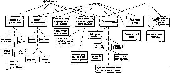
To the group of toxicoses with a tendency to repetition belong also some dermatoses of pregnancy (see Dermatoses), namely herpes gestationis and impetigo herpetiformis (major dermatoses according to Wechselmann's terminology). The question of S. can be raised only in impetigo herpetiformis, a dermatosis that repeats as a rule, proceeds significantly more severely upon repetition, and gives a rather significant mortality percentage; in herpes gestationis, a significantly milder disease, the question is raised only about the termination of pregnancy in more severely proceeding and more sharply expressed cases. As for other individual toxicoses, e.g., eclampsia, chorea, osteomalacia, etc., indicated S. can be only in osteomalacia, eclampsia, the recurrence of which cannot be absolutely excluded (but contrary to the opinion of some authors, e.g., Stroganov, its repetition is quite rare); nevertheless, in the opinion of the majority of authors, eclampsia is not an indication for sterilization (in the literature there is only 1 case of sterilization by Rissmann in repeatedly recurring eclampsia with a large number of seizures in several pregnancies), life, divorce or death of the husband, a new marriage, the death of an only child, etc.). Obstructions from the birth canal (narrow pelvis, stenoses, vaginal atresia) can also serve as an indication for the surgical sterilization of a woman. The majority of authors nevertheless believe that with these complications, S. should be performed as an accompanying operation during a Cesarean section, and not as a "prophylactic" operation, especially in young women, Postpartum period, DISORDERS AND COMPLICATIONS OF PREGNANCY AND LABOR IN WHICH THE QUESTION OF STERILIZATION MAY ARISE.
In neoplasms of the female genital sphere (the question can only be of benign neoplasms), sterilization may take place only in exceptional cases. In fibromyomas, neither fibrotomy during pregnancy nor the performance of Caesarean section is an indication for it. Not so rarely, in the presence of a neoplasm, there may be a necessity for subsequent supravaginal amputation of the uterus (Porro operation), or sterilization may be indicated during the enucleation of several fibromatous nodes or in the presence of a number of additional factors, e.g., a large number of previous pregnancies, advanced age, social conditions, etc. Even more rarely in clinical practice is there a necessity to perform sterilization in ovarian neoplasms. The use of sterilization during the first Caesarean section can be indicated only in exceptional cases; the majority believe that repeated Caesarean section, given the current state of surgical technique, does not present such a danger to a woman as in past days, and all authors, along with Winter, object to the formula «Ein Kaiserschnitt — nie wieder ein Kaiserschnitt». The decisive word must, of course, belong to the woman, but it is also the duty of the physician and his authority to point out to her the consequences to which sterilization during the first Caesarean section can lead (possible change in circumstances, age) (the question of whether sterilization should be performed for a narrow pelvis at the very first Caesarean section should be resolved in the plane indicated above, which, however, does not exclude the possibility of the presence of such conditions under which surgical sterilization of a woman will find full justification when performing a Caesarean section for the first time). — The question of surgical sterilization of a woman may also arise in various kinds of hemorrhage associated with pregnancy; however, as literature data show, it takes place extremely rarely in such cases. Although the recurrence of such complications as placenta previa, premature detachment of the placenta, hypo- and atonia of the uterus after childbirth, or hydatidiform mole is not excluded, almost all authors who have touched upon this issue have pointed out that in view of the fact that the recurrence of these complications cannot be stated definitively (normal subsequent births have been repeatedly observed after them), the question of surgical sterilization of a woman should be resolved negatively (however, cases are not excluded in which, in the presence of a number of conditions — a large number of previous pregnancies, the presence of several living children — sterilization can be performed with full justification). (Sterilization in ectopic pregnancy — see Pregnancy). Cases of «obstetric trauma» (uterine ruptures, fistulae, injuries to the bone ring) may serve as an indication for surgical sterilization of a woman when they are expressed to a significant degree and, by virtue of this, subsequent births may threaten new complications, sometimes dangerous to life. This applies equally to injuries of the soft birth canal, as well as the pubic symphysis and the bone ring in general. Of particular attention are cases of significant divergence of the pubic symphysis during pregnancy (occurring, it is true, infrequently), which should be considered not as local processes, but as general diseases, as changes in the initial forms of osteomalacia. In these cases, as clinical observations show (Selitsky), sterilization can be performed with full justification. — The question of performing surgical sterilization in other cases of obstetric pathology, such as after difficult labor, after postpartum psychoses, postpartum diseases — «obstetric shock», appears to be still far from resolved. Although the recurrence of these complications is not excluded, all obstetricians without exception believe that they do not provide sufficient grounds for performing sterilization, since after all these complications, normal and even repeated births have been observed. The performance of surgical sterilization of a woman in various kinds of female diseases usually finds its place during a concomitantly performed gynecological operation and depends both on the degree of severity of the disease and a number of additional conditions, such as age, social conditions, the presence of living children, etc. The question of sterilization may arise in prolapses, descents, malpositions of the uterus, large perineal tears, and fistulae. — In diseases of other organs, surgical sterilization of a woman is performed when the disease is expressed so sharply that the onset of pregnancy can significantly worsen the pathological process or even threaten life. These may include organic heart diseases (e.g., myocarditis, cardiosclerosis, chronic endocarditis, sharply expressed aneurysms, etc.), kidney diseases, blood diseases (e.g., progressive malignant anemia), some sharply expressed endocrinopathies (Basedow's disease, Addison's disease, diabetes mellitus), pulmonary tuberculosis (in the cavitary form), laryngeal tuberculosis, otosclerosis, albuminuric retinitis, congenital feeblemindedness, idiocy, progressive paralysis, spastic paralysis, tabes dorsalis, etc.
S. Olitsky.
Related articles
Cite this page
“Sexual Sterilization.” Soviet Medical Encyclopedia. English translation of Bolshaya Meditsinskaya Entsiklopediya, 1st ed. (Moscow, 1928–1936), ed. N. A. Semashko. https://sovietmedicalencyclopedia.pages.dev/article/sexual-sterilization/