Castration
Historical document, translated for reference. It reflects medical knowledge of the 1920s–30s and is not medical advice.
Summary
Castration is the surgical removal of the gonads, historically used in agriculture and studied for its physiological effects. The article examines its impact on animals and humans, including changes in development, metabolism, and sexual characteristics.
Encyclopedia article (1928–1936)
Castration (from Latin castratio - sterilization), the artificial removal of the gonads from the body. This operation, since time immemorial, has been used in agriculture for the economic improvement of certain properties of domestic livestock and poultry. The scientific study of the phenomena of castration has shown its extraordinarily high importance as a method for clarifying the role of the gonads (rather, the hormones secreted by them) in both the general physiology of the organism and its morphology. The question of the relationship between the 'soma' of the body and its gonads has been little studied to this day, despite the enormous number of works that have appeared. Complete removal of the gonads in many invertebrates apparently leads to no consequences in the body: the glands are restored (e.g. in planarians and tunicates) to their full size. For some animals, a difference in the body's reactions before and after the maturation of the gonads has been proven; thus, regeneration processes proceed at different rates: better outside the sexual period and worse during it (tunicates). The development and final formation of the gonads in most organisms is connected with the cessation of growth (but not necessarily: for example, in fish growth continues throughout life). This connection is clearly visible in annual plants, in which after flowering (i.e. formation of sexual elements) drying up and death occur. To what extent these two phenomena are connected with each other is evident from the experiments of gardeners, who by means of castration can not only delay the time of flowering by several months but also extend the life of the plant for several years. This is achieved, for example, in our mignonette by removing the flower buds, and it grows into a single-trunked tree up to 2 m high with a dense crown of branches at the top. The greatest interest castration presents in those forms in which the gonads are connected with the secretion of special hormones that stimulate the formation of sexual differences (see Dimorphism). This connection has not been found everywhere, and in some cases it has not been firmly established which elements of the gonads secrete these hormones (see Interstitial gland). Thus, in the type of arthropods, castration of insects has absolutely no effect on either the behavior of the male or the secondary sexual characteristics [experiments of Oudemans, Meisenheimer and Koree on butterflies Limantria dispar (nun) and Pieris (cabbage white)]. On the other hand, in decapod crustaceans (Sternorrhyn-chus, Eupagurus, etc.) in the so-called parasitic castration, caused by the penetration of parasitic crabs (Saccuiina, Pelmatogaster) into their gonads, males take on the external appearance of females, while females remain almost unchanged (only the abdominal legs decrease). Castration of vertebrate animals. Experiments on castration of fish (minnow and stickleback), according to the research of Kopez and Bok (Vosk), lead to underdevelopment of their secondary sexual characteristics. In amphibians, sexual dimorphism is mainly of a seasonal nature, which is why the results of castration are somewhat contradictory. Removal of the testes in frogs leads to the destruction of the callous formation on the big toe, which forms in males during the sexual period, but the ability to clasp the female with the front legs, as in normal coitus, does not disappear. The latter may depend on the fact that this is an act of a neuro-reflex nature, not hormonal. On the other hand, transplantation not only of the testis but also of the ovary again causes the formation of a callus. In newts, removal of the testes leads to the destruction of those sexual differences that occur in the male before mating: bright pigment, crest, etc. (Bresca).-The phenomena of castration have been most studied in birds, mainly by Pezard, Goodale, Paul and M. Zavadovsky. According to their data, castration of roosters leads to a decrease or disappearance of the comb, wattles and earlobes; a change in the posture of the body and the position of the tail, which changes from vertical to horizontal; after the first molt, the feathers and tail become even longer; the skull decreases, the tubular bones increase, the sexual instinct disappears, singing although sometimes continues, but the voice is strongly changed - becomes hoarse; obesity appears. Castration of hens, on the other hand, leads to the appearance of a rooster's plumage in the female: spurs grow on her, and thus the castrated male and female become similar to each other. The same results were obtained on pheasants, wild and domestic ducks. Castration in mammals, particularly in domestic animals, is used mainly for economic reasons. Castration leads to animals becoming calmer in their behavior, more capable of fattening: more fat is deposited in the meat; the unpleasant specific smell disappears from it (pigs, sheep); in cows, milk secretion increases, the content of casein and fat in it increases, the milking period is extended, etc. Castration in mammals affects, above all, the growth of the limb bones: according to Poncet, in a castrated rabbit, the tibia and femur are longer than in control animals; in female dogs castrated at 3.5 months of age and killed at one and a half years, the spine is 10 cm longer than in controls; the tubular bones are elongated, the pelvis of castrated males and females becomes average and shows no sexual differences; the skull decreases, and accordingly the brain decreases (according to Luschka, the average weight of the brain of a stallion is 534.8 g, of a gelding - 519.62 g). In the family Cervidae, the age at which castration is performed has a great influence on the development of antlers. With castration before the formation of the frontal processes, neither antlers nor frontal processes are formed at all; with castration during the appearance of the processes, irregular club-shaped antlers grow; with castration after the formation of antlers, they fall off, new ones grow, never taking the form of the old ones; the skull becomes similar to the skull of a female. According to the observations of M. Zavadovsky, males of the antelope (portax pictus Antilope cervicapra) and the horned Ukrainian cattle acquire the coloration of the female after castration. Castration in humans. Self-castration was often performed and is still performed in many countries, both ancient and modern, for religious purposes (in the USSR - the Skoptsy sect). Castration as a means of revenge and humiliation (castration of prisoners) has been preserved from ancient times, when it was widespread, to the present day (among some tribes of Central Africa). Castration of a person, especially when performed at a young age, leads to a number of significant changes. The skeleton becomes disproportionate, mainly in the sense of inconsistency in the length of the limbs relative to the trunk (the so-called eunuchoid growth: above-average height, disproportionately long limbs, small head) due to the slowing down of the process of ossification in the area of the epiphyseal sutures of the long tubular bones. The hair cover lacks male characteristics. In addition to the cessation of the growth of mustaches and beards, the absence of hair or its insufficiency on crines pubis, in the perineal area, on the trunk and in the armpits, the psyche also changes: early male eunuchs differ in sluggishness of temperament, lack of aggressiveness, courage and enterprise. Later castration, of course, no longer affects the skeleton due to the completion of its formation by the time of puberty and causes changes mainly in metabolism and psyche (in the above-mentioned sense). However, the sexual apparatus also undergoes quite sharp changes in the sense of its reduction. As for libido sexualis, this is preserved to a greater or lesser degree for quite a significant time in a number of cases, so to speak, by inertia of once established functions, in other words - due to the already existing eroticization of the higher psycho-sexual center and other parts of the sexual nervous mechanism.-From the side of metabolism, generally speaking, a change is noted in the direction of decreasing its intensity depending on the decrease in the energy of oxidative processes (mainly the data of Levy and Richter), although a certain share of participation in this must also be attributed to the change in the temperament of the individual, its less mobility compared to the norm; but as for the fat metabolism specifically, in this regard two types are still observed among early male eunuchs in humans: the type of simple eunuchoid growth without a tendency to obesity and the type of obese castrate, apparently depending on constitutional features. More detailed deviations in metabolism in castrates are as follows: male castrates not only show a tendency to obesity, but fat is deposited in them according to the female type (lower abdomen, thighs, chest circumference); in female castrates, however, no particularly sharp changes are observed in this regard. In protein metabolism, a number of authors have not noted special deviations; others speak of a decrease in nitrogen excretion. In carbohydrate metabolism, the following two facts attract attention: according to Stolper, castrated rabbits show an increased tendency to alimentary, and according to Cristofoletti-to adrenalin glycosuria.
The excretion of calcium and phosphorus from the body under the influence of Castration is apparently delayed, judging by the favorable effect of this operation in osteomalacia. Castration affects almost the entire endocrine system, causing hypertrophy of the adrenal cortex, delaying the involution of the thymus, and increasing the weight and volume of the pituitary gland due to an increase in its eosinophilic cells. The thyroid gland, according to most authors, decreases in size in castrates, and only Engelhorn speaks of its increase, but this discrepancy may be explained by the difference in the duration of observation (initially hypertrophy, later atrophy); in the pancreas, an increase in the islets of Langerhans was found; in the pineal gland, according to some authors, no significant changes were found, according to others, an acceleration of regression was found. The generally accepted opinion about the reduction in the lifespan of castrates is refuted by data on the lifespan of famous castrato singers, most of whom died between the ages of 60 and 97 (Barbieri). The ability to coitus in castrates sometimes not only does not disappear but is greatly increased, and if the operation is performed before the period of maturity, coitus is longer and more frequent, of course without ejaculation of semen. These peculiarities of castrates led in ancient times in Italy to the existence of brothels of castrats for women. The social position of castrats in the East does not differ from the normal, but in the West the Catholic Church waged a sharp struggle against them, mainly against their marriages. As early as the 9th century, a priest who performed such a marriage was deprived of his rank, but apparently this did not achieve the goal, since in both the 16th and 17th centuries there existed similarly strict decrees of the ROMAN popes.
N. Bogoyavlevsky, G. Sakharov. Castration of women. The first indications of the application of castration to women are found in remote times; even the ancient Greeks and Romans applied castration to women to preserve the supposed youth and beauty of the body. Among the Abyssinians and Hindus, there was a custom of castrating women to obtain hardy, strong maids. Robert describes 25-year-old Bechuana eunuchs who were castrated in childhood; they were tall, muscular and strong. Hair on the pubis and under the arms was absent. The mammary glands were underdeveloped, with small atrophic nipples. They had no menstruation at all. The pelvis was narrow or flattened, with the pubic bones close together. The labia majora were flabby, dry. The entrance to the vagina was narrowed, the perineum was low. Castration is considered complete if both ovaries are removed entirely. There are semi-castrates or eunuchoid women with various forms of functional insufficiency of the ovaries after partial resection of them or other factors damaging the generative and endocrine parts of the ovaries. When assessing the changes that occur after surgical castration, the age at which the ovaries are removed is of great importance, and the time at which the post-castration changes are evaluated is also important. After late castration, there may be no noticeable external changes, but after castration in young individuals, a series of characteristic morphological and functional changes occur. After castration in women, a clear transition to an asexual (sexless) type develops, which vividly confirms the significance of sex hormones. The degree of such post-castration changes in young women is more sharply expressed. Clinically, in women who have undergone complete castration, a characteristic symptom complex develops. Menstruation ceases, although according to some data, removal of both ovaries causes cessation of menstruation only in 60%. This is explained by the possible existence of additional areas of ovarian tissue in the broad ligaments or in other places in the abdominal cavity, and sometimes it may also depend on minimal remnants of ovarian tissue left during the operation. According to Artmann's data, in women, additional ovaries are observed in 4%, which cause preservation of menstruation after castration. Sometimes in castrates after late castration, replacement of menstruation with nasal, intestinal bleeding or periodic serous secretions is observed. However, such bleeding more often depends on the presence of uterine polyps and malignant growths. Furthermore, as a consequence of castration, atrophy of the mammary glands, external genital organs and uterus occurs, in which not only the muscular elements but also the endometrium atrophies until the glands completely disappear; where the endometrium is preserved, the uterine cycle still disappears. The external genital organs show various degrees of atrophy and arrested development. The clitoris is reduced, the entrance to the vagina is narrowed, the labia majora and minora are flabby, underdeveloped, somewhat dry, with defects in pigmentation. The vagina is small, the transverse folds are smoothed out, the mucous membrane is atrophic, fragile and easily injured. Early castration causes special changes in skeletal development, and the shape of the skull, pelvis, and limbs changes. The essence of the bone changes consists in enhanced growth of bones in length and delayed ossification in the epiphyseal areas, which is associated with a disturbance in the regulation of calcium metabolism, which is regulated by the ovaries together with other endocrine glands. Castrates are characterized by elongated growth, long limbs, a flattened pelvis in which the curvature of the sacrum and coccyx is altered. Castrates have a small skull in relation to the total length and a correspondingly reduced brain compared to normal women. Bone changes in the limbs and thickening of the supraorbital ridges show in castrates signs of acromegaly. Early castration does not directly lead to the development of infantilism; it only delays the differentiation of the sexual type. In castrates, the larynx lengthens, the arytenoid cartilages enlarge, the voice changes, acquiring a low timbre. The general appearance acquires partly masculine features. As a result of the absence of endocrine influence of the ovaries, a series of nervous, vasomotor and secretory disorders occur. The developing symptoms depend on the excitation of the autonomic nervous system as a result of the loss of the inhibitory influence of the ovary. Removal of the ovaries creates conditions for autointoxication in castrates. Castration has a significant effect on the entire psyche of a woman, lowering her intellect, depriving her of emotional liveliness and making her sluggish, depressed and irritable. Sexual desire with early castration usually does not exist. If castration is performed in mature age, sexual desire and satisfaction are rarely weakened. There are observations that in the first period after surgical castration, an increase in sexual desire may be observed, but later its intensity decreases. It can persist and appear in castrates in cases where there are special conditions eroticizing the nervous system. Where libido persists, the implementation of sexual life in castrates is difficult due to the painfulness, dryness and easy injury of the genital organs. As a result of surgical castration and the complete absence of endocrine influence of ovarian hormones, a disturbance of the basic metabolism occurs. After castration, obesity with infiltration of fatty tissue is observed in 13-15%. Fat is deposited in typical places: in the area of the mammary glands, abdomen, upper third of the thighs, forming fatty 'gallopes', and around the pelvic girdle, from which characteristic fatty folds are noticeable on the lower back and sides of the body. Fat accumulation in women is not constant, whereas in men after surgical castration in post-pubertal age, obesity is almost always expressed. There are two types of castrates - fat and thin. The first is characteristic of castrates of mature age, and the second - muscular - is characteristic as a metamorphosis of the female type; it is observed after early castration. In addition to typical bone and constitutional changes, in castrates and eunuchoid women, changes in the skin and hair are expressed to varying degrees. Hair may be completely absent, be sparse, resemble the child type, i.e., be distinguished by scarcity and uniform arrangement. The hairiness most often has the appearance of down. Weak hairiness is especially sharply expressed in the axillary pits, on the pubis, around the clitoris and labia majora. Hair on the head is dry, brittle, prematurely grays and easily falls out. Much more rarely with early castration, as an expression of false masculinization, mustaches and beard may appear, and hair becomes thicker. The skin in young castrates becomes lighter, striking by its pallor, and lacks density of pigment. In elderly castrates, skin changes come down to dryness, tendency to peeling and increased number of wrinkles, as is characteristic of natural menopause or old age. Clinically, the phenomena of the consequences of castration in mature women, in addition to the above mentioned, are expressed by painful moli-mina menstrualia, headaches, hot flashes, disturbances of cardiac tone, sweating, unbalanced mood of depressive nature, weakening of memory. Sometimes pains in the bones appear. All these symptoms proceed more acutely and painfully than in natural menopause. The application of castration for therapeutic purposes before the development of endocrinology and the identification of the endocrine role of the ovary was quite widely admitted and often without sufficient etiological grounds. The first exact data on the removal of healthy or somewhat enlarged ovaries date back to 1869, when Koberle, during an operation for fixation of the uterus for technical reasons, removed the ovary. Incidental castration was applied to ovaries found in hernial sacs. The castration previously used in underdevelopment of the uterus, in its incorrect positions complicated by parametritis, in dysmenorrhea, in parametritis atrophicans, in chronic endometritis is now abandoned. In some of these forms, e.g. hypoplasias, to enhance the growth of the uterus and functions, on the contrary, additional transplants of ovaries are now used together with the anterior lobe of the pituitary gland. Experimental and chemical data show that it is rational to combine castration with the removal of malignant tumors, in particular cancer, deciduoma, as such a combination helps to reduce the chances of recurrence. Removal of the ovaries was used for the treatment of fibromyomas, as besides the symptomatic stopping of bleeding castration can also cause the reverse development of these tumors. Among the modern methods of treatment of uterine fibromyomas, simple extirpation of the ovaries has been abandoned, but X-ray castration, or irradiation of the ovaries with radium, has become widespread. To finally eliminate the glandular elements of the ovaries, to obtain persistent amenorrhea, an average dose of 100-120 N.E.D. is required. The symptoms of deficiency after X-ray castration are the same as after extirpation of the ovaries. According to Fuchs, in this case the vasomotor symptom complex is encountered 5 times more often than heart changes; nervous-psychic phenomena - from 10 to 14%, weakening of sexual desire and satisfaction - in 21.4%, weakening of memory - in 35% (2 times more often than after surgical castration). The assessment of the frequency and degree of deficiency phenomena after X-ray castration and surgical castration is by no means the same.
After X-ray castration—a vasomotor symptom complex in the form of heavy sensations: periodically alternating heat, sweat, rush of blood to the head, dizziness, insomnia, general excitement, feeling of fear, seeing spots before the eyes, fainting; heart palpitations are expressed more mildly than with extirpation of the ovaries. This is explained by the fact that even castration doses of X-rays do not completely destroy the entire internal secretory apparatus of the ovaries. However, in individuals particularly sensitive to X-rays, after X-ray castration, the phenomena of deficiency sometimes proceed more acutely and painfully than with extirpation of the ovaries. In such individuals, the usual symptoms of deficiency are joined by changes on the part of the heart in the form of tachycardia, arrhythmia, and the subjective sensations resemble the picture of cardiospasm.—Long inflammatory-degenerative lesions of the ovaries with suppuration and a tendency to constant exacerbations still constitute indications for surgical castration, where prolonged conservative therapy remains without result.—The application of castration in women for certain mental diseases (dementia praecox, circular psychosis), nervous diseases (epilepsy, hysteroneuroses, nymphomania) has always been characterized by uncertainty of indications and little justification. To this day there are no firm grounds for complete castration in these diseases without the presence of a patho-anatomical substrate. Partial resection within the limits of healthy ovarian tissue in cystic degeneration and tumors of the ovaries may prove to be causal therapy in epilepsies of ovarian origin.—A serious indication for surgical castration is puerperal osteomalacia. To this day the etiological essence of it has not been precisely clarified, but it is undoubtedly that the disturbance of mineral exchange in the bones and loss of lime assimilation are closely connected with the changed function of the ovaries. It has been established that in cases of osteomalacia in women with sharply expressed phenomena not yielding to conservative therapy, castration is rational therapy. The operation of castration in women is technically simple, if there are no special complicating conditions (adhesions, interligamentous location of ovarian tumor, intimate connection with neighboring hollow organs). After opening the abdominal wall or vaginal vault, ligatures are applied to both ovarian ligaments. The ovaries are then removed. Subsequent peritonization and suturing of the abdominal cavity.—Treatment of the consequences of castration has the character: 1) symptomatic therapy, in particular with ovarian organ preparations, but it, just like medicinal therapy, gives little success. The best effect is obtained from the combination of organ preparations with salts of calcium, potassium, magnesium and preparations of digitalis; 2) causal replacement therapy by hetero- or homoplastic transplantation of ovaries, which for a prolonged period restores the disturbance of the endocrine status, ensuring clinical recovery.
M. Serdjukov. Castration of men. (For the consequences of castration of men, see above.) Indications for removal of the testicle are: 1) malignant neoplasms of the testicle, appendage and spermatic cord (sarcoma, cancer, chorioepithelioma, etc.), 2) tuberculosis of the testicle and appendage, 3) purulent, septic processes leading to gangrene of the testicle, and 4) its necrosis after injury. In some cases, benign tumors can also be a reason for removal of the testicle, if they are very large, cause the patient great inconvenience and disrupt his ability to work. It is most expedient to perform the operation under local anesthesia. First, local anesthesia of the inguinal canal is performed, as in the operation for herniotomy, and then infiltration (with solution) of the root of the scrotum, skin and septum is added to this. With the left hand the surgeon grasps the scrotum and tenses the skin. The incision of the skin and subcutaneous tissue is made from the external inguinal ring downward, along the convex surface of the testicle to the lower edge of the scrotum. In the presence of purulent fistulas or adhesions of the skin with the affected testicle, incisions are made within healthy tissues so as to form an oval flap remaining on the testicle. To prevent the transfer of infection, the edges of the flap are sutured over the fistulas. After cutting through the skin with subcutaneous tissue, the dartos membrane and the common vaginal tunic (tun. dartos, tun. vag. com.) are exposed and the spermatic cord is isolated directly at the external inguinal ring, and if necessary also higher, thereby opening the inguinal canal. The vas deferens lying in the posterior part of the spermatic cord is isolated (fig. 1). The arteries and veins of the spermatic cord are ligated with two or three ligatures, and then cut. Subsequently the testicle is enucleated bluntly from the scrotum and is dislocated from the wound, which usually easily succeeds. Only in the lower
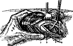
Figure 1. Isolation of the spermatic cord (3); 1 - tunica dartos; 2 - tunica vagin. commun.; 4 - vas deferens. part of the scrotum is it necessary to resort to cutting the testicular-scrotal ligament [lig. scrotale testis, s. gubernaculum testis (fig. 2)]. The isolated testicle remains hanging on the vas deferens, which is as much as possible drawn into the wound, after which it is cut. After examination of the wound and careful stopping of bleeding, the wound is closed tightly with sutures. In a whole series of cases the healing process proceeds aseptically. In extensive purulent processes with involvement of the scrotum and in gangrene, the wound is left open for secondary healing. Since in tuberculosis of the testicle and appendage the process extends high up along the vas deferens, for more thorough removal of foci of infection Büngner (Bilng-peg) proposed to remove the greater part of it. This is achieved by strong traction on the vas deferens after its thorough isolation. At this time
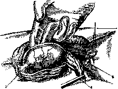
Figure 2. Castration: 1 - lig. scrotale testis; 2 - vessels of the spermatic cord; 3 - vas deferens,
usually the greater part of the duct (about 4/5 of its length) tears off. According to the opinion of Schede and Kocher, the vas deferens can easily tear off at the place affected by the tuberculous process, and in this case it is possible for tuberculous pus to flow into the surrounding parts. Therefore it is recommended to abandon the method of tearing off (evulsio) in view of the possible danger, and better through an inguinal incision to isolate the duct high up, ligate it and cut it with a thermocauter in the area of healthy tissues. Individual authors recommend in castration for tuberculosis to sew into the wound the central end of the vas deferens and subsequently inject into it an iodoform emulsion for the treatment of the seminal vesicle, usually also affected. In those cases when the operation of castration is performed for malignant neoplasm of the testicle, appendage or spermatic cord, it is necessary to examine and remove the retroperitoneal lymph glands, as well as the inguinal
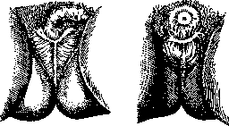
Figure 3.
Figure 4.
and femoral ones. Kocher for this purpose uses an oblique incision along Poupart's ligament, separates the aponeurosis of the external oblique muscle directly above the ligament, raises the aponeurosis of the internal and transverse from the groove of the ligament, and then splits the transverse fascia. After this the muscle together with the spermatic cord is pulled upward, the peritoneum is dissected upward, and along the vessels the affected glands are exposed and removed.—A more extensive intervention can be performed if the incision along Poupart's ligament is extended outward and the abdominal muscles are separated from the iliac crest. In such cases it is possible to dissect the peritoneum in more extensive dimensions. For more thorough cleansing of the inguinal and femoral region, a vertical incision is added to the transverse one, running downward along the large vessels (T-shaped incision). The peritoneum is then dissected far upward, the glands located along the iliac vessels, as well as the overlying ones, are well exposed. After removal of the glands all tissues are sutured; special care should be taken to join the edges of Poupart's ligament; if it was cut during this. The extended operation with the help of a T-shaped incision is incomparably more traumatic than Kocher's method, but it gives better and more extensive access. After this operation in patients elephantiasis of the scrotum and the corresponding leg often develops. Great medico-legal interest is presented by the operation of castration performed for religious purposes. In the USSR castration is used by adherents of the religious sect of the Skoptsy, which appeared as early as the 18th century. In the early period of the existence of Skoptism they used the so-called 'fiery baptism', which consisted in the fact that to an adept of this sect the removal of the testicles with part of the scrotum was performed with the help of a heated iron. Subsequently for this operation various cutting instruments began to be used, and the heated iron only for stopping bleeding. This operation of removal of the testicles according to the Skoptist doctrine represents the 'first purification', or 'small seal' (fig. 3). The 'Great', or 'royal seal' (fig. 4) represents a combination of removal of the testicles with the taking away of the penis. This operation is usually performed in a sitting position and after preliminary ligation of the base of the scrotum and the penis. After these operations extensive and very characteristic scars remain.
Ya. Brusvin.
Related articles
Mentioned in
Cite this page
“Castration.” Soviet Medical Encyclopedia. English translation of Bolshaya Meditsinskaya Entsiklopediya, 1st ed. (Moscow, 1928–1936), ed. N. A. Semashko. https://sovietmedicalencyclopedia.pages.dev/article/castration/