Visceral Skeleton
Historical document, translated for reference. It reflects medical knowledge of the 1920s–30s and is not medical advice.
Summary
This article describes the visceral skeleton (splanchnocranium) of vertebrates, which forms the framework of the oral cavity and pharyngeal region. It details the evolutionary transformation of these skeletal arches from the gill-supporting structures in fish to the jaw apparatus and auditory ossicles in mammals and humans.
Encyclopedia article (1928–1936)
VISCERAL SKELETON, or splanchnocranium, is the skeleton of the head that surrounds the oral cavity and the pharyngeal region of the digestive tract in vertebrate animals; it is contrasted with the axial skull, or neurocranium, which is the skeleton of the braincase and sensory organs. In lower vertebrates in the adult state, and in higher vertebrates in the embryonic state, the visceral skeleton consists of a series of skeletal arches between the gill slits. The cartilaginous visceral skeleton is most typically represented in sharks (see Figure 1). On the ventral side of the shark's skull is located a series of
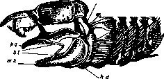
Figure 1. Head skeleton of the shark Scyllium canicula (from R. Wiedersheim after T. Parker): 1 br–5 br—five gill arches; hm—hyomandibulare; hd—hyoideum; li—labial cartilages; mh—Meckel's cartilage; pq—palatoquadratum.
visceral arches, of which the 5 posterior ones are true gill arches, encompassing the pharynx on the right and left and supporting the gill filaments seated upon them. Each arch is segmented into 4 sections; the right and left arches are connected to each other by a series of unpaired cartilaginous elements on the ventral side. The anterior two arches are modified into the jaw apparatus—an apparatus for grasping food, consisting of the jaw and hyoid arches; the jaw arch is segmented into two sections: the upper—the palatoquadrate cartilage (palatoquadratum, upper jaw) and the lower—Meckel's cartilage (cartilago Meckeli, lower jaw). The hyoid arch is also segmented into 2 sections: the upper, the so-called suspensorium (hyomandibulare), connects to the skull and suspends the jaw arch from the skull; the lower section—the hyoid arch proper (hyoideum)—adjoins Meckel's cartilage from behind.
Between the jaw and hyoid arches, a vestigial gill slit is preserved—the so-called spiracle (spiraculum). In front, on the jaw arch, sit small, so-called labial cartilages, which, according to data from comparative anatomy (Gegenbaur)
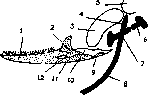
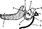
Figure 2. Diagram of the relationship of the articulation of the lower jaw and auditory ossicles in reptiles and birds (according to Reichert's theory): 1—dentale; 2—coronoid; 3—quadratum; 4—proc. dorsalis; 5—proc. paroticus; 6—stapes; 7—extracolumella; 8—hyoideum; 9—goniale; 10—articulare; 11—angulare; 12—surangulare. and developmental history (A. N. Severtsov), are reduced anterior visceral arches. In bony fish, the visceral skeleton ossifies. The gill arches ossify without changing. The jaw arch ossifies with many bones of various origins: chondral, or replacement, and dermal, or investing. In place of the palatoquadrate cartilage, through its ossification or the formation of dermal bones, the following bones are produced: the palatine (palatinum), 3–4 pterygoids (pterygoideum), and the quadrate (quadratum); the lower jaw attaches to the latter. The lower jaw—Meckel's cartilage—is partially preserved in the form of a cartilaginous column, covered by the dermal bones, the dentary (dentale) and the angular (angulare), and is partially transformed into the articular bone (articulare). Furthermore, in front of the jaw arch, secondary jaws appear, consisting of the premaxillary (praemaxillare) and maxillary (maxillare) bones. Of these bones, only the quadrate, the posterior pterygoid, and the articular are chondral; the others are dermal, and teeth sit upon them. In terrestrial vertebrates, in connection with the loss of gill respiration, the visceral skeleton undergoes significant transformations: the palatoquadrate cartilage grows onto the skull, and its ossifications connect with the cranial box, while the premaxillary and maxillary bones, upon which the teeth sit, begin to perform the function of jaws. The suspensorium (hyomandibulare) loses the function of suspending the jaw and gradually acquires a new Figure 3. Diagram of the relationship of the articulation of the lower jaw and auditory ossicles in mammals (according to Reichert's theory): 1—dentale; 2—cart. Meckeli; 3—proc. articularis; 4—malleus (articulare); 5—incus (quadratum); 6—stapes; 7—goniale; 8—hyoideum; 9—tympanicum (angulare). function, transforming into the auditory ossicle (columella auris, or stapes), located in the cavity of the middle ear, which was formed from the cavity of the fish's spiracle. The lower section of the hyoid arch (hyoideum) transforms into the anterior horns of the hyoid bone. The remaining visceral arches,
pes; 7—proc. folianus
in connection with the loss of gill respiration, undergo complete reduction, and of the entire complex skeleton, only one hyoid bone remains. In mammals and humans, the visceral skeleton undergoes further transformation. The lower jaw receives a new articulation to the skull by means of the coronoid process (proc. coronoideus). The former articulation via the quadrate bone is disrupted. The number of auditory ossicles increases from one to three, due to the incorporation of new bones of the visceral skeleton into the tympanic cavity. Figure 4. Derivatives of the visceral arches in humans (diagram from R. Wiedersheim): I—from the jaw arch comes the lower jaw and the ear ossicles—the malleus (ml) and the incus (in); II—from the hyoid arch—part of the hyoid bone (bs), the styloid process (ps), and the ear ossicle—the stapes (fs); III—from the 1st gill arch—the hyoid bone (bs); IV—from the 2nd gill arch—the anterior part of the thyroid cartilage (thy'); V—from the 3rd gill arch—the posterior part of the thyroid cartilage (thy''). Figures 2 and 3 present their transformation according to Reichert. To the auditory ossicle (stapes) are joined three ossicles from the jaw arch: the incus (incus), which originates from the quadrate bone (quadratum), the malleus (malleus), which forms from the articular bone (articulare), and the tympanic bone (tympanicum), which develops from the angular bone (angulare). Figure 4 presents the transformation of the visceral skeleton in humans, where, in addition to the above, one can also see the transformation of the gill arches: the 1st arch—into the hyoid bone, the 2nd and 3rd—into the cartilages of the larynx (cart. thyreoidea).
B. Matveyev.
Related articles
Mentioned in
Cite this page
“Visceral Skeleton.” Soviet Medical Encyclopedia. English translation of Bolshaya Meditsinskaya Entsiklopediya, 1st ed. (Moscow, 1928–1936), ed. N. A. Semashko. https://sovietmedicalencyclopedia.pages.dev/article/visceral-skeleton/