Skeleton
Historical document, translated for reference. It reflects medical knowledge of the 1920s–30s and is not medical advice.
Summary
The skeleton is a system of relatively dense formations that form a more or less durable framework for an animal or its parts. It protects softer tissues and organs from pressure and damage, and serves as an attachment point for muscles and support during movement.
Encyclopedia article (1928–1936)
SKELETON (from Greek skeletos - dried) in animals represents a system of relatively dense formations that constitute a more or less durable framework of the animal or its parts. On one hand, skeletal formations protect the more delicate tissues and organs from pressure and damage from neighboring parts and the external environment, while on the other hand, they serve as the site of muscle attachment and provide support to the animal during its movements. According to position, one distinguishes the external S., a product of the integuments, from the internal skeleton, not directly connected with the integuments. The external S. may have different origins and reaches quite different degrees of development in various animals. Its simplest form is the "cuticle" of many invertebrates - a dense and usually quite elastic outer body covering, representing a product of secretion by the epithelium (e.g., in the earthworm). In arthropods, the cuticle exists in the form of a strong layered substance - "chitin"; in crustaceans, this chitin is often impregnated with lime and then reaches even greater hardness. The chitinous skeleton of arthropods develops in areas corresponding to the segments and articles of their body, provides excellent protection for internal organs, and at the same time serves as the site of attachment for the musculature of the body and limbs. An even harder calcareous external skeleton is observed in coral polyps. The formations of the same kind also include the calcareous shells of mollusks. In other animals, the external skeleton is formed in deeper layers of the skin. Among invertebrates, such a skeleton reaches particularly significant development in echinoderms, where it appears as a shell made of closely connected calcareous plates. In vertebrate animals, the external skeleton is never cuticular. Its most primitive form is the placoid scale or skin teeth of sharks. The scales of fish and the skin ossifications of terrestrial vertebrates, which sometimes reach very significant development (crocodiles, turtles, armadillos), provide certain protection to the animal's body, but are always supplemented by a powerful internal skeleton. The internal skeleton is usually poorly developed in lower animals and appears in them as a system of acellular membranes or connective tissue formations, sometimes supplemented by denser horn-like ("spongin" in sponges) fibers, or siliceous or calcareous spines (in sponges, corals). Cartilage occurs as skeletal tissue only in rare cases in some annelid worms and in mollusks (especially in cephalopods). In vertebrate animals, the body is always supported by a complexly developed system of internal skeleton. In lower fishes, it is almost entirely (cyclostomes, sharks, rays, chimeras) or for the most part (sturgeons) cartilaginous, while in higher fishes and terrestrial vertebrates, it is partly supplemented by skin bones (the dermal bones of the skull and shoulder girdle of fishes and terrestrial vertebrates), and partly replaced by bone, which more or less completely displaces the original cartilage (the "primary" bones of the skull, spine, and limbs). The internal skeleton of vertebrates consists of several sections independent in origin: the central flexible axis - the axial skeleton, supplemented in the head region by the skeleton of the anterior part of the intestine - the visceral skeleton, and the skeleton of the limbs. In the head region, the anterior part of the axial skeleton forms together with the visceral skeleton the skull. The axial skeleton is represented in some lower vertebrates throughout life (cyclostomes, chimeras, sturgeons, dipnoans), while in others only in the early stages of embryonic development as a cellular cord with a strong elastic sheath - the notochord, or chord. In higher vertebrates, it is replaced by the spine (see). At the head end of the axial skeleton, the skull is formed (see). The visceral skeleton (see) has an origin independent of the axial skull. The skeleton of the limbs is represented in fishes by cartilaginous or bony rods supporting the unpaired fins, as well as by a more complexly segmented cartilaginous or bony skeleton of the paired fins with their girdles. The anterior or pectoral girdle is covered in higher fishes by a series of skin bones connecting it with the roof of the skull. In terrestrial vertebrates, the skeleton of paired limbs is complexly segmented and their girdles reach considerable power (see PELVIC GIRDLE, PECTORAL GIRDLE).
I. Shmal'gauzen. The soft skeleton of vertebrates and humans is a connective tissue supporting apparatus, to which they refer to the adipose tissue (lower form) and the connective tissue coverings of tendons and muscles - fasciae (higher form). The bony skeleton is divided into the skeleton of the head - the skull, the skeleton of the trunk - the spine, ribs, sternum, the skeleton of the pectoral girdle and free upper limbs, the skeleton of the pelvic girdle and free lower limbs (fig. 1). The total number of bones is determined differently by various authors depending on, for example, whether they include the auditory ossicles, the hyoid bone in the composition of the skeleton or do not take them into account. Thus, the number of bones of the skeleton is determined to be 228 (without sesamoid bones) (Hyrtl), over 200 (Rauber and others). In general, for the skeleton of an adult, one can accept the number of bones as 207. This number can be increased due to the appearance of additional bones (e.g., XIII pair of ribs), bones in the sutures of the skull, in the place of fontanelles, etc. In the course of development and formation of the skeleton, the number of bones changes, especially if one counts as separate bones the appearing bone areas in the cartilaginous skeleton.
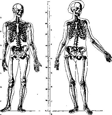
Figure 1. Skeleton of a human: a - male; b - female.
According to Scammon, the distribution (number) of bone areas in different periods of human life is as follows: Centers of ossification is tr o rr o 2 № o k IS! e o o e £ go N o V a e g o « W c* a ю co a M B upper limb « i И d S щ k k in O En tki o [-. O R Total number of centers (approx.) . of which appear before birth ..... after birth Number of separate centers at 14 years of age . . . Number of separate elements of adult skeleton . . . The weight of the bony skeleton in relation to the total body weight is xjb-x/7. Bischoff gives the following numerical data: Sex and age Total weight (in kg) Skeleton weight (kg) Male 33 years . » 16 years . Female 22 years . 69,668 35,54? 55,400 11,080 8,436 8,390 The weight of cartilaginous and bony skeleton at birth is 15-20% of total body weight (A. Hasselwander). The bony skeleton in the human body and mammals serves as support for soft tissues, in particular as the site of attachment of the so-called
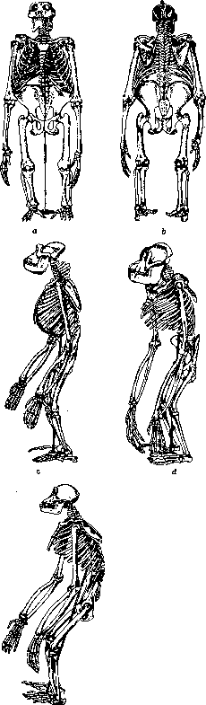
Figure 3.
Figure 2. [Skeletons of anthropoid apes: a - gorilla (front view); b - same (rear view); c - same (side view); d - orangutan; e - chimpanzee. (From K. Günther.)
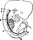
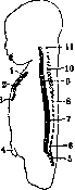
Figure 3.
Figure 4. Figure 3. Development of the skeleton; stages of the mesenchymal period. Embryo 11 mm in length: 1- occipital vertebra - plate of the base of the skull; 2-1st cervical vertebra; 3-1st thoracic vertebra; 4-1st lumbar vertebra; 5-1st sacral vertebra; 6-1st coccygeal vertebra. Figure 4. Sagittal section of the torso of a child (newborn) - age features of the thoracic cage and curvatures of the spine: 1- manubrium of sternum; 2- body of sternum; 3- xiphoid process; 4 - navel; 5- sacrum; 6- V lumbar vertebra; 7- XII thoracic vertebra; 8- spinous process; 9- body of vertebra; 10- VII cervical vertebra; 11- atlas.£,!(From I. Kollmann.) of skeletal muscles. Representing in its individual parts (mainly in the limbs) systems of bone levers (I, II and III class), connected mostly with each other movably, S. together with the connecting elements (joint capsules, ligaments, cartilages, etc.) is the passive part of the system of organs of movement. But at the same time, being the place of origin of muscles, distributed and developed partly depending on the method of connection and character of the «66 individual bone levers, S. to a certain extent is artificially isolated from the general osteo-muscular system. The supporting function of S. in the moment of standing or displacement of the body and its parts in space is possible only with the active participation of the musculature. Individual parts of S, taking upon themselves the load (weight) of the overlying sections, e.g. the spine, feet, have besides the passive "tension" (ligamentous apparatus) an active "tension", represented by the muscles of the corresponding area. Relaxation of these tensions gives sharp changes in S. (curvatures and length of the spine, degree of expression of the arches of the foot, etc.). Thus the bone S. can be isolated only on dead material, while in the living the bone system is inseparable from the muscular and constitutes with it one whole. The soft S., in particular fasciae (especially of the limbs), plays an important role in creating support for the soft parts of the body. Approximately 2/3 of the muscles take their origin and are attached not only to bones but also to fasciae (Bardeleben). The size, shape, internal structure of bones, the relief of their surface are determined to a large extent by the supporting function of S. All bones are distinguished: long bones (ossa longa), flat bones (ossa plana), short bones (ossa brevia), pneumatic bones (ossa pneu-matica), bones of irregular shape and mixed. The internal structure of individual bones is characterized by its peculiarities (e.g. most bones of the limbs are long, tubular). The second no less important function of S.-the protective role of its individual parts. Such are: the cerebral part of the skull for the brain and organs of sense, the facial part for the initial sections of the respiratory and digestive systems (see Visceral skeleton), the spine for the spinal cord, the thoracic cage for the organs of the thoracic cavity, the pelvis for the urinary bladder, uterus, etc.-Phylogenetically S. has undergone a number of changes of its individual bone links, which especially sharply manifested themselves in man in connection with the transition of the body to the vertical position, with the freeing of the upper limbs from the functions of support and displacement of the body in space and the transfer of the entire load to the lower limbs; From this point of view the comparison of the skeleton of quadrupeds, especially anthropomorphic, and man seems very important. E.g. already in anthropoid apes the appearance of the lumbar lordosis of the spine is noted, which is very characteristic for the human spine; in anthropoids the upper limbs although they perform a supporting function (hence their length) but already not to such a degree as in other animals (fig. 2). .Way of life, environment along with other factors play an essential role in the formation of S. of various vertebrates (reduction of the weight of S. by pneumatization of bones in birds, peculiarities of the structure of the limbs, etc.). An exclusively connective tissue skeleton (membranous S.) exists in the early period of development (approx. until the beginning of the 2nd month of intrauterine life) (fig. 3), in the subsequent stages of ontogenesis the skeleton of man in its main mass is represented by cartilaginous (cartilaginous S.). Of the bones, not passing through the cartilaginous stage, only the "covering" bones of the skull (bones
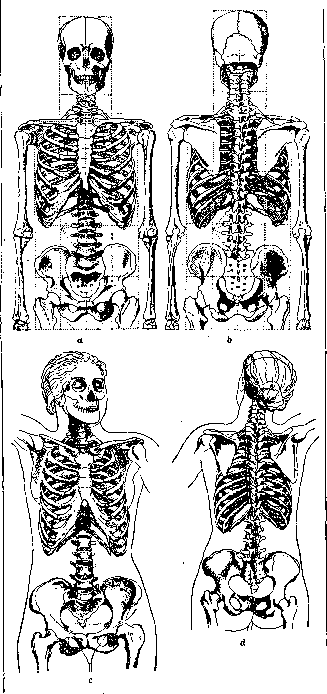
Figure 5. Skeleton of the torso: a and b of a man; c and d- of a woman.
of the cranial vault and most of the bones of the face) and the clavicle are noted. Later in all parts of S. the so-called processes of ossification occur, in place of cartilage and connective tissue areas of bone tissue appear. This process of replacement of the soft basis by bone continues comparatively long after birth (until the cessation of the processes of growth of the body in length to 25-26 years). Cartilage remains only on the articular ends of articulating bones. The growth and formation of individual parts of S. at different ages proceed unevenly. Hence the presence of age features of S. (fig. 4). The size of the entire S. and its individual links determine the size of the body, the ratios of its parts to each other and to the body as a whole-proportions of the body. Together with the musculature, subcutaneous fat and other tissues S. influences the relief of the body surface. The bony prominences of S. are used in anthropology and medical practice as recognition points
^^-^^
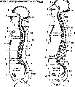
Figure 6.
Figure 7.
Figure 6. Sagittal section of the torso of a woman (25 years) (features of the spine, thoracic cage and pelvis): 1 - frontal sinus; 2 - hard palate; 3 - I thoracic vertebra; 4- manubrium of sternum; 5- body of sternum; 6- xiphoid process of sternum; 7- navel; 8- pubic symphysis; 9- perineum; 10-anus; 11- coccygeal vertebrae; 12-V sacral vertebra; 13-V lumbar vertebra; .16. and 20- vertebral canal; 17 - XII thoracic vertebra;. 18- spinous process; 19- area of scapula; 21- VII cervical vertebra; 22- axis; 23- atlas. (From I. Kollmann according to Braune.) Figure 7. Sagittal section of the torso of a man (21 years) (features of the spine, thoracic cage and pelvis): 1- frontal sinus; 2- hard palate; 3- lower jaw; 4- manubrium of sternum; 5- body of sternum; 6- xiphoid process; 7- intervertebral disc; 8- navel; 9- pubic symphysis; 10- perineum; 11-anus; 12-IV coccygeal vertebra; 13-II coccygeal vertebra; 14- sacrum; 15- V lumbar vertebra; 16- I lumbar vertebra; 17- XII thoracic vertebra; 18 and 21-vertebral canal; IV- area of scapula; 20- IV thoracic vertebra; 22- axis; 23- atlas. (From Kollmann according to Braune.) anthropometric research). Applying various methods of research, the physician establishes on the patient the projection on the skeleton of the boundaries and position of internal organs. S. is subject to significant sexual, racial, typological [constitutional (see Constitution)] and individual fluctuations in the sense of size, proportions, influence on the external forms of the body, etc. Thus e.g. S. of a woman is distinguished by thinner and lighter bones, a sharper curvature of the lumbar part of the spine, a short and wide sacrum, a low and wide pelvis, a relatively greater width and roundness of the skull, a longer and narrower thoracic cage, etc. (fig. 1, 5- 7). Violation of the normal course of ossification processes, deposition of lime salts, growth of bones, etc. in various diseases, in particular of the internal secretion organs (see Internal secretion), sharply affects the appearance, size and proportions of the human body (and animals) (see Marie's disease, Acromegaly, Dwarfism, Eunuchoidism, Castration, Goiter, Thyroid gland, Osteomalacia, Rickets, Osteogenesis imperfecta, Achondroplasia}.-Bones, little subject to destruction, are almost the only material that has made it possible to reconstruct the appearance of prehistoric man. A large number of finds of skeletons during special excavations or accidentally, during geological and construction (earth) works gives valuable data (paleoanthropological) for the doctrine of the origin of man. The relative incorruptibility of the skeleton also has significance in forensic-medical practice. The skeleton of man and animals, specially treated (see Maceration), on natural ligaments or artificially tied with wire and correspondingly mounted, is an important visual aid in the study of morphology and a valuable exhibit of museums (anatomical, anthropological, zoological, health protection, etc.). Artificially prepared (assembled) S. is called sceleton artificiale in contrast to the natural S.-sceleton naturale.
Related articles
Mentioned in
Cite this page
“Skeleton.” Soviet Medical Encyclopedia. English translation of Bolshaya Meditsinskaya Entsiklopediya, 1st ed. (Moscow, 1928–1936), ed. N. A. Semashko. https://sovietmedicalencyclopedia.pages.dev/article/skeleton/