Cubitus Varus
Historical document, translated for reference. It reflects medical knowledge of the 1920s–30s and is not medical advice.
Summary
This article from the 1928–1936 Soviet medical encyclopedia discusses cubitus varus, a deformity of the hand and forearm. It covers the anatomy, causes, and treatment of the condition, including its congenital forms and association with other deformities.
Encyclopedia article (1928–1936)
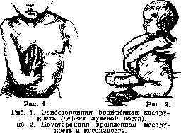
CUBITUS VARUS [manusvara and manusvalga, Klumphand (German), main bote (French)], deviation of the hand with respect to the axis of the forearm. The deviation of the hand can be volar, ulnar, radial, or dorsal. In most cases, a radial-volar or ulnar-volar deformity is observed. Deviation in the dorsal direction is extremely rare. C. occurs significantly less frequently than, for example, foot deformities in the form of clubfoot, with which C. is occasionally combined. Hoffa encountered cubitus varus once in 1,414 deformations; the data of Panzeri and Fischer approximately coincide with the numerical data of Hoffa. C., just like clubfoot, is more often bilateral and congenital and is, in all probability, genotypic. However, there are very few precise genealogical data. Blencke described a case where, among nine children of healthy parents, four had C. It is probable that C. is inherited as a recessive trait. In rarer cases, it may be acquired. - In congenital C., radiopalmar deviation is mainly encountered. All cases of congenital C. can be divided into C. without changes in the skeleton and C. with incomplete development of the skeleton. In terms of etiology, the first type of C. approaches typical clubfoot, which arises without changes in the skeleton, and this type of C. is more often observed in the form of palmar or ulnar-palmar deviation. Rosenkranz found approximately 50 cases of C. without bony defects in the literature. Such C. can have as its cause 1) a malposition in the uterus; 2) dysplasia of the forearm muscles; Potel draws attention to congenital partial muscular dystrophy; 3) joint dysplasia; congenital rigidity of the wrist joints is usually combined with limited mobility of other joints. In such cases, C. arises not because dynamic factors in the form of muscles partially fail, but because the development of the joints is delayed in a certain embryological phase. Changes in the muscles occur secondarily (Rocher). - The main theories of the origin of clubfoot (see) remain in force for C. as well, namely: 1) formation of the deformation due to mechanical causes (the role of the amnion); 2) neurogenic theory; 3) C. is the result of vitium primae formationis. If the theory about the role of the amnion can explain a known 10% part of cases of C. without defects of the skeletal system, then the theory, which attaches importance to vitium primae formationis, explains the predominant number of cases of cubitus varus with various defects of the skeleton. C. has defects in the development of the radius much more frequently. Data from various statisticians (K. Kato, Rabaud, Hovelacque) indicate a predominant frequency of defects in the development of the radius on both sides, in connection with which "bilateral congenital C. with deviation towards the radial side (manus valga) is more often observed. Such deviation occurs in men twice as often as in women. Pathological anatomy. With complete or partial absence of the radius, defects are most often noted simultaneously on the side of the carpal bones: absence of the os lunati and multanguli maj., atrophy, underdevelopment or complete absence of individual phalanges and metacarpal bones, sometimes ankylosis of interphalangeal joints. The remaining ulna is usually curved. Changes are also noted on the side of the humerus: it is often shortened, there is aplasia of the outer part of the lower epiphysis. All these details are well revealed on the X-ray. - Anatomical defects in radial deviation are also observed in muscles, vessels, and nerves. In muscles, both complete absence of individual groups and absence of differentiation into individual groups are noted, especially on the forearm. Thus, on many preparations carefully studied by Stoeffel and Stempel, the m. biceps had only a short head, the long one being absent; on some muscles there were variants of muscle origin and defects of differentiation. These defects are explained by delay in a certain period of embryonic development. This also explains defects in the vascular system: a. radialis is often preserved only in the form of a thin branch of the ulnar artery; changes in the topographic location of other arteries on the shoulder are noted. Defects of innervation in patients with C. are related to defects in the development of the bone-muscular system. A sharply expressed underdevelopment of the p. radialis, as well as some anomaly in the development of the p. musculo-cutanei, attracts attention. The latter is sometimes absent. - Pathological-anatomical changes in the deformity type manus vara with ulnar and ulnar-palmar deviation present a different picture. In these cases, there is a complete or partial defect of the ulna and often a defect of the carpal bones, which form from the ulnar ray. The radius may be normal, sometimes ankylosed with the humerus, or the head of the radius may be luxated. In the shoulder joint, limited mobility is often present, in the elbow - complete ankylosis or limited mobility. The clinical picture of C. is very characteristic. Deviation of the hand both towards the radial side (manus valga, figure 1) and towards the ulnar side (manus vara) is usually combined with volar flexion. Defects on the side of the carpal, metacarpal bones, and phalanges are often simultaneous, as a result of which the grasping ability of the hand is disturbed. With the absence of muscles of the shoulder girdle or forearm, significant functional disorders are noted. - In one case of congenital C. in combination with bilateral clubfoot in a 16-year-old girl (observations of Chaklin), the muscles of the shoulder girdle and individual groups of muscles of the shoulder and forearm were paralyzed. Nevertheless, her grasping ability was preserved. Congenital C. is sometimes combined with clubfoot (fig. 2); in some cases, other deformities are observed: congenital dislocation of the hip, congenital scoliosis, hare lip, spina bifida, etc. - It should be noted that despite the good adaptability of persons with congenital defects in general, bilateral C. makes the patient more of an invalid than many other deformities, for example, clubfoot.
Treatment of C. in its main features has something in common with the treatment of clubfoot. Methods of treatment of C. should be chosen depending on whether there is C. with a defect in the bony skeleton, C. without a bony defect but with pronounced muscle and fascial contracture, or C. on a neurogenic basis. - In C. with a predominance of muscular or fascial retraction, careful reducing manipulations and massage are indicated. The achieved correction is fixed each time with a splint or a light plaster bandage. Orthopedic devices with the possibility of gradual correction in the wrist joint have also been proposed. The Redard and Claude Martin apparatus is based on correction with elastic traction. - Bloodless manual reduction and staged bandages, used in clubfoot, give a good result also in C. If individual tendons represent a serious obstacle, tenotomy is indicated. Roche obtained good cosmetic and functional results by resection of the wrist bones with subsequent hypercorrection with a plaster bandage. - Thus, the task of orthopedic therapy in C. of the described type consists in correcting the deformity of the hand, acting primarily on the soft tissues. In C. caused by a bony defect, the task of therapy is more complex: not only to correct the deformity but also to maintain the correct position of the hand with respect to
Figure 3. Romano's method.
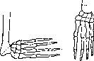
to the forearm. In early childhood, contracture is corrected by manipulations and massage, and the hand is held in a slight dorsal flexion by means of a leather apparatus until the moment when surgery can be resorted to. Chaklin applied twisting for correcting the deformity in children. There is a series of osteotomies and resections of the ulna for correcting radial and radio-palmar deviation. Osteotomies in the lower third of the ulna, even with soft tissue elongation, do not guarantee against recurrence in the presence of a bone defect. The method of Romano, consisting of wedge resection of the lower third of the ulna with reinforcement of the carpus by a metal or silk suture (Fig. 3), is very convenient. Sayre creates a new joint between the wrist and the ulna by performing a two-stage operation. In the first stage—resection or osteotomy of the ulna with reduction. After several months—the second stage: partial resection of the wrist with insertion of the thinned lower end of the ulna into the defect. Another type of operation consists in replacing the defect of the radius with free bone grafting. Antonelli and Ryerson took a free transplant from the radius, implanting it into the soft tissues at the place where the radius should be, after preliminary correction of the hand. The usual fate of transplants implanted into muscles is resorption, as a result of which such interventions lose practical interest. Albee takes a plate from the tibia for plastic purposes, fixing its upper end in the ulna and its lower end in the carpus (Fig. 4). The fate of such an intervention is more favorable because the transplant fuses with the bone, which is subject to mechanical and functional influences, which consequently extend to the transplant. In all interventions on the hand and forearm, it is extremely important to preserve full freedom of movement of the fingers, which increases the functional effect of the operation. Prolonged wearing of an apparatus that corrects the faulty position prevents recurrence. Like paralytic clubfoot, there is also cubitus varus caused by disorders in the neuromuscular apparatus. A hand deformity of this origin is possible on the basis of both flaccid paralysis (due to damage to the peripheral neuron) and spastic paralysis (damage to the central neuron). In addition, Roche distinguishes another group of cubitus varus based on reflexive contracture. Cubitus varus with signs of flaccid paralysis occurs with traumatic damage to the radial nerve, with obstetric paralysis, and as a consequence of poliomyelitis. Cubitus varus with spastic paralysis is characterized by a tense ulnar-radial position of the hand and often involuntary movements in muscles of athetoid or choreoathetoid type. Spastic cubitus varus can be based on encephalitis suffered in early childhood, as a consequence of Little's disease, and also of hysterical origin. In addition to cubitus varus with radial deviation, there are rarer cases of dorsal deformity of the hand. Such cubitus varus is noted in childhood paralysis (Dejerine, Menciere), in syringomyelia, and in adult hemiplegia. Various hand deformities of paralytic origin, although French authors (Roche) classify them as cubitus varus (main bote), differ sharply from congenital cubitus varus both in etiology and pathological anatomy, as well as in therapy. For the treatment of paralytic cubitus varus, in addition to apparatuses which improve mainly the cosmetic aspect, there is a series of operations aimed at improving hand function. If the flexor group is preserved, it is necessary to set the hand in a dorsal flexion position convenient for function. In such a position, tenodesis of the extensors or arthrodesis of the wrist joint is performed. With good strength of the flexor group, an excellent effect is obtained by transplanting active muscles to inactive extensors and the abductor of the thumb. For this purpose, the methods of Perthes and Stoffel are most suitable. Transplantation of flexors through the interosseous interval is less successful. Of the arthrodesis methods, the method of Albee with transplantation of a tibial transplant to the dorsal surface of the wrist joint is convenient. Interventions on nerves in paralytic cubitus varus have only theoretical significance. For the treatment of spastic cubitus varus, the following methods exist: 1) neurotomy (Stoffel) or alcoholization (Sicard), 2) posterior radicotomy (Forster), 3) ramisectio cervicalis (Royle), 4) hyperneurotization (Spitzy), 5) partial resection (reduction of cross-section) of the median and ulnar nerves (Vreden). Of these methods, neurotomy according to Stoffel is shown for milder cases, posterior radicotomy for the most severe cases. The other methods are in the stage of clinical study. In two severe cases of spastic cubitus varus, Chaklin applied the following method: tenotomy of the palmaris longus muscle, correction of contracture by staged bandages, and then intraosseous tenodesis of the hand extensors. A fully good cosmetic and relatively favorable functional effect was obtained. Summarizing the methods of treating cubitus varus, one can say that in congenital cubitus varus, when function is not particularly disturbed, orthopedic manipulations and operations can achieve a good cosmetic effect and preserve satisfactory function. In flaccid paralytic cubitus varus, especially traumatic, good cosmetic and functional results can be obtained by transplanting active muscles to inactive ones.
Related articles
Mentioned in
Cite this page
“Cubitus Varus.” Soviet Medical Encyclopedia. English translation of Bolshaya Meditsinskaya Entsiklopediya, 1st ed. (Moscow, 1928–1936), ed. N. A. Semashko. https://sovietmedicalencyclopedia.pages.dev/article/cubitus-varus/