Dental Cysts
Historical document, translated for reference. It reflects medical knowledge of the 1920s–30s and is not medical advice.
Summary
Dental cysts are cavity formations located in the alveolar processes and jaw bones, etiologically related to dental diseases. They are classified as simple (follicular) or root (periodontal) cysts, with distinct origins and characteristics.
Encyclopedia article (1928–1936)
DENTAL CYSTS, cavity formations located in the thickness of the alveolar processes, and partly in the body of the jaw bones, and etiologically related to diseases of the teeth. They have a connective tissue wall lined on the cavity side with a layer of epithelium. The latter is as a rule constructed according to the type of the covering epithelium of the oral cavity, but on the upper jaw it is sometimes represented by cylindrical ciliated epithelium of the type of the mucous membrane of the maxillary sinus. They are distinguished: 1) simple, or follicular cysts and 2) root (periodontal, periosteal) cysts. The formation of follicular cysts is connected with a disturbance in the development of the germs of permanent teeth or of excessively laid tooth germs, with fluid accumulating in the tissues of the tooth follicle. It is assumed that the basis of the process is the degeneration of cells of the enamel pulp, and according to some views, the so-called debris epitheliaux paradentaires undergo cystic changes. As a result of the process, the cavity described above is formed. The contents of the cyst consist of transparent, serous or sero-mucous, sometimes bloody fluid, usually with an admixture of cholesterol crystals. Often a tooth is found in the thickness of the wall of a follicular cyst, the crown of which protrudes into the cavity of the cyst, and the root is immersed in the thickness of the wall. Teeth located in follicular cysts are often rudimentary or consist of only a crown, which depends on the time of onset of the pathological process: if the tooth germ undergoes pathological change at an early stage of development, the further differentiation of its tissues may completely stop; however, follicular cysts containing fully developed teeth are also noted. The question of the genesis of follicular cysts should be considered insufficiently clarified. New clinical and roentgenological observations give reason to assume their inflammatory origin, with the starting point of the process being a disease of the milk tooth, and the germs of permanent teeth are involved in the process only secondarily (Lapidus). Root cysts are inflammatory neoplasms developing from epithelial and cyst-like granulomas by degeneration and breakdown of their tissue, gradually passing into the contents of the cyst cavity. As a result of the death of granuloma tissue, the apex of the root may be exposed, which thus turns out to be enclosed in the cyst cavity, and the contents of the latter gain access to the root canal (see separate table, fig. 4). The further growth of the formed dental cyst depends: 1) on the accumulation of inflammatory exudate as a result of the action of infection continuing to enter from the root canal, and 2) on the proliferation of the epithelial lining in the form of strands immersed in the surrounding granulation tissue; at the same time, degeneration and splitting of the epithelial strands occur. The cavity of such a cyst is filled with a viscous transparent liquid with a pearly luster (cholesterol); a suppurated cyst contains a turbid sero-purulent or purulent liquid, and sometimes a cottage cheese-like or mush-like mass due to a significant accumulation of cholesterol, products of protein breakdown, mucus, etc. With hemorrhage, the contents acquire a chocolate-brown color. Dental cysts, developing at the apices of teeth in the depth of the alveolar process, as they grow, produce significant destructive changes not only in the area of the alveolar process but also of the jaw body. Under the influence of pressure exerted by the growing dental cyst, the bony wall of the jaw, most often the vestibular one, first bulges in the form of a clearly defined hard tumor with a smooth surface, and then as the dental cyst grows, it thins out, becomes pliable, and on pressure with the finger crepitates (the parchment crackle)-the so-called Dupuytren's symptom. Further, as the bony wall becomes thinner and thinner, it disappears completely, and then the capsule of the dental cyst directly adjoins the soft tissues covering the jaw. In this case, the cystic tumor, maintaining the same clearly defined character, becomes soft, fluctuating; the mucous membrane covering the tumor is usually unchanged, of normal color. Bulging of the bony wall or cystic sac can be observed simultaneously from both sides: vestibular and palatal (lingual). Dental cysts often, in addition to the usual path mentioned above, also develop toward the maxillary sinus and into the nasal cavity, displacing and narrowing these cavities. Growing into the maxillary sinus and developing further, the cyst expands its boundaries to significant dimensions, capturing almost the entire upper jaw. On the lower jaw, a dental cyst, spreading along the length of the jaw, can also involve the ascending ramus. Dental cysts are often multiple, i.e., in the same patient several dental cysts of different sizes are observed in different areas of the jaws. The presence of teeth or roots with dental cysts is not necessary, since the development and growth of cysts can continue even after the removal of the offending tooth. Dental cysts grow very slowly and painlessly, and have no tendency to malignant degeneration. Sometimes after treatment and filling of the root, and sometimes on their own, the growth of the cyst ceases or even, if it is small, its reverse development begins, i.e., the processes of breakdown in the wall of the cyst stop, the accumulation of contents of the cavity decreases and its nature changes: previously turbid due to the admixture of cellular elements-leukocytes, desquamating epithelial cells and elements of granulation tissue in various stages of degeneration-it gradually becomes clear; sometimes the liquid contents are absorbed, and then cholesterol together with mucus and products of protein breakdown forms a mush-like mass. The capsule becomes more and more fibrous and shrinks. Along the periphery, new formation of bone trabeculae occurs (see separate table, fig. 5). It should be noted that cystic formations, being etiologically related to teeth, are in essence diseases of the jaw bones; from this point of view, follicular and root cysts, along with multilocular cysts developing from adamantinum, should be considered in the section of jaw cysts. The diagnosis of dental cysts is based on the presence of a limited bulging (swelling), in later stages elastic, crepitating or fluctuating. There is no pain on pressure, the growth is slow, painless, the mucous membrane, if there is no suppuration of the cyst, is usually unchanged. The decisive significance is the roentgenogram, on which the shadow of the dental cyst has a clearly contoured, round or oval shape, and often the displacement of the roots of neighboring teeth is visible. The frequency of dental cysts can be judged from the following data: at the Berlin Dental Institute according to data for 12 years (Becker) almost every 50th patient of those who came to the institute had a cyst. According to the data of my State Institute of Stomatology and Odontology (GISO) on 57 thousand patients who passed through the surgical department of the institute's outpatient clinic for the period 1923-29, 754 dental cysts were operated on, i.e. 1 cyst per 76 people. The treatment of dental cysts is exclusively surgical-extirpation of the cystic sac with subsequent hermetic suturing of the surgical wound or partial removal of the anterior wall of the cyst and formation of thus from the cyst as it were an additional cavity vestibuli oris. Attempts at such interventions as the injection of tincture of iodine, alcohol, etc., in the hope of obtaining subsequent shrinkage, obliteration and reverse development of the dental cyst, have not been successful and are now completely abandoned.
At the present time, there are 2 methods of operating on dental cysts—according to Partsch and according to the State Institute of Stomatology and Odontology (proposed by Danilevsky). Partsch's method consists in that, by means of an arc-shaped incision in the paradental tissues (the convexity of the incision is turned toward the gingival margin), the cystic tumor is exposed, and if the cyst does not exceed 2-3 cm, it is completely enucleated, after which the operative wound is tightly sutured. This is so-called Partsch II. However, if there is a cyst larger than 2-3 cm, it is not enucleated, but only the partial removal of its anterior wall is performed, after which the flap of paradental tissue that covered the cystic tumor is not returned to its place, but is sutured to the free edge of the capsule of the dental cyst, and thus an additional cavity vestibuli oris is created. This is so-called Partsch I. The cavity obtained in this way, sometimes very large and deep, subsequently gradually flattens out. Since Partsch's second method (Partsch II) gives good results when operating on small cysts, the application of Partsch's first method (Partsch I) is not advisable, since the deep pit remaining as a result of such an operation for a long period, with food getting stuck and rotting in it, is a distressing condition for the patient. The method of operating according to GISO consists, first, that dental cysts are extirpated in all cases regardless of size and the wound is tightly sutured, and second, that exposure of the cystic tumor is achieved by separating a trapezoidal flap of paradental tissue, starting from the gingival margin; the incisions in this case are made outside the boundaries of the cyst. The lines of these incisions begin at the transitional fold or, if necessary, even higher and end on the midline of the necks of the teeth, and in the absence of teeth—on the crest of the alveolar border. Then a horizontal incision is made along the edge of the gum between the teeth (in the absence of teeth—along the crest of the alveolar process), and thus the ends of the vertical incisions are connected. The resulting trapezoidal flap of paradental tissue is easily separated and, being pushed aside, exposes the entire cellular process with the focus of the disease in the center. After removal of the protruding bony wall, if it exists, and enucleation of the cyst, the flap is placed back in position and fixed with several sutures; first the lateral sutures are applied, and then between the teeth (see figure). The roots and teeth affected by the cyst, if they have functional value and are firmly in place, are not removed. In the case of intact teeth, appropriate treatment of them and filling of the canals with a dense filling material is performed. The part of the root protruding into the cyst cavity is resected. A dense root canal filling carried out to the apex is in such cases the main requirement for successful results of the operation.
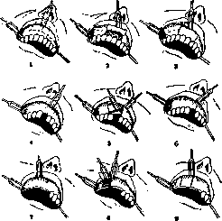
The roots and teeth affected by the cyst, if they have functional value and are firmly in place, are not removed. In the case of intact teeth, appropriate treatment of them and filling of the canals with a dense filling material is performed. The part of the root protruding into the cyst cavity is resected. A dense root canal filling carried out to the apex is in such cases the main requirement for successful results of the operation.
Related articles
Mentioned in
Cite this page
“Dental Cysts.” Soviet Medical Encyclopedia. English translation of Bolshaya Meditsinskaya Entsiklopediya, 1st ed. (Moscow, 1928–1936), ed. N. A. Semashko. https://sovietmedicalencyclopedia.pages.dev/article/dental-cysts/