Granulomas
Historical document, translated for reference. It reflects medical knowledge of the 1920s–30s and is not medical advice.
Summary
Granulomas are tumor-like processes in tissues associated with productive inflammation, occurring in infectious diseases like tuberculosis and syphilis, and as specific entities like granuloma annulare. They vary in size and form, undergo characteristic changes including necrosis and fibrosis, and can sometimes mimic malignant tumors.
Encyclopedia article (1928–1936)
GRANULOMAS, GRANULOMATOSIS (from Latin granum-grain and -oma suffix for tumors), terms denoting neoplastic processes in tissues associated with the so-called productive inflammation (see) of them; consequently, granulomas are not tumors in the proper sense of the word, but inflammatory tissue proliferations. Granulomas are most frequently observed in chronic and acute infectious diseases (tuberculosis, syphilis, leprosy, rhinoscleroma, actinomycosis, glanders, abdominal and typhus fevers, malaria, rabies, acute rheumatism, etc.). Certain designations have become established for certain granulomas; thus, tuberculous granulomas are called tubercles, syphilitic granulomas-syphilomas, gummas. Sometimes granulomas bear the names of their describers; thus, one speaks of Aschoff's nodules in the myocardium in acute rheumatism, of Durck's granulomas in the brain in tropical malaria, of Babes' rabic nodules in rabies, etc.; the term "granuloma malignum" some apply to lymphogranulomatosis. In addition to such infectious granulomas, characteristic of various general infections and not having independent significance, pathology also recognizes several special types of granulomas that represent to some extent isolated conditions, i.e., as it were, independent nosological units; thus, one speaks of g. (mycosis) fungoides, g. teleangiectodes (see Botryomycosis), g. inguinale tropicum, spinal granuloma, apical granuloma (of the tooth root apex), etc. The size of granulomas varies from barely noticeable, and sometimes even imperceptible to the naked eye, points (miliary and submiliary granulomas) to the size of an apple or a child's head. The shape of granulomas is most often round, sometimes irregular, branched. Granulomas usually appear denser compared to surrounding tissues, and this density is directly proportional to the duration of the granuloma and is explained by the significant development of dense connective tissue at and around the site of the granuloma. The mechanism of development (formal genesis) of granulomas varies depending on the nature of the tissues and the etiological factor. Usually, there is first a lesion of the vessel wall (capillaries, small veins, arteries, lymphatic vessels), and then around the latter, from local tissue elements, a granuloma develops (see Inflammation, Granulation tissue). In rarer cases, granulomas arise entirely in epithelial complexes (for example, tubercles in the lumens of seminiferous tubules, in the follicles of the thyroid gland), while the involvement of vessels and generally mesenchymal elements is manifested only later and to a minor extent. COURSE AND OUTCOME OF GRANULOMAS. The newly formed cells, over time (varying for different granulomas), undergo fatty degeneration, necrobiosis, and disintegration, especially in the centers of the granulomas; this includes the so-called caseous disintegration in tubercles, gummas. On the other hand, dense connective tissue develops, especially at the periphery of the granuloma; sometimes the entire granuloma undergoes such fibrous transformation. Necrobiotic processes in granulomas are often caused by sharp changes in the vessels supplying the area of the granuloma (see Gumma). Parts of the organ surrounding the granuloma may experience compression, constriction, strangulation, and associated atrophic and degenerative processes in the parenchyma, phenomena of stenosis, etc. Often the central parts of the granuloma undergo purulent melting (actinomycosis, glanders). The indicated stages of development are accompanied by changes in the macroscopic properties of the granuloma: from a gray or gray-red nodule (initially) it turns into a pale-yellowish focus (lipoid infiltration, necrosis), and then into a pale-gray or white focus (scar). With abundant development of blood vessels (e.g., in epulides, in botryomycosis), granulomas retain a pink-red color, resembling an angioma; however, the similarity to an angioma can be very significant even microscopically. The causal genesis of granulomas is studied by applying various methods of their processing for bacteria; microscopic, and sometimes bacteriological, methods in some cases are all the more necessary because histologically granulomas can be similar or even identical in very different diseases (e.g., in tuberculosis, syphilis, leprosy). Granulomas can sometimes take an unusual course, simulating by their size, rapid growth, multiplicity, or systemic spread (for example, to lymph glands, reticuloendothelial apparatus), as well as by general phenomena (changes in the blood, cachexia) - typical malignant tumors. A classic example of such a malignant granuloma is lymphogranulomatosis (see). For granulomas in various diseases - see these diseases themselves.
I. Davydovsky. Granuloma annulare, ring-shaped granuloma, lichen annularis, helodermia simplex et annularis, celluloma annulare, a chronic skin disease characterized by the formation of dense nodules, the size of a millet grain to a pea, of flesh-colored or dull-rose color; these nodules are arranged in circles or in the form of rings due to eccentric growth and the appearance of new elements. G. annulare represents sharply delimited, flat-elevated, round or oval-shaped, deeply seated in the skin, not fused with underlying tissues nodules, which most often develop on the back of the hands and fingers, especially in the area of joints, but can occur on elbows, forearms and feet, shins, thighs, buttocks, ears, occiput and as an exception on the face. The nodules never ulcerate. The skin in the center of the circle or ring is usually normal, rarely atrophy is observed. Subjective sensations are absent. The disease occurs more often in women and children. G. annulare develops slowly and lasts for months and even years. Spontaneous, without treatment, resolution of nodules without scar formation and the appearance of recurrences has been observed. - The erythema elevatum diutinum described by Crocker and Williams is a chronic skin disease characterized by the presence of sharply delimited, concave nodules that do not form ring-shaped forms and apparently represents a rare variety of G. annulare. Histological examination reveals in the deep and middle layers of the skin a focally located cellular infiltrate consisting of fibroblasts, epithelioid and lymphoid cells. The presence of giant cells has been noted in individual cases; necrosis of collagen fibers and cells of the granuloma are observed much more frequently. The vessels are dilated, thickened, around them - small-cell infiltration. Elastic tissue is absent in the nests of the infiltrate. - The etiology of G. annulare is unclear. Most authors see the cause of the disease in tuberculosis, however, without providing sufficiently convincing evidence for this. No one has succeeded in finding tubercle bacilli in sections, and inoculations of affected tissue into pigs gave negative results. For therapeutic purposes, radium, mesothorium, X-rays, arsenic, salvarsan, salicylic preparations, resorbing and peeling ointments, cryotherapy (cauterization with snow carbon dioxide - 40 seconds with moderate pressure) are recommended.
M. Roaentup. Dental granuloma (syn. root granuloma), a limited area of granulation tissue, usually located at the apex of a tooth with a necrotic pulp. The dental granuloma represents a manifestation of chronic inflammation arising in the tissues surrounding the root apex, under the influence of penetration of microorganisms and their toxins, pulp tissue decay products, food residues, as well as filling materials and medications into these tissues from the root canal. The dental granuloma arises in the periodontal tissue (root periosteum) in the area of the apical foramen in the form of a round-cell focus; the zone of cellular proliferation gradually expands, capturing new areas of periodontal tissue and also spreading to the bone marrow in the area of the alveolar floor. Due to resorption of bone trabeculae, an area of bone rarefaction first forms in the area of the alveolar floor, and later a focus of granulation tissue devoid of bone crossbars; in other words, there is a limited rarrifying osteitis (see separate table, fig. 3). A mature dental granuloma consists of granulation tissue proper and a peripheral fibrous capsule; the fibers of the latter directly pass into the periodontal tissue, so that during root extraction the granuloma is often also removed. The tissue of the dental granuloma proper is characterized by an abundance of plasma cells, as well as the presence of large "macrophages"-cells whose protoplasm, in the order of resorptive obesity, is packed with small grains of doubly refractive lipoids (cholesterol esters). Among these elements are located various cells of histiogenic and hematogenous origin: in long-existing granulomas, under conditions of a subsided inflammatory process, - predominantly fixed elements of the fibroblastic series [see color plate (vol. III, pp. 679-680), fig. 3], and in cases of exacerbation of inflammation - with a predominance of leukocytic infiltration, often transforming the granuloma into an abscess. Besides simple dental granulomas consisting only of connective tissue elements, epithelial granulomas are distinguished, the tissue of which is more or less permeated with strands of epithelium (see separate table, fig. 1), and cystic granulomas, characterized by the presence of epithelium-lined cavities (see separate table, fig. 2). The latter, usually appearing in the center of the granuloma, may arise as a result of a) degeneration of central areas of massive epithelial strands with their subsequent splitting (see separate table, fig. 1) and b) decay of granulation tissue, for example in the process of suppuration; in this case, the resulting cavity is successively lined with epithelium by the growth of the epithelial strand closest to the cavity. As a result of regressive processes in the granuloma tissue, protein and fat detritus appears. When the detritus decays, cholesterol crystals precipitate, which are a characteristic component of the contents of the cavities of cystic granulomas and root cysts (see separate table, fig. 2). The epithelium in the granuloma is constructed according to the type of the Malpighian layer of the covering epithelium of the oral cavity; an exception is the occasionally found cylindrical epithelium in the granuloma of the upper jaw, appearing as a result of inflammatory proliferation of the mucous membrane of the maxillary sinus. Regarding the origin of the epithelium in granulomas, two theories exist: 1) the theory of proliferation of the so-called "debris epithelia paradentaires", islands of embryonic epithelium lying in the periodontium of all teeth, and 2) the theory of proliferation of the covering epithelium of the oral cavity. The first theory is considered the most substantiated. Under favorable conditions, created as a result of root treatment, and sometimes spontaneously, dental granulomas undergo reverse development. In the process of organization of the dental granuloma tissue, new bone crossbars are formed, and restoration of normal architecture in the area of the alveolar floor may occur. From the point of view of the doctrine of the so-called oral sepsis, dental granulomas are considered as hidden infectious foci from which microorganisms can enter the general bloodstream. Epithelial and cystic granulomas are a preliminary stage in the development of dental cysts (see).-Clinically, dental granulomas as a rule do not manifest themselves or give indefinite symptoms of chronic inflammation of the root periosteum; most often they are detected only during exacerbations of the inflammatory process.-The diagnosis of dental granuloma is usually made on the basis of an X-ray, which gives a picture of the mentioned
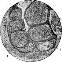
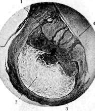
Figure 1.
Figure 2.
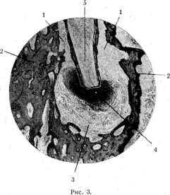
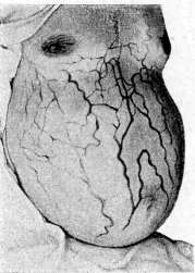
Figure 4.
Figure 1. Epithelial granuloma: / splitting of the epithelial strand and formation of a slit-like cavity: 2 strands of epithelial tissue; 3 islands of granulation tissue. Figure 2. Cystic granuloma: / - strands of epithelium; 2 granuloma cavity containing a large amount of cholesterol crystals; 3 epithelial lining of the cavity; 4 tooth root (from the author's specimen). Figure 3. Dental granuloma in situ (post-mortem material): / periodontium; 2 bone walls of the alveolus; 3 fibrous capsule of the granuloma, passing into the periodontium (due to the presence of the granuloma and resorption of bone tissue in the area of the floor of the alveolus in the periapical spongy bone substance, a space devoid of bone trabeculae has been formed, filled with granuloma tissue); 4 - granuloma; 5 - tooth root. Figure 4. Head of a jellyfish (according to Mohr-Stehelin) - TO ST. Granulomas, Head of a jellyfish. above the limited periapical (at the root apex) rarrifying osteitis.-Treatment of dental granuloma: a) conservative - according to the general rules for treating gangrenous tooth roots, little reliable, and b) predominantly used, surgical - operation of resection (see) of the root apex, removed together with the dental granuloma.
Related articles
Mentioned in
Cite this page
“Granulomas.” Soviet Medical Encyclopedia. English translation of Bolshaya Meditsinskaya Entsiklopediya, 1st ed. (Moscow, 1928–1936), ed. N. A. Semashko. https://sovietmedicalencyclopedia.pages.dev/article/granulomas/