GUMMA
Historical document, translated for reference. It reflects medical knowledge of the 1920s–30s and is not medical advice.
Summary
A gumma is a type of infectious granuloma observed in syphilis during the tertiary period, characterized by nodular lesions that can affect various tissues and organs. The article describes the macroscopic and microscopic appearance of gummas, their development, and differentiation from tuberculous lesions.
Encyclopedia article (1928–1936)
GUMMA (from Latin gummi-gum), or syphiloma, is a type of infectious granuloma observed in syphilis in various tissues and organs during the late stages of the disease, in the so-called tertiary period [see separate table -(pp. 739-740, 2nd table), fig. 4]. Some French authors use the term G. also in relation to other granulomas, which is to some extent correct, since in many, especially chronic infectious diseases (tuberculosis, leishmaniasis, leprosy, mycotic lesions, etc.), morphologically similar formations can be observed.-G. (see separate table, fig. 2) is a node of round or irregular shape, from barely noticeable sizes (miliary G.) to the size of a chicken egg (solitary G.), more or less dense consistency depending on the duration of the process and location of the G. The color of G. varies depending on the same factors; usually its central parts have the appearance of a homogeneous gum-like, pale gray or yellowish mass, sometimes of a drier, cottage cheese-like appearance and dense consistency. Toward the periphery, the tissue of G. has the appearance of granulation tissue of gray-red color due to the abundance of newly formed blood vessels. In old G., this zone represents a coarse scar-like, very dense whitish tissue, which sometimes penetrates into the central parts, which gradually resorb, more rarely undergo calcification. Tissues adjacent to G. undergo dual changes; on the one hand, an inflammatory infiltrate penetrates here in the form of rays, on the other hand, the resulting scars have the property of strongly tightening surrounding parts, causing deformation, constrictions, stenoses, etc. On microscopic examination of G. (see separate table, fig. 3) in their initial development stages, an intense inflammatory infiltrate is found, consisting of lymphocytes and plasma cells with a small number of leukocytes, eosinophils, and sometimes giant cells. When (later) necrosis occurs in the center of G., its peculiarity is noted, which is that the outlines (shadows) of former tissues, e.g. vessels, scar strands, elastic fibers,
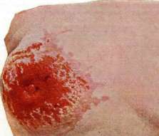
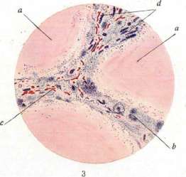
Figure 1. Paget's disease (after Jacob) >. Figure 2. Multiple gummas (y) of the liver, partially with sclerosis and atrophy of liver tissue (/); calcification of some gummas; b - right adrenal gland; c - vena cava mr.; d - Spiegelian lobe of the liver; e - left lobe of the liver. Fig. 3. Gumma of the liver: a - gummatous masses; b - round cell infiltrates along the course of fibrous septa; c - here well-developed vascular network and strands of liver cells (l4). (Figs. 2 and 3 - according to preparations from the Anatomical Institute of 1st Moscow State University.) To the stt tyMMU, GrOUpin '." t i- gg. in contrast to tuberculosis, at the site of caseous necrosis, all structures quickly disappear. The necrosis itself is usually explained by the presence of vascular changes in the area of the gumma of the endarteritis and endophlebitis type. To distinguish G. from tuberculous foci, it is important to consider the weak participation of epithelioid and Langhans-type giant cells in gummatous infiltrates, the abundant development of blood vessels, as well as the more diffuse nature of the necrosis itself, which occurs after the development of fibrous tissue, and not before it, as in tuberculosis. However, it should be remembered that the differential diagnosis (between syphilitic G. and tuberculum) can sometimes present from a purely histological side almost insurmountable difficulties (e.g. gummas in the form of nodules of epithelioid cells with the presence of Langhans-type giant cells, etc.), and then the question is resolved with the help of serological, bacterioscopic examination, inoculation of animals, etc. It should however be borne in mind that pale spirochetes are found in G. very rarely. In congenital syphilis in newborns, G. are observed less frequently and often differ by their insignificant sizes (miliary G.), sometimes having the appearance of miliary abscesses [see vol. VII (pp. 183-184, fig. 5) and article Hypernephritis]. The gummatous process can also be observed not in the form of limited G. nodes, but in the form of gummatous infiltration, i.e. a diffuse specific infiltrate, which first increases the volume of the affected organ or its part by delamination and separation of its normal tissue by newly formed cellular elements, sometimes along with the formation of small blood vessels, and then, due to fibrous transformation of the infiltrate with uneven wrinkling and contraction, decreases its volume and distorts its shape with radiant depressed scars, sunken linear, meshy or formless strands and disks, which can capture the entire organ (syphilitic fibrosis). See also Syphilis (experimental), Granulomas.
Related articles
Mentioned in
Cite this page
“GUMMA.” Soviet Medical Encyclopedia. English translation of Bolshaya Meditsinskaya Entsiklopediya, 1st ed. (Moscow, 1928–1936), ed. N. A. Semashko. https://sovietmedicalencyclopedia.pages.dev/article/gumma/