Heart Block
Historical document, translated for reference. It reflects medical knowledge of the 1920s–30s and is not medical advice.
Summary
This historical article from the 1928-1936 Soviet Medical Encyclopedia discusses heart block (heart-block), detailing its neurogenic and cardiogenic forms, types of conduction disturbances, and clinical manifestations such as Stokes-Adams syndrome.
Encyclopedia article (1928–1936)
HEART BLOCK (heart-block; the unfortunate term "block" should be abandoned), an interruption of the excitation running through the heart from its sinus node down to the terminal ramifications of the atrioventricular bundle (see) of His - Tawara (His - Tawara), the so-called Purkinje fibers (see diagram). Depending on the site where the excitation block occurred, as well as on the cause that provoked it, both the clinical picture, the main symptom of which is
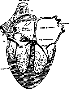
Diagram of the intracardiac regulatory apparatuses according to Aschoff - Koch: N.S. - sinus node of Keith-Flack, W. - Wenckebach's bundle; v. E. - valvula Eustachii; v. Th. - valvula Thebesii; S.a. - Septum atriorum; S.v. - Septum ventriculorum; a - atrial part of the atrioventricular node of Aschoff-Tawara; b - ventricular part of the node; T - common trunk (truncus) of His's bundle; S - left "branch" of the bundle; B - right "branch" of the bundle; P - terminal ramifications of the conduction system (Purkinje fibers); P.M. - papillary muscles. disturbance of the cardiac rhythm, and the prognosis of the disease change. On the basis of pathogenesis, a distinction is made between: a) the neurogenic form of heart block, in which the propagation of the impulse through the intracardiac nerve apparatuses is inhibited under the influence of excitation of extracardiac nerve apparatuses (central or, more rarely, peripheral irritation of the vagus nerves) and b) the cardiogenic form with anatomical lesions (inflammation, scars, gummas, etc.) of the heart itself in the region of its regulatory apparatuses (Pletnev, Nagaio). Depending on the localization of the interruption and excitation, four types of heart block are distinguished. 1. Sino-auricular heart block is characterized by a single or intermittent dropout of contractions of all divisions, depending on the activity of the Keith-Flack sinus node (see table, Fig. 1), most often under the influence of over-irritation of the vagus nerve, sometimes, possibly, depending on anatomical lesions around the sinus node or the artery supplying it. The pause is approximately equal to 2 normal contractions; the arterial pulse and heart sounds are absent. With the dropout of every second contraction (Lewis), bradycardia is detected (30-40 beats), which (upon cessation of heart block) can suddenly be replaced by a normal number of contractions. Subjective sensations are usually absent, work capacity is not reduced. Prognostically, this form of heart block is quite favorable, since it is only rarely based on anatomical damage. A therapeutic effect in this form (disappearance of dropouts) is produced by the use of atropine preparations. 2. Atrioventricular heart block, or transverse dissociation: anatomical or functional separation of the Keith-Flack node from the Aschoff-Tawara node, as a result of which the atria contract in their usual rhythm (60-80 beats), while the ventricles, subordinate only to the 2nd-order center (with lesser automatism), produce 40-30 contractions per minute and less. Several types of atrioventricular heart block are distinguished: a) impairment of conductivity along the bundle of His-Tawara (elongation of the PR interval of more than 0.25 sec.; see figure 4); b) so-called Wenckebach periods: gradual elongation of PR until the complete cessation of conductivity and isolated dropouts of ventricular systoles. Forms "a" and "b" are called relative heart block, in contrast to absolute, or complete heart block with permanent separation of the ventricles and atria. In the latter, generally characterized by a rare arterial pulse, the following are noted: c) cases with a preserved, although reduced, reaction of the ventricles to sympathicotropic excitations (acceleration of the pulse from movement, temperature, atropinization) and d) cases with a constantly rare systolization of the ventricles, which, under the influence of muscular exertion, can either slow their rhythm down even more or even stop for some time, thereby causing the clinical symptom complex of Morgagni-Adams-Stokes: loss of consciousness with a pale face and epileptiform convulsions (brain ischemia). In cases "c", the Aschoff-Tawara node apparently is not damaged, but only isolated from the atria by a connective tissue cuff (Monckeberg); in cases "d", the node itself or the artery feeding it has suffered anatomically [fibrous scars, rheumatic myocarditis, inflammation of the medial cusp of the tricuspid, gummas (see figure on p. 567), neoplasms, wounds; see figures 2, 3, and 5]. Without electrical or phlebographic analysis, transverse dissociation can be suspected in the presence of a very rare arterial pulse that does not show noticeable acceleration from atropine (0.001 g subcutaneously, according to Degio), and upon auscultation of the so-called "cannon sounds" (Stokes, Strazhesko): with a regular ventricular rhythm, some sounds are so intensified that they indeed have the character of gunshots. In Fig. 3, which depicts this phenomenon, it can be seen that it appears only when the atrial contraction almost coincides with (slightly precedes) the ventricular contraction (Zelenin, Fogelson). Sometimes a doubling of sounds is determined: the atrial gallop rhythm (Fogelson-Rozhkova), since, due to the discrepancy in the activity of the atria and ventricles, the atrial sound, separated by an interval greater than normal from the 1st ventricular sound, is heard as an independent sound. The course and prognosis of atrioventricular heart block depend (according to Zelenin) on the state of the myocardium and
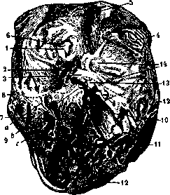
The His-Tawara system from the side of the right ventricle and its relation to gummatous formations. The ramifications of the system are schematically depicted: b - right branch of the bundle and the Tawara node, located near the membranous septum; 1 - fossa ovalis; 2 - valvula Thebesii; 3 - pars membranacea septi; 4 - musculi pectinati (auriculae atriorum); 5 - vena cava superior; 6 - vena cava inferior; 7 - septum ventriculorum; 8 - dotted line approximately indicating the ostium atrioventriculare dextrum; 9 - valvula tricuspidalis: a) v. posterior externa, b) v. interna, c) v. anterior externa; 10 - entrance to the pulmonary artery; 11 - musculi papillares; 12 - uneven surface of the epicardium covered with false membranes, after removal of the outer layer of the pericardial sac; 13 - gumma in the ventricular septum that has also damaged the valve tissue; 14 - gummas seated in the tissue surrounding the origin of the pulmonary artery and aorta and protruding into the cavity of the right atrium (a large gumma is sectioned).
vessels: in young subjects, heart block can remain without effect on the heart's work capacity for many years. The Morgagni-Adams-Stokes syndrome most frequently occurs at moments of transition from a normal rhythm to a pathological one. Treatment can be etiological only on a syphilitic basis of the condition, but in general it reduces to the organization of regimen. Digitalis is contraindicated in the relative and neurogenic forms, because, due to its effect on the vagus nerve, relative heart block can turn into absolute (Zelenin), or even the entire heart can be arrested in toto. In persistent, organic heart block (form "d"), in the stage of compensation failure, the administration of digitalis can be permitted, because with it the ventricles are almost excluded from the influence of the vagus nerve. According to recent observations (Zelenin), squill has an even greater effect on the conduction system than digitalis. The comparatively rare cases of transient heart block of rheumatic origin (Mackenzie) either pass spontaneously or yield to treatment with salicylates. The foregoing refers predominantly to the cardiogenic form of heart block. In the neurogenic form, there may occur either transverse dissociation, which passes from atropine, or cardiac activity is associated, and only from time to time does a sudden arrest of the ventricles occur with the above-described Morgagni-Adams-Stokes syndrome (see Figure 6). The use of atropine, transition from a horizontal position to a vertical one, movement (exclusion of the vagus nerve or excitation of the sympathetic nerve) can completely stop the attack and return a normal rhythm to the heart. Full recovery can be counted upon only in syphilitic lesions of the central nervous system. 3. Bundle-branch heart block. Sometimes the blocking process can damage only one (more often the right) branch of the His-Tawara bundle: the activity of the atria and ventricles is associated, but the ventricular complex of the electrocardiogram waves has a peculiar appearance (see Figure 7). The heart rate is not only not decreased, as in all other forms of heart block, but, on the contrary, is more frequently increased. A gallop rhythm is frequently auscultated as a result of the asynchronous contraction of the ventricles (longitudinal dissociation). Diagnosis can be established only electrocardiographically. A history of syphilis is frequently noted. The prognosis is doubtful in block of the right branch and completely unfavorable in block of the left branch. One must be careful with digitalis preparations, and especially with squill, since they can also block the second branch of the His-Tawara bundle. 4. Block of the terminal branches of the His-Tawara bundle [arborisation-block (Oppenheimer, Rothschild)] is also diagnosed only electrocardiographically: the ventricular complex (QRS) is widened, the magnitude of the waves is very small, the R wave is notched, the T wave is frequently absent or has a negative direction (see Figure 8). Pathologically and anatomically, widespread fibrosis of the ventricular wall adjacent to the endocardium is discovered, mainly in the region of the branching of the descending branch of the left coronary artery. The prognosis is extremely poor: patients do not live longer than 1 year. Forms 3 and 4 require further study.
V. Zelenin.
(I
I' II
1 II I II
I II
I II
I- II
1 II
I II
I II
т « P
T
P
T P
T * P
T
P
T P
I

Figure 1. Sinoauricular block (I - tones, II - electrocardiogram). Blocking occurs at the places marked with an asterisk. II!
I
и И „ I
П

Figure 2. Wenckebach periods, progressive exhaustion of conductivity function: with a PR interval > 0.3 sec, ventricular contraction drops out. On the phonogram, it is seen how the atrial tones move away from the ventricular ones, creating conditions for a gallop rhythm. At a1a2, there are only atrial tones.

se...
Figure 3. Complete transverse dissociation (electrocardiogram and phonogram). On the phonogram, both atrial tones (a1) and ventricular tones (I, II) are separately indicated. At *, the phenomenon of cannon sound is noticeable. On the electrocardiogram, complete independence of the atrial and ventricular rhythms.
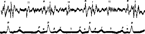
Figure 4. Impairment of impulse conduction along the His-Tawara bundle; prolongation of segment a.

a c v a
c* a
Figure 5. Venous pulse in complete block: the atrial wave (a) is not connected with the ventricular elevations; in places it is superimposed on the carotis wave (c).

P
PPPPPPppp
TRRp ft
a
\PR.\PlTP\ TR y p p p p p p Figure 6. Neurogenic form of block: after several associated contractions, the ventricles stop beating (only the atrial wave and atrial tones are visible). 1C
I II
1 II
i п
i и

wmmMNmi
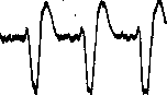
Figure 7. Right bundle-branch block and gallop rhythm (splitting of the 1st tone). 1_ Юсе* Figure 8. Terminal branch block (Arborisation block).
Related articles
Mentioned in
Cite this page
“Heart Block.” Soviet Medical Encyclopedia. English translation of Bolshaya Meditsinskaya Entsiklopediya, 1st ed. (Moscow, 1928–1936), ed. N. A. Semashko. https://sovietmedicalencyclopedia.pages.dev/article/heart-block/