Cardiac Arrhythmias
Historical document, translated for reference. It reflects medical knowledge of the 1920s–30s and is not medical advice.
Summary
This historical article from the 1928-1936 Soviet Great Medical Encyclopedia covers the physiological basis, classification, and electrophysiological mechanisms of cardiac arrhythmias, detailing the role of the sinoatrial and atrioventricular nodes as well as the conduction system.
Encyclopedia article (1928–1936)
Cardiac arrhythmias (from the Greek a- negative particle and rhythmos - rhythm) include not only disorders of cardiac activity in which all regularity in systole is completely lost, but also conditions that lead to the emergence of pathological rhythms (allorhythmias), as well as simple accelerations and decelerations of the heart rate: tachycardias (Gerhardt) and bradycardias (Eichhorst). The classification of arrhythmias is constructed on a topographical principle, according to the location of the morbid disorder within the system of intracardiac regulatory apparatuses. The basis of the subsequent exposition is the classical theory advanced by the German school and by no means shaken in its main features by the works
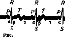
I. Above - phonogram, below - electrocardiogram.
of French investigators (Vaquez, Donzelot, Bard, Gerodel). The histological relations of the various parts of the heart, necessary for understanding both the physiology and pathology of the cardiac rhythm, are assumed to be known, which is why they are omitted (see Atrioventricular bundle, nodes). Experimental studies by Lewis through local derivation of the heart's action currents proved that excitation arises in the Keith-Flack sinus node, which is the highest regulatory center (of the 1st order), which is why the normal heart rhythm is called the sinus rhythm. The excitation, moving further through the atria, reaches the atrioventricular node (center of the 2nd order) and then through the His-Tawara bundle penetrates into the papillary muscles of the ventricles of the heart via Purkinje fibers (center of the IIIrd order). On the electrical curve of the heart (see Electrocardiography), the individual phases of normal cardiac activity find expression in the following form (see figure 1). Following the excitation of the sinus node, contraction of the atria occurs (wave P), lasting on average about 0.15 seconds; then the impulse penetrates into the atrioventricular node and the His-Tawara bundle with its branches, spending about 0.1-0.15 seconds on passing through these formations [segment pq of the electrocardiogram, according to A. Hoffmann (A. Hoffmann), and segment ac of the phlebogram], after which the almost synchronous contraction of both ventricles occurs (segment RT, on average about 0.25 seconds). The straight line ty (of varying length depending on the heart rate) coincides with the diastole of the heart, ending with the presystole (segment PR, which at present is measured entirely, and not just pq, to form a judgment about the conductivity function).-The relation of the various phases of cardiac activity, finding their expression on the electrocardiogram, to sound phenomena in the heart is presented in the Hoffmann-Zelenin scheme (see figure 2).-The dominating, leading significance of the sinus node (pace-maker, Schritt
"macher) is determined by its higher automaticity in comparison with subordinate centers (of the 2nd and 3rd orders): while the sinus node at rest already "discharges" 60-70 times per minute, and under conditions continuing to be physiological, up to 80-90 times, the Aschoff-Tawara atrioventricular node possesses great inertia (30-40 "discharges" per minute) and, moreover, an almost complete absence of reaction to physiological stimuli. As a consequence of this, the centers of the 2nd and IIIrd orders can become sources of cardiac contraction either at a very slow sinus rhythm ("passive extrasystole") or in the case when pathology increases
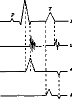
Figure 2. Hoffmann-Zelenin scheme: 1-electrocardiogram; 2-phonogram; 3-heart apex beat; 4-carotis.
if their activity ("active extrasystole"). Such is the classical theory of the mechanism of cardiac activity. Recently, a new theory of the independence of atrial and ventricular contractions has been put forward, mainly by the French (Vaquez, Donzelot, and other authors). This theory presents much of interest; however, it is not sufficiently substantiated experimentally. Regarding the question of what constitutes the primum movens in the chain of impulse transmission to contraction, there is no exhaustive information. Of primary importance is the physical and chemical state of the intracardiac regulatory devices themselves, in connection with their blood supply, which is why the property of the nourishing fluid (its temperature and chemical composition, hormones, blood flow velocity) serves as the carrier of the charge (impulse). However, extracardiac neural devices play no small role here. As part of the autonomic nervous system, which is so closely connected in terms of its tone with the endocrine apparatus, the vagus and sympathetic nerves exert a profound effect on the action of the intracardiac devices, thereby establishing a reactive connection between the functional state of various internal organs and the cardiovascular system. In particular, the vagus nerve exerts a depressing effect on all cardiac functions, in contrast to the sympathetic nerve (accelerans), which enhances the vital properties of the heart (trophic influence?). The investigations of many authors have, moreover, established functional differences between the right and left neural devices. According to the data of Meyer, Rothenberger, and others (in dogs and horses), Colin and others (in humans), the right vagus and accelerans have a greater relation to the Keith-Flack sinoatrial node, while the left have a greater relation to the Aschoff-Tawara node and the bundle of His. On the basis of a disturbance in the relationship between the intra- and extracardiac regulatory apparatuses, pathological conditions develop that are grouped together under the term arrhythmias. Disturbances of sinus rhythm. Tachycardia. The frequency of heart contractions, as is known, has age-related fluctuations approximately within the following limits: up to 10 years the pulse ranges between 90 and 100 beats, by 10-15 years it reaches 80-90, and between 15 and 70 years it remains at figures of 60-80. This latter, rather considerable amplitude, which could be expanded even further in both directions, depends on purely constitutional features of the organism. The heart responds with tachycardia of a greater or lesser degree to a whole series of physiological stimuli. Orthostatic tachycardia: in the standing position (depending on muscle tension, a drop in peripheral blood pressure, and an increase in sympathetic nerve tone), the number of heartbeats usually increases. It increases even more under the influence of active movement—working tachycardia, which depends, apparently, on a simultaneous increase in sympathetic nerve tone and a decrease in vagal tone. Of interest are cases of spontaneous (volitional) influence on the accelerans. In a hypnotic state, depending on the picture of the suggested movement, tachycardia may also arise. Disruption of the tone of autonomic innervation under the influence of affect is usually accompanied by an accelerated pulse (more rarely slowed). Of great practical importance are prolonged tachycardias in so-called soldier's hearts during periods of military operations. Here, two factors apparently take part: 1) the influence of emotions and 2) physical overstrain. The latter factor, one must think, is of a physical-chemical nature and determines the state of so-called myoerethism (Hering). Among "myoerethic" hearts, and consequently those predisposed to tachycardia, are also hearts affected by one or another organic disorder that creates conditions for functional insufficiency of the muscle. On this principle is built the functional diagnostics of the heart using dosed work (Martinet et al.). Sexual intercourse, as a rule, is accompanied by an accelerated pulse: both affect (individually), purely muscular tension, and possibly perturbations in the hormonal system play a role here. Toxic tachycardia: atropine, adrenaline, caffeine, alcohol, etc., cause either excitation of the sympathetic nerve (adrenaline), paresis of the vagus (atropine), or affect the blood supply to the higher regulatory center (amyl nitrite). Basedow's (hormonal) tachycardia is a sinus tachycardia, that is, it is based predominantly on the shortening of the diastolic phase. However, when analyzing the electrocardiogram, an unusually high final T wave is noted as a rule. Conversely, the deficiency of thyroxine in myxedema, along with a relatively slow pulse, is characterized by the almost complete absence of the T wave (see Basedow's disease). Tachycardia of a toxic-bacterial nature is observed in various febrile diseases, and sometimes in afebrile forms of infection (some cases of pulmonary tuberculosis). The determining factor here is, chiefly, the temperature of the blood; as for the toxic factor, it may even act in reverse (e.g., typhoid fever). Rhythm changes in meningitis depend on central irritation of the vagus nerve. Reflex tachycardias may appear with various pain symptoms. Mention should be made in particular of meteorism, which, in addition to the reflex via the vagosympathetic system, alters the purely mechanical conditions of the heart's work. The treatment of tachycardia should first of all be directed at eliminating the most probable cause. Bradycardia. Like tachycardia, slow beating of the heart can exist on a purely constitutional basis (Vaquez). Practically, it is important to note that slowly beating hearts are more robust than hearts prone to tachycardia (Wenckebach). Bradycardia from starvation occurs alongside hunger edema and other signs of disruption of the body's vital functions. Convalescent bradycardias are closely related to these. Between these two forms and the constitutional type lies the so-called hypotonic bradycardia of Herz-Hocke (M. Herz, E. Hocke), which is associated with functional insufficiency of the adrenal glands. Treatment of bradycardia is in accordance with the etiology. Bradycardias based on missed ventricular systoles due to conduction disorders belong to heart block (see). Sinus arrhythmias. Already physiologically, a certain inequality in the length of the diastolic segments is observed, which disrupts the regularity of the rhythm and depends on fluctuations in the tone of autonomic innervation (partly in connection with reflex influences, partly hormonal). Respiratory arrhythmia. In this condition, a noticeable acceleration of the pulse occurs with inspiration, and a slowing with expiration. In addition to a simple fluctuation in rhythm, some hemodynamic deviations can also be noted: thus, expiratory engorgement of the veins is revealed on the phlebogram. The electrocardiogram registers a change in the dynamics of both the atria and the ventricles: during the long pause, the P waves (atrial) almost disappear, and the R waves (ventricular) increase significantly (see Figure 3). Pathogenesis. At the root of the respiratory fluctuations in rhythm lies, according to Hering's opinion, a reflex disturbance of the tone of the vagus nerve; Fredericq, however, believes that the process plays out centrally, in the nucleus of the vagus nerve, which is closely connected with the respiratory center, while Albrecht sees in the described phenomenon a direct response on the part of the myocardium, irritated by increased blood filling during the inspiration phase. There is an undoubted psychic influence: thus, during sleep (physiological vagotonia?), respiratory fluctuations are expressed particularly sharply.
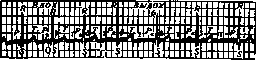
Figure 3. Case of respiratory arrhythmia (electrocardiogram).
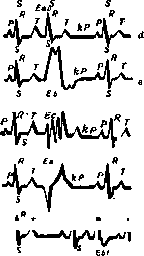
Respiratory arrhythmia is frequently observed in animals (dogs). In newborns, it is entirely absent due to the high heart rate; in children, it occurs as a rule (juvenile arrhythmia of Mackenzie); among adults, it is more frequent in women than in men. The clinical significance of respiratory arrhythmia is minor. Arrhythmia in Cheyne-Stokes respiration (acceleration in the apneic phase and deceleration during hyperpnea) is of central origin and is associated with periodic oversaturation of the blood with carbon dioxide (Henderson), as it responds to oxygen administration (Roth, Wassermann). Sinus arrhythmias not related to respiration depend on an imbalance between the vagus and sympathetic nerves (intermittent sinus acceleration of de-Meyer) and occur in young asthenic subjects, possibly in connection with thyroid hypofunction (Wenckebach), especially after physical exertion. Extrasystolic arrhythmia. If variations in the sinus rhythm in the above-described disorder patterns differ only quantitatively from the normal rhythm, being "nomotopic" or "homogenetic" (according to Lewis), then extrasystole, which is based on excitation arising outside the sinus node (heterotopic, heterogenetic), is a phenomenon of a purely pathological nature. An extrasystole is a contraction of the heart that occurs prematurely and is often accompanied by an elongated, so-called compensatory pause. This pause arises because the contraction following the extrasystole, originating from the sinus node (nomotopically), catches the heart in the refractory phase (the stage of inexcitability): the nomotopic systole drops out, and thus the total number of heartbeats is restored, which is why the pause is called compensatory. An extra-contraction is the result of extra-excitation that activates the automatism of one of the intracardiac nervous apparatuses. In this respect, it differs from the passive rhythm disturbances described by Lewis (escaped beats), of which see below. The mechanism of extrasystole generation is determined by the topography of the additional excitation. A distinction is made between extrasystoles originating from the atria, the atrioventricular node, and from one or the other branch of the His-Tawara bundle. The electrocardiogram, as a method for recording extrasystoles, has the advantage over all other methods in that it allows the determination of the site where the extra-excitation arises. This is well illustrated by Nicolai's diagram (see Figure 4). S Cs S a Normal rhythm. R R
Es sinuses, extrasyst.; no compensatory pause. Eai - auricular (atrial) extrasystole; kP - compensatory pause. Eao - atrioventricular extrasystole. Eb - right ventricular extrasystole. c f Ee - mixed ("median") form of ventricular extrasystole. d Ea - left ventricular extrasystole. ", "z RL 1 rk TL Pi h Ebl - right ventricular inserted ("interpolated") extrasystole without a compensatory pause. Figure 4. Diagrams of extrasystoles (after Nicolai). Electrocardiographic differentiation of extrasystoles into right- and left-ventricular is possible because the impulse, arising ectopically, distorts both the course of the excitation wave and the mechanism of contraction itself: an allodromic or aberrant systole is produced, which has its own electrical equivalent. Under the name "escaped beats", Lewis described passively arising ectopic contractions of the heart resulting from a decrease in the activity of the Keith-Flack node during a long pause. They can be single or form the basis for the appearance of a pathological rhythm (nodal rhythm, idioventricular rhythm). Most frequently, one has to observe "escaped beats" originating from the atrioventricular node, and according to the character and location of the P wave on the electrocardiogram, a distinction is made: 1) supranodal extrasystole (shortening of the PR interval or a negative P wave in the normal place with a "coronary" extrasystole), 2) proper atrioventricular extrasystole (the P wave coincides with the RT ventricular complex), and 3) infranodal extrasystole (negative P wave after R). Other recording methods (sphygmography, cardiography, phlebography, cardiophonography) make it possible to judge the mechanism of circulatory disturbance caused by extrasystole. The arterial pulse is either small or entirely absent (pulsus deficiens), and its filling decreases from top to bottom depending on the degree of chamber filling: in an extrasystole originating in the atria, the contents of the latter are poured into the ventricles in a normal manner, which throw a sufficient amount of blood into the periphery; if, however,
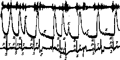
Figure 5. Stenosis et insuff. mitralis; insuff. tricuspid. relativa. Upper curve - phonogram; middle - sphygmogram; lower - electrocardiogram. The 1st extrasystole E on the left side of the figure is of atrial origin: the P wave of the electrocardiogram is pushed closely against the preceding T; the 2nd extrasystole originates from the right ventricle: the wave on the sphygmogram is almost absent; the same is noticeable on the right side of the figure: the 1st extrasystole is atrial (P superimposed on T), the 2nd is ventricular (left), the 3rd is also ventricular (right). On the tone curve, atrial extrasystoles are more clearly expressed, which also yield a higher arterial pulse wave (according to Zelenin).
the extrasystole arises in the ventricle, especially at the beginning of its diastolic phase, the ventricle may turn out to be completely empty (see Fig. 5). The absence of an arterial pulse can also result from the weakness of the extra-contraction, as a result of which the semilunar valves do not open at all Figure 6. Phonocardiogram: E - extrasystole, the contraction period is significantly reduced compared to normal; the 1st tone after the compensatory pause is amplified (according to Zelenin). (Hochhaus and Quincke): upon auscultation, only the 1st tone will then be determined. Usually, however, two clear short tones are heard during an extrasystole; sometimes the post-compensatory first beat is also accompanied by loud tones (see Figure 6). Large atrial waves (a) are frequently noted on the phlebogram, which depends on the coincidence of ventricular and atrial systoles ("Vorhofpfropfung" of Wenckebach), as a result of which the latter throw their contents back into the veins (see Figure 7). ac v ac vac v ocv ac v ee v oc v ac v ac v ac v ac v \Mч^^ч^^NJЧ^^/ч-ч^ч^- Figure 7a. Phlebogram and sphygmogram, pulse is rhythmic (according to Kabakov). Allorhythmias. Extrasystoles can be either single, or randomly scattered in large numbers (pulsus irregularis extrasystolicus), or grouped into groups,
[vqCv\ %rf«*X ^чгОчлчМ4 creating conditions for a pathological rhythm (allorhythmia). A distinction is made between pulsus bigeminus, trigeminus, quadrigeminus, etc., depending on the number of heart contractions grouped together. Allorhythmia can be temporary, prolonged, or permanent. According to modern views, prolonged allorhythmia indicates a constant focus of extra-excitation causing a continuous struggle (interference) of two rhythms—normal and pathological. Under the name of parasystole or parasystolic rhythm, Rothberger, together with Kaufmann and Singer (Roth-berger, Kauffmann, Singer), described a double rhythm that is often indistinguishable due to the fact that the activity of the pathological excitation focus differs little from sinus automatism, as a result of which extrasystoles, existing continuously in potency, manifest themselves only during periods of long pauses (see Fig. 8). Under certain circumstances, Figure 7b. After pilocarpine injection, extrasystoles E appeared: on the phlebogram, the coincidence of atrial and ventricular contractions is noticeable: a+c (according to Kabakov).
Figure 8. Parasystole; interference of two rhythms: normal—
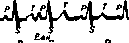
the R wave is directed upwards, and the ectopic one points downwards (according to Winterberg), instead of single, scattered, or grouped extrasystoles, an entire attack of extrasystolic arrhythmia arises, lasting from several seconds (pulsus trigeminus) to several hours, days, and even months (1-2). These attacks have a peculiar clinical picture and are described separately (see Paroxysmal Tachycardia). Rhythm disorders can also arise on the basis of a impairment of conductivity function, when the impulse on its way from the atria to the ventricles is blocked, and ventricular systoles drop out sporadically or allorhythmically (ventricular bradysystole). Correspondingly, the arterial pulse is absent, and heart sounds are not auscultated (see Heart Block). Subjective sensations in extrasystole vary: some patients are completely unaware of the existence of extrasystoles or intermissions in their heart action, while the majority (predominantly neuropathics) complain of jolts in the heart (enhanced postcompensatory beat), sinkings, and dizziness (long pauses). These phenomena are especially pronounced in individuals suffering from aortic valve insufficiency (homo pulsans). Etiology. It should be noted that in 55% of individuals suffering from extrasystoles, no signs of organic lesion of the cardiovascular apparatus are detected (Wenckebach). Moreover, these signs are so rare (for example, in septic endocarditis) that their appearance serves as a bad omen (Liebmann). In valvular defects, they are encountered more frequently in mitral than in aortic lesions. Hypertensive states in themselves do not serve as a cause for extrasystoles, whereas fluctuations in blood pressure in connection with physical exertion or vegetative crises (angina pectoris) may be accompanied by extrasystoles. Experimentally, Pletnev induced extrasystole by increasing the pressure inside the left ventricle through compression of the aorta or irritation of the vasomotor centers. Using the first method, he always obtained separate extrasystoles at first, and upon continuation of the experiment, prolonged extrasystole. With the second method, which is less crude and therefore more suited to natural conditions, the fluctuations in blood pressure and the number of extrasystoles did not depend on either the absolute or the percentage increase in blood pressure. Consequently, it was largely determined by the biological individuality of the experimental animal. The most frequent cause of extrasystoles is neuropsychic influences. The mechanism of these phenomena is fully explained by the abundance of the heart's connections with the autonomic nervous system. A whole series of diseases, predominantly of organs located below the diaphragm, can serve as a source of reflex extrasystoles (enteroptosis, meteorism, chronic cholecystitis, stomach ulcers, phimosis, various women's diseases). The reflex is apparently carried out with the participation of the vagus nerve (pneumogastricus). Can extrasystoles arise under the direct influence of the vagus nerve? A positive answer to this is given by the experiments of Cyon—with alternating simultaneous stimulation of the vagus nerve and accelerator nerves—and the experiments of Pletnev—with faradic and galvanic stimulation of the cervical branches of the vagus nerve. In humans, extrasystoles can be observed in some cases during deep breaths (see Figure 9), during swallowing (Mackenzie), and upon distension of the stomach (Lyass and Levin). By increasing the tone of the vagus nerve with injections of pilocarpine, it is possible to induce the appearance of extrasystoles (see Figure 6) and to achieve their complete and prolonged disappearance through the systematic application of atropine (see Figure 10). Sometimes, however, conversely, atropine may apparently cause a predisposition to extrasystoles. The effect of physical exertion is also ambivalent: with moderate loads, extrasystoles present beforehand may disappear, whereas overstrain may serve as a cause for the onset and prolonged existence of extrasystoles. Obviously, factors predisposing to the occurrence of extra-stimuli exist. In addition to constitutional features and climatic influences (hot climate, as well as hot procedures, promote extrasystoles), nutrition conditions
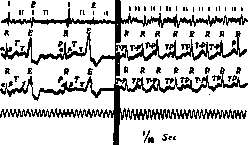
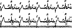
Figure 10. Left half of the figure: cardiac bigeminy; extrasystolic sounds are less pronounced. Right half: extrasystoles have disappeared due to tachycardia after atropine.
and various intoxications play a significant role: alcohol, coffee, tobacco, and among pharmacodynamic agents, primarily digitalis, can cause single and even grouped extrasystoles; the secretion of the thyroid gland acts in the same direction. The physicochemical state of the myocardium is the main factor predisposing to extrasystoles ("myoeretism"). These "functional" changes may lie on the border of already anatomically discernible lesions. Thus, in most infections, extrasystoles are extremely rare, whereas diphtheria, which easily causes myocarditis, is conversely frequently accompanied by extrasystole. Moreover, on the basis of a prolonged attack of grouped extrasystole arising immediately after status anginosus, a local diagnosis could be made: myomalacia of the heart in the region of the left side of the interventricular septum (Zelenin), which was confirmed at autopsy (see Figure 11). Hemodynamic conditions and the associated hypertrophy of one or the other ventricle can be the cause of a homonymous ventricular extrasystole. Right-sided extrasystoles are observed in emphysema, and left-sided ones in aortic valve insufficiency (Zelenin). Still, according to statistics, the greater half of all cases of extrasystoles proceeds in the absence of organic lesions of the cardiovascular apparatus. We are most often dealing with neurasthenics (especially on a sexual basis),
/io sec
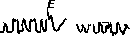
Figure 11. Bigeminus continuus originating from the left ventricle (after Borisova). In asthenics, angioneurotics, persons with impaired metabolism, or those suffering from diseases that are particularly easily complicated by reflex extrasystoles. Treatment and prognosis. Psychotherapy stands in the first place: sometimes a simple reassurance of the patient regarding the non-serious nature of the affliction is sufficient, sometimes only psychoanalysis gives the key to a cure. In the presence of damage to the heart itself (myocarditis) and disturbances of coronary circulation, attempts are made to influence the latter (theobromine preparations, iodine, strychnine); attention is drawn to the organs serving as sources of reflexes. Of greatest importance (after mental influence) is the organization of the regimen (an abdominal binder for splanchnoptosis, etc.). Drug therapy is the same as for sinus tachycardia (bromine, valerian, digitalis in small doses, quinine with strychnine). In stubborn cases, quinidine can be tried, which sometimes has a beneficial effect even after the first doses (Wenckebach). The prognosis in the majority of cases is completely favorable quoad vitam; as for the disappearance of extrasystole as such, due to the multitude of factors taking part in its occurrence, success does not always accompany even competent and persistent treatment. As a rule (according to Zelenin), long-term allorhythmias (bigeminia continua) are regarded poorly. The prognostic significance of parasystole has not been sufficiently clarified. Arhythmia perpetua (seu absoluta), pulsus irregularis perpetuus, delirium cordis. The main feature of this form of arrhythmia is the complete absence of any regularity whatsoever in pulse elevations: small and large waves alternate haphazardly with pauses of various durations (see Figure 12). Auscultation reveals some incoherent mumbling: loud tones are replaced by quiet ones outside of any system completely unexpectedly ("delirium of the heart", delirium cordis). Figure 12. Sphygmogram in pulsus irregularis perpetuus. Pathogenesis. Experimental studies by Rotberger, Winterberg, and Lewis established that at the heart of arhythmiae perpetuae lies a peculiar disorder of atrial activity. They do not produce normal contractions, but are in a state of some undulation: either fairly large waves (200–300 elevations per minute) or small twitches (up to 600 times per minute) are noticeable, to which the ventricles respond with chaotic, fairly frequent contractions (100–180 per minute). Rarer and more rhythmically arising waves were named flutter by the English, Flatter by the Germans, and trepetanie (fluttering) in Russian (Lang); more frequent and chaotic twitches—fibrillation, Flimmern, flickering. There are a number of theories explaining both the state of the atria themselves and the peculiar reaction of the ventricles. Hering assumes multiple foci of excitation in the atria, which was refuted by Rotberger and Winterberg. At the present time, the theories of de Boer and Lewis-Mines enjoy the greatest distribution. According to these authors, the cause of the described phenomenon is a metabolic disturbance in the atrial muscle, as a result of which, along with an increase in its excitability, the conductivity of the impulse is, conversely, reduced: the excitation wave, having arisen somewhere, quickly closes within individual sections of the atria, without involving them in a full systole and without reaching the atrioventricular node. In this case, a single circular wave, from which a number of secondary waves depart, is characteristic of atrial flutter; during their fibrillation, there are many independent circular waves. Therefore, the entire phenomenon is called the circus rhythm. Flutter, resp. fibrillation, of the atria does not pass to the ventricles, in all probability, because the latter are equipped with an independent apparatus for conducting excitations, while the atria lack special conductors. The comparatively low frequency of ventricular contractions is explained by the fact that the limit of conductivity for the His-Tawara bundle is about 180 impulse waves originating from the atria. The main symptom, namely the arrhythmia of ventricular contractions, depends on the chaos of the excitation waves sent by the fluttering atria. Diagnosis. Sphygmography does not always make it possible to distinguish circus rhythm from pulsus irregularis extrasystolicus. On the phlebogram, in place of the disappeared wave a, one can sometimes notice a number of small zigzags; the character of the arrhythmia stands out particularly clearly on the electrocardiogram (see Figure 13): instead of a single wave, there is a series of zigzags (/\) or more pronounced waves (p), the ventricular complex has a normal appearance, which speaks in favor of a supra-atrioventricular origin of the impulse. Etiology. Under experimental conditions, atrial flutter, resp. fibrillation, can be caused either by the direct application of induction shocks of greater or lesser frequency, or by the combined excitation of the sympathetic and vagus nerves; both, apparently, affect myocardial metabolism. In humans, disorders of intramuscular metabolism can occur as a result of cardiosclerosis, myocarditis, focal lesions of the heart muscle, disease of the artery supplying the sinus node; such diseases were in a number of cases discovered at autopsy. More often, however, the basis of metabolic disturbance is apparently a "functional", resp. toxic factor. Among heart defects, circus rhythm is most often encountered in mitral lesions (overstretching of the atria). Intoxications, apparently, affect both the muscle and the autonomic nervous system: thus, direct stimulation of the vagus nerve, the use of muscarine, physostigmine, thyroid secretion, digitalis in certain doses promote circus-type arrhythmia. A predisposing factor is infectious diseases (Aryev), possibly due to a direct effect on the heart muscle. 7 I
I II I II
I II E IJ
1 II
I Ч 1 II I П I » ii' I
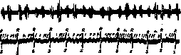
Figure 13. Pulsus irregularis perpetuus. Polymorphism of tones is noticeable on the phonogram. On the electrocardiogram, along with atrial tachysystole (p), flutters (l) are also noticeable.
Reflex influences from organs below the diaphragm (flatulence) may also take place here (cf. extrasystole). Clinical picture. Two forms of absolute pulse irregularity are distinguished: slower and faster. In the first form, subjective sensations are poorly expressed, and cardiac performance is satisfactory; in the second form, patients complain of palpitation, sinking, jolting, and general weakness. The pain symptom occurs as an exception. The effect on hemodynamics and the exhaustion of all cardiac functions are very great: the atria do not systole at all, and the ventricles in severe cases, without any regularity and sometimes without benefit to the circulation, are exhausted by constant tachycardia. Treatment and prognosis. Notwithstanding some theoretical premises and experimental data (transformation of flutter into fibrillation), digitalis has an excellent effect in this form of arrhythmia; this was noted by Mackenzie, who treated circular rhythm (under the guise of nodal) with large doses of digitalis (to the point of vomiting). The beneficial effect of this agent is based both on blocking excitations in the bundle (His-Tawara) and, consequently, slowing down ventricular activity, and on the effect on the contractile ability of the myocardium. Does digitalis lead to the disappearance of atrial flutter (resp. fibrillation)? There are observations speaking in favor of this (Zelenin, Edens, Wenckebach). However, only with the introduction of quinine and especially quinidine into therapy did the restoration of normal rhythm to the heart become accessible (according to some statistics in 50% of cases). Treatment with quinidine requires caution, since its therapeutic effect is based on a profound suppression of myocardial functions, which is why quinidine is contraindicated in cardiac decompensation, in endocarditis, in heart defects proceeding with embolisms (upon a sudden transition of the heart from a pathological rhythm to a normal one, the possibility of detachment of thrombotic masses increases). The typical therapeutic methodology, according to Aryev, boils down to the following: after preparing the patient with rest and sedatives (bromine, opium preparations) until complete compensation, quinidine is given (Chinidinum sulfuricum): 1st day—0.2, 2nd—0.2 x 2; if there are no complications, on the 3rd day—0.4 x 2; on the 4th—0.4 x 3 and they are kept at this dose until the onset of a regular rhythm. On the 12th–15th day, if there is no effect, a break of 1–2 weeks is made, and the course is repeated. Often the normal rhythm is restored on the 4th–5th day, while sometimes only after repeated prolonged Figure 14. Flutter of the entire heart (after Kahn). treatment. Throughout the duration of quinidine therapy, camphor is administered (2 cubic centimeters of a 20% solution). Caffeine should be avoided, as it increases the propensity for fibrillation. If digitalis had to be resorted to in order to restore compensation, it is better to wait 1–11/2 weeks with quinidine.—Prognosis. Often the circular rhythm disappears within a few days and even hours after its appearance (transitory form): this indicates a transient, benign nature of the cause affecting the metabolism of the heart muscle. The constant and, especially, prolonged existence of delirium cordis always indicates profound changes in the heart and worsens the prognosis. Cases that do not respond to quinidine therapy or give relapses are poor in prognostic terms. In the latter point, the information of various authors does not coincide, since some failed to obtain positive results in the slow form of arrhythmia, i.e., more benign, at least in the sense of the effect on hemodynamics and exhaustion of heart functions.—Ventricular flutter already belongs to the realm of prethanatology: once arisen, it leads to the destruction of the heart. On the periphery, it manifests as the disappearance of the arterial pulse. Its electrocardiographic equivalent is analogous to the atrial one (Fig. 14). Intermittent disorder of cardiac activity (pulsus alternans, systolia alternans). Pulsus alternans was first described by Traube in 1872, and the named author considered it a subspecies of pulsus bigeminus. Most of the authors who worked initially, and among them such luminaries of cardiology as Huchard, Wenckebach, and Mackenzie, adhered to Traube's view, who saw no principal difference between p. alternans and p. bigeminus. However, Riegel and Wenckebach singled out p. alternans into an independent form, while those cases of p. bigemini where the extrasystole occurs too late, Hering proposed to call p. pseudoalternans. Thus, for the diagnosis of pulsus alternans at the present time, besides the intermittent appearance of a small wave, it is required that the latter appears not prematurely. Usually, on the contrary, it is somewhat delayed (Extrapulsverspatung), which already excludes any

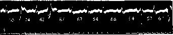
,Figure 15. Pulsus bigeminus depending on regularly occurring extrasystoles. The numbers show in tenths of a second the duration of each pulse period, and it is seen that the longest pauses occur after small contractions (after Mackenzie).
possibility of confusing it with an extrasystolic one (Figs. 15, 16). Experimental alternation of cardiac contractions is obtained during poisoning with cardiac poisons [antiarin, aconitine, El-tortoxin, glyoxylic-acid sodium, digitalis (Zelenin)], and in general upon bringing the heart into a "hypodynamic state" (Hoffmann). In humans, pulsus alternans occurs under various conditions. Most often, with high blood pressure (in nephritics), during attacks of tachycardia; with a slow pulse—in cardiosclerotics, after

Figure 16. Pulsus alternans. The numbers show a slight lengthening of the pause before smaller contractions, in contrast to that depicted in Fig. 15 (after Mackenzie).
exhausting diseases. It is especially often observed in patients with Spanish flu (with a poor outcome). Most authors, in agreement with experimental data, consider pulsus alternans to be an expression of exhaustion of the contractility function. In order to form a judgment about cardiac activity in the presence of p. alternans in all its complexity and versatility, a single graphic image of the pulsating artery is not enough, and the use of other methodologies is necessary. Examination of extensive material allows us to conclude that, as a rule, the following relationships exist between registration methods. The larger wave of pulsus alternans also corresponds to a stronger systole, since during this are observed: a larger amplitude of the cardiac shadow on the X-ray screen (A. Hoffmann), a more energetic heart beat (Hering), increased intraventricular pressure (Cahn and Starkenstein), a greater sweep of the suspension curve (Rihl), louder tones upon auscultation (Mackenzie, A. Hoffmann, etc.).—As for the recordings of tones using a galvanometer, a hidden form of p. alternans (false bradycardia, hemisphyxia) can be noted on the phonogram, when the semilunar valves do not open and, as a consequence, arterial waves and the 2nd sound drop out. The electrocardiogram in this case does not show significant deviations from the norm: its appearance is usual, only in the T waves is alternation sometimes noticeable. This circumstance, by the way, speaks against Hering's assumption of partial ventricular asystole, since in this case the electrocardiogram would have an atypical ("aberrant") appearance.—From the etiology of pulsus alternans flows the serious prognostic significance of the described phenomenon. Pulsus paradoxus. In 1854, Griesinger made the following observation on a patient: the pulse completely disappeared upon deep inspiration, only to reappear upon expiration. This phenomenon was named (in 1873) by Kussmaul the paradoxical pulse and was linked to adhesive pericarditis (non-obliterating), which was later confirmed by many authors. However, Sommerbrodt, Cloetta, and others showed that even under physiological conditions in the inspiration phase the heart receives less blood, and consequently, together with the drop in blood pressure and a decrease in stroke volume, the pulse elevations become lower, while upon expiration the opposite relations exist. Thus, there is nothing paradoxical in the phenomenon itself; only the amplitude of physiological fluctuations in the blood filling of the arteries is accentuated in it.—One must always keep in mind that with certain chest deformations p. paradoxus can also be observed without accretio cordis (Lewis, Wenckebach).
Related articles
Mentioned in
Cite this page
“Cardiac Arrhythmias.” Soviet Medical Encyclopedia. English translation of Bolshaya Meditsinskaya Entsiklopediya, 1st ed. (Moscow, 1928–1936), ed. N. A. Semashko. https://sovietmedicalencyclopedia.pages.dev/article/cardiac-arrhythmias/