Atrioventricular Bundle
Historical document, translated for reference. It reflects medical knowledge of the 1920s–30s and is not medical advice.
Summary
The atrioventricular bundle is a system of pale muscle fibers in the heart that establishes the anatomical and physiological connection between the atria and ventricles. It is part of the conducting system responsible for cardiac automatism.
Encyclopedia article (1928–1936)
ATRIOVENTRICULAR BUNDLE - ATRIOVENTRICULAR NODES, the name of a system rich in nerve fibers of pale muscle fibers of the heart with barely noticeable transverse striations; through these fibers, the anatomical and physiological connection between the atria and ventricles is established (syn.: bundle of His or His-Tawara). The entire physiological system controlling cardiac automatism consists of the sinoatrial node and the atrioventricular bundle.
The sinoatrial node, or sinus node, described in 1906 by Keith and Flack, is located in the wall of the right atrium at the mouth of the superior vena cava and is built from a plexus of thin (atypical) pale muscle fibers with barely noticeable transverse striations. This node is connected with branches of the sympathetic and vagus nerves, surrounded by connective tissue, and receives blood from a separate arteriole or arterial arch.
The atrioventricular bundle, described by His in 1893 and thoroughly studied by Tawara in 1906 and Monckeberg in 1908, originates from the atrioventricular node (Aschoff-Tawara), located at the base of the atrial septum near the sulcus coronarii and the posterior point of attachment of the medial sail of the tricuspid valve (proximity to the valve). The Aschoff-Tawara node is a bizarre network of the same atypical muscle fibers as in the sinus node. From this node, the atrioventricular bundle (main trunk) extends forward, dividing into right and left crura, runs under the endocardium of the ventricular septum, and connects with the papillary muscles (see Monckeberg's scheme).
The terminal branches of the crura, which disappear into the thickness of the heart muscle, are, like the bundle itself, composed of poorly differentiated cells (thicker), also described by Purkinje. In the main trunk of the atrioventricular bundle, the fibers run parallel, while in both crura, especially the left, they form a bizarre network. The main trunk and crura are enveloped in connective tissue along their course and are separated from the myocardium by it. The Aschoff-Tawara node has its own arterial branch.
Histologically, the sinus node, Aschoff-Tawara, and the atrioventricular bundle represent a continuous sarcoplasmic network of pale fibers, with continuous bundles of cross-striated fibrils running through them, mainly in the peripheral parts, and are essentially only a specially differentiated part of the muscular framework of the heart. The sarcoplasm of Purkinje fibers contains a significant amount of glycogen.
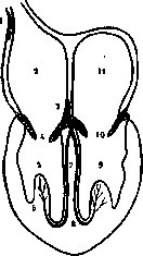
The previously prevailing view that Purkinje fibers are simply muscle elements of the heart that have stopped at the embryonic stage of development has not been confirmed. It turns out that they are laid down very early and immediately differ sharply from the usual elements of cardiac muscle. From comparative anatomical data, it is known that these fibers are clearly expressed not in all, but only in large mammals, for example, the elephant, horse, etc. In humans, they are much less developed. In some birds, they can be easily found. They cannot be found at all in all small mammals, for example, in rodents. In them, the fibers forming the atrioventricular bundle differ very little from ordinary cardiac muscle fibers.
At present, it is firmly established that the coordinated contractions of individual parts of the cardiac muscle depend on a special 'conducting irritation system' (Reizleitungssystem), which is the atrioventricular bundle. The motor impulse arises in mammals in the aforementioned sinus node (Keith-Flack), from here the impulse passes to the atria to another muscle atrioventricular node (Aschoff-Tawara), and then along the atrioventricular bundle is transmitted to the muscle of the ventricles. Cases of impaired coordinated contractions of the heart sections due to compression or destruction of the atrioventricular bundle by pathological processes are known (see Heart Block).
Related articles
Mentioned in
Cite this page
“Atrioventricular Bundle.” Soviet Medical Encyclopedia. English translation of Bolshaya Meditsinskaya Entsiklopediya, 1st ed. (Moscow, 1928–1936), ed. N. A. Semashko. https://sovietmedicalencyclopedia.pages.dev/article/atrioventricular-bundle/