Sphenoid Bone (the main bone located posteriorly - foramen)
Historical document, translated for reference. It reflects medical knowledge of the 1920s–30s and is not medical advice.
Summary
The sphenoid bone is a central cranial bone with complex anatomy including greater and lesser wings, pterygoid processes, and various foramina. It forms part of the base of the skull and contributes to the orbits and nasal cavity.
Encyclopedia article (1928–1936)
SPHENOID BONE - the main bone located posteriorly - foramen spinosum, piercing the posterior acute angle of the wing (spina angularis) and transmitting the a. mening. media and n. spinosus. The wing has three surfaces: the inner, facing the cranial cavity; the anterior, orbital; and the outer-temporal. The inner, cerebral surface (facies cerebralis) is concave, bearing on it juga cerebralia and impressiones dig-itatae; the orbital surface (facies orbitalis) is a smooth, rhomboid-shaped area; the temporal surface (facies temporalis) is divided by a crest (crista infratemporalis), which ends anteriorly in a well-developed tubercle (tuberculum spinosum), into two parts: the upper-larger, participating in the formation of the fossa temporalis, and the lower-smaller (facies infratemporalis), participating in the formation of the fossa infratemporalis. The upper edge of the greater wing, in its medial segment is smooth and pointed, together with the lesser wings forms the superior orbital fissure (for the vv. ophthalmicae, nn. oculomotorius, trochlearis, ophthalmicus, abducens); the outer portion of the upper edge is serrated, connects with the frontal bone (margo frontalis, s. parieto-frontalis) and posteriorly passes at an angle (angulus parietalis) into a serrated edge, connecting with the squamous part of the temporal bone (margo squamosus) and further posteriorly with its petrous part (margo petrosus). The posteromedial edge of the wing, forming the posterior semicircle of the oval foramen, at the same time participates in the formation of the foramen lacerum. The anterior edge in its upper portion is serrated and connects with the zygomatic bone (margo, s. crista zygomatica, s. crista malaris, s. jugalis), in the lower portion is smooth, and externally limits the inferior orbital fissure (for v. ophthalm. inf., a. infraorbitalis, nn. zygomaticus et infraorbitalis and branches of the ganglion sphenopalatini).
Pterygoid processes (processus pterygoidei) depart from the body of the sphenoid bone at the root of the greater wings and are directed vertically. Through the base of the process passesch a horizontally directed canal (canalis pterygoideus Vidii) for vessels and nerves of the same name, opening into the pterygopalatine fossa (see). Below this canal continues into a groove (sulcus pterygoideus), which by the palatine and maxillary bones is closed into a canal (canalis pterygo-palatinus). The process consists of two plates: the outer (lamina lateralis) and the inner (lamina medialis), which by their planes form an angle open posteriorly (fossa pterygoidea). In the lower part there is no complete closure of the plates, as a result of which a fissure (fissura pterygoidea) is formed. The medial plate ends in a hook-shaped projection (hamulus pterygoideus), through which the tendon of the tensor veli palatini is thrown, leaving a trace in the form of a smooth groove (sulcus hamuli). At the base of the plate posteriorly there is a downward-facing fossa and groove (fossa scaphoidea et sulcus tubae auditivae) t containing the cartilaginous part of the Eustachian tube (see). At the base of the plate is mentioned above processus vaginalis, on the lower surface of which is located the opening of the pharyngeal canal (canalis pharyngeus), and between it and the lower surface of the body of the sphenoid bone-the opening of canalis basipharyngei for the branches of the pterygopalatine ganglion.-The posterior free edge of the outer plate approximately in the middle of its length has a projection, from which to the "spina angularis" a bony bridge (lamina pterygo-spinosa) is sometimes thrown, representing a ossified ligament (ligamentum pterygo-spinosum Civinini), as a result of which an opening (foramen pterygo-spinosum) is formed. The sphenoid bone develops from cartilage with the exception of conchae sphenoidales and lamina medialis; processus pterygoidei, which originate from connective tissue. Ossification begins at the end of the 2nd month and occurs" from two merging points in the anterior part of the body, two in the posterior half, one for each of the wings, in addition the medial plate of processus pterygoidei, lingula sphenoidalis and conchae sphenoidales have their own points of ossification. The sphenoid sinus develops in the third year of life.-Pathology-see Paranasal sinuses.
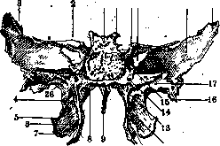
Figure 3. Sphenoid bone from behind: 1-dorsum sellae; 2-ala parva; 3-ala magna; 4-proc. pterygo-spinosus; 5-proc. pterygoideus; 6-lamina med.; 7-lamina lat.; 8-canalis pharyngeus; 9-rostrum sphenoidale; 10-hamulus pterygoideus; 11-fissura pterygoidea; 12-fossa pterygoidea; 13-fossa scaphoidea; 14-canalis pterygoideus; 15-lingula sphenoidalis; 16-spina angularis; 17-sulcus tubae auditivae; 18-rangulus parietalis; 19-facies cerebralis; 20-fissura orbitalis sup.; 21-proc. clinoideus ant.; 22-proc. clinoideus post. (According to Spaltenholz.)
Figure 4. Sphenoid bone from the front; 1-corpus; 2-ala parva; 3-ala magna; 4-proc. pterygoideus; 5-crista sphenoidalis; 6-rostrum sphenoidale; 7-canalis basipharyngeus; 8-proc. vaginalis; 9-canalis pharyngeus; 10-canalis pterygoideus; 11-sulcus pterygo-palatinus; 12-lamina med.; 13-hamulus; 14-sulcus hamuli; 15-fissura pterygoidea; 16-lamina lat.; 17-facies sphenomaxillaris; 18-foramen rotundum; 19-spina angularis; 20-facies infratemporalis; 21-crista infratemporalis; 22-facies temporalis; 23-facies orbitalis; 24-upper wall of fissurae orbitalis sup.; 25-foramen opticum; 26-apertura sinus sphenoidalis; 27-concha sphenoidalis. {According to Spaltenholz.)
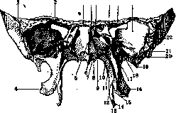
1 27 26 25 24 23
The sphenoid bone develops from cartilage with the exception of conchae sphenoidales and lamina medialis; processus pterygoidei, which originate from connective tissue. Ossification begins at the end of the 2nd month and occurs" from two merging points in the anterior part of the body, two in the posterior half, one for each of the wings, in addition the medial plate of processus pterygoidei, lingula sphenoidalis and conchae sphenoidales have their own points of ossification. The sphenoid sinus develops in the third year of life.-Pathology-see Paranasal sinuses.
Related articles
Cite this page
“Sphenoid Bone (the main bone located posteriorly - foramen).” Soviet Medical Encyclopedia. English translation of Bolshaya Meditsinskaya Entsiklopediya, 1st ed. (Moscow, 1928–1936), ed. N. A. Semashko. https://sovietmedicalencyclopedia.pages.dev/article/sphenoid-bone/