Orbit
Historical document, translated for reference. It reflects medical knowledge of the 1920s–30s and is not medical advice.
Summary
This article from the 1928–1936 Soviet medical encyclopedia describes the anatomy of the human orbit, detailing its bony structure, openings, surrounding cavities, and the fascial system including Tenon's capsule.
Encyclopedia article (1928–1936)
The ORBIT (orbita) has the shape of a quadrilateral pyramid with rounded edges, the base of which faces forward, and the apex backward and inward. The axes of both orbits intersect in the region of the sellae turcicae at an acute angle, which varies individually. The upper bony wall of the orbit is formed in its anterior and middle part by the horizontal plate of the frontal bone and in the posterior part by the lesser wing of the sphenoid bone. The lower wall is formed mainly by the orbital surface of the maxilla, to which are joined in front and outside
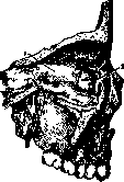
a part of the zygomatic bone and posteriorly the orbital process of the palatine bone. The outer wall is formed anteriorly by the orbital plate of the zygomatic bone and posteriorly by the greater wing of the sphenoid bone, between which a small intercalary bone is sometimes found. The inner wall is formed by the lacrimal bone, the lamina papyracea of the ethmoid bone, and the anterior part of the body of the sphenoid bone (see Figure 1). The anterior margin of the upper, outer, and lower walls of the orbit is thickened in the form of a bony ring protecting the eye (margo orbitalis). The inner margin does not form such a thickening, and here the eye is protected only by the wall of the nose. The upper wall of the orbit appears concave and has an oblique direction backward and downward; in front, on it
there are two depressions: externally for the lacrimal gland and internally for the trochlea of the superior oblique muscle of the eye. The lower wall has a slope from behind and inside to the front and outside and is somewhat concave in its anterior part. The outer wall is directed from the outside inward and backward. The inner wall runs approximately straight from front to back and is almost parallel to the same wall of the other orbit. Figure 1. Inner bony wall of the orbit: 1- lamina papyracea; 2- os lacrimale.
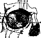
Figure 2. Right orbit: 1-fissura orbitalis sup.; 2-spina musculi recti lateralis; 3-canalis zygomatico-orbitalis; 4-fissura orbitalis inf.; 5-canalis zygomatico-facialis; 6-canalis infraorbitalis; 7-foramen infraorbitale; 8-fossa lacrimalis; 9-arcus superciliaris; 10-foramina ethmoidalia; 11-incisura supraorbitalis; 12-foramen opticum. In its anterior part there is a depression - the fossa for the lacrimal sac. At the apex of the orbit is an opening - the foramen opticum, leading into the middle cranial fossa, for the passage of the optic nerve and the ophthalmic artery. At the border of the upper and outer walls of the orbit, in its posterior third, there is the superior orbital fissure, fissura orbitalis superior, leading into the same middle cranial fossa (see Figure 2). Through it enter the nerves of the eye muscles and the first branch of the trigeminal nerve, and the veins of the orbit exit (see Figure 3). At the border of the outer and lower walls of the orbit, in its posterior half, there is the inferior orbital fissure, fissura orbitalis inferior, leading into the pterygopalatine and infratemporal fossae, through which enter the nerves of the second branch of the trigeminal nerve and where venous anastomoses exit. The largest of these nerves, the infraorbital nerve, together with the artery of the same name, passes through the sulcus infraorbitalis present on the lower wall of the orbit and the corresponding foramen infraorbitale to the facial surface of the maxilla. Through the incisura supraorbitalis, present on the upper margin of the orbit,
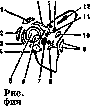
Figure 3. Topography of the circumference of the foramen opticum and fissura orbitalis superior (left): 1-m. levator palpebrae superioris; 2-m. obliquus superior; 3-m. rectus internus; 4-m. rectus inferior; 5-n. opticus; 6-a. ophthalmica; 7-n. oculomotorius; 8-n. abducens; 9-m. rectus externus; 10-v. ophthalmica; 11-n. nasociliaris; 12-n. lacrimalis; 13-n. supraorbitalis; 14-n. trochlearis; 15-n. rectus superior.
the supraorbital nerve with the artery of the same name emerges onto the forehead. Externally, the orbital cavity communicates with the temporal fossa through the canalis zygomatico-facialis, internally through the foramina ethmoidalia with the cranial cavity, and internally-inferiorly through the canalis nasolacrimalis with the nasal cavity. The orbit is surrounded by other adjacent cavities, the diseases of which can be transmitted to it. These are the nasal cavity with its appendages - the frontal, maxillary sinuses, and the cells of the ethmoid bone (see Figure 4). The bony walls of the orbit are lined internally with the periosteum - the periorbita, which is loosely connected to the underlying bones and more strongly fused only at the sutures. In the place where the periosteum spans the inferior orbital fissure, numerous smooth muscle fibers are woven into it, the so-called m. orbitalis (Müller's muscle), innervated by the sympathetic nerve. At the margin of the orbit, the periorbita passes on one side into the periosteum of the face, and on the other into the fascia tarsoorbitalis, which attaches to the upper margin of the eyelid cartilage and to the inner and outer ligaments of the eyelids, thus forming, as it were, the anterior wall of the orbit, the septum orbitale, which, when the eyelids are closed, closes the orbit from the front and contains its contents. Posteriorly, the periorbita passes through the foramen opticum into the dura mater of the brain. The orbital cavity, the average volume of which is approximately 30 cubic cm, is lined with loose adipose tissue, the individual lobules of which are enclosed in loose connective tissue that holds them in their places. This adipose tissue, condensing due to the fasciae, forms sheaths for the nerves, vessels, and muscles of the orbit. Around the eyeball, a system of fascial plates, located generally parallel to the sclera and reinforced by a significant amount of elastic fibers (Levitsky), forms a fibrous capsule - the Tenon's (or Bonnet's) capsule. Between Tenon's capsule and the surface of the eyeball, a slit-like lymphatic space is formed, communicating posteriorly with the lymphatic space that surrounds the outer vaginal sheath of the optic nerve (Fuchs, Lange). Anteriorly, Tenon's capsule extends to the conjunctival fornix and the beginning of the bulbar conjunctiva. Tenon's capsule represents a kind of peculiar articular surface in which the eye rotates. But not all authors recognize the existence
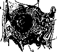
Figure 4. Frontal section through the orbit: 1-lacrimal gland; 2-maxillary sinus; 3-nasal cavity; 4-sinus ethmoidalis; 5-sinus frontalis.
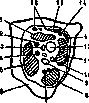
Figure 5. Frontal section through the right Orbit near the optic nerve: 1-rectus superior muscle; 2-nasociliary nerve; 3-ophthalmic artery; 4-abducens nerve; 5-rectus lateralis muscle; 13-rectus superior muscle; 14-trochlear nerve; 15-levator palpebrae superioris muscle; 16-lacrimal and frontal nerves. Tenon's space: some assert that the space between the eye and Tenon's capsule is filled with loose connective tissue; others speak of numerous adhesions between the capsule and the sclera. In all connective tissue formations of the Orbit, elastic fibers participate. The ocular muscles passing through the Orbit are clothed in fasciae. Somewhat posterior to the point where the rectus muscles enter Tenon's capsule, from their muscular sheaths, here tightly fused with the muscle, bundles of connective tissue depart, the so-called orbital ligaments lat. periorbita; (according to Levitsky their four bundles), directed toward the ciliary ganglion, the cartilages of the eyelids, the conjunctiva, the sclera and the edges of the cornea. The physiology, role of these ligaments is to serve together with Tenon's capsule as a support, preventing the eyeball from shifting and sinking in its immobile position (Levitsky). From the fasciae covering the ocular muscles, processes extend, which connect them with the walls of the Orbit and with each other (Fuchs). The lacrimal gland, according to some authors, has its own ligament, beginning from the periosteum of the upper wall. Arterial vessels of the Orbit originate from the ophthalmic artery, entering the Orbit through the optic foramen. Venous blood is carried away through the superior ophthalmic veins and through the optic foramen. The veins of the Orbit are connected by numerous anastomoses with the veins of the face, which facilitates the spread of inflammatory processes from various areas of the face into the Orbit. No lymphatic vessels were found in the Orbit. In the Orbit, besides the optic nerve, there are also motor nerves-for the eye muscles, sensory-the first and second branches of the trigeminal nerve, and sympathetic nerves (see figures 5 and 6). Closer to the optic foramen, on the outer-lower side of the optic nerve, lies the ciliary ganglion, receiving its motor fibers from the oculomotor nerve (short root); sensory-from the trigeminal nerve, namely from the nasociliary nerve (long root), and sympathetic-from the plexus surrounding the carotid artery. The nasociliary nerve gives branches not only to the ciliary ganglion directly to the eye in the form of long ciliary nerves, but these nerves also have sympathetic fibers arising from the same plexus. Short ciliary nerves, numbering from 3-6 to 8-10 (Lange), originating from the ciliary ganglion, enter the eye around the optic nerve together with the long ciliary nerves. The largest part of the Orbit is occupied by the eyeball. Its position in the middle is such that a ruler applied vertically to the upper and lower edges of the Orbit does not noticeably press on the cornea. In the usual position of the eyeball, it is weakly protected from the outside, since the bony wall of the Orbit reaches here only to the posterior border of the ciliary body. The position of the eye in the Orbit is influenced by the peculiarities of the structure of the skull, fluctuations in the amount of adipose tissue in the Orbit, changes in the degree of filling of the vessels of the latter. Under pathological conditions, both protrusion of the eye forward-exophthalmos, up to its complete dislocation from the Orbit, and sinking of the eye into the Orbit-enophthalmos are possible.
Related articles
Mentioned in
Cite this page
“Orbit.” Soviet Medical Encyclopedia. English translation of Bolshaya Meditsinskaya Entsiklopediya, 1st ed. (Moscow, 1928–1936), ed. N. A. Semashko. https://sovietmedicalencyclopedia.pages.dev/article/orbit/