Orthophoria
Historical document, translated for reference. It reflects medical knowledge of the 1920s–30s and is not medical advice.
Summary
Orthophoria refers to the ideal muscular balance of the eyes in a state of rest, where both eyes are directed straight forward with parallel visual axes. This condition is rare, with most people exhibiting some degree of heterophoria.
Encyclopedia article (1928–1936)
ORTHOPHORIA (from Greek orthos- straight and phoros- tendency), a term proposed in 1886 by Stevens for denoting the ideal muscular balance of the eyes in a state of rest. The position of the eyeballs in the orbit is determined by mechanical and nervous factors. The former are due to certain topographic-anatomical relationships—shape, size of the orbit, direction of its axis, attachment and length of muscles, size and shape of the eye, and the relationship of the eye to surrounding tissues—conjunctiva, eyelids, orbital fat tissue, etc. Nervous-psychic influences on the position of the eyes consist of a number of factors: 1) volitional impulses, 2) fusional tendency existing independently of will, 3) association of accommodation and convergence; 4) muscle tone, the causes of which are extremely varied and the magnitude of which is extremely different. By orthophoria or normal rest of the eyes or their muscular balance is understood such a state when the eyes, regardless of nervous influences, and exclusively due to anatomomechanical factors, when looking into the distance are directed straight forward, when the vertical meridians of the retina are perpendicular, both visual lines are directed straight forward and parallel to each other. This ideal muscular balance, as numerous researchers teach, represents an exception, since extremely rarely is complete symmetry observed in the anatomical relationships of both orbits and eyes. The rule, as Bielschowsky says, is heterophoria. In studying the muscular balance of the eyes, the matter usually concerns the determination of the physiological or functional rest of the eye muscles, understanding by it such an eye position which depends not only on anatomomechanical conditions but also on the muscle tone of the eye muscles with the exclusion of binocular vision. In such determinations, made on a large amount of material, Bielschowsky found 20-25% orthophoria with binocular vision present, whereas in cases where binocular vision had been disturbed long before the examination, orthophoria is encountered incomparably less frequently (Bielschowsky's cases with unilateral aphakia). Among recent Russian researchers, Sergeevsky, Luzhinsky, and others generally confirm these conclusions. Luzhinsky out of 1,449 cases had only 175 cases of orthophoria, Sergeevsky out of 270 had 51 cases of orthophoria. The methods for establishing orthophoria are different: they are essentially the same as in determining heterophoria; of these, the main one is observation of the so-called adjustment movements for distance and near with the exclusion of binocular vision by closing one eye. The Graefe test is based on excluding one eye from binocular vision by applying a prism of 10° to it (base upward or downward). When fixing an object (black circle on white paper or candle flame) at a distance of 5-6 m, a double image is obtained. If there is orthophoria, these images will be located one under the other along a vertical line, but there will be no (as in heterophoria) lateral divergence of images. The Maddox method. The object of fixation is a bulb of the Maddox tangent scale (see Strabismus); in front of one eye is placed the so-called Maddox rod, so that to the eye the light source will appear as a red line going horizontally or vertically (depending on the position of the 'rod'). With orthophoria, this red line will pass through the very center, not deviating to the sides. With the small Maddox scale, the examination is carried out at a distance of 25 cm. If a prism of 12° (angle downward) is placed in front of the eye, an impression of 2 scales will be obtained, but with orthophoria both arrows will be located strictly on a vertical line. Based on the same principles is constructed the apparatus-orthophorimeter by Stock, which is used in studying muscular balance in various cases.
ON I
I C
COOHCH3 orthoform
neoorthoform O. white crystalline powder, barely soluble in water, soluble in 5-6 parts of alcohol and in 50 parts of ether. Melts at t° 141-143°. Obtained as a result of the study of cocaine and tropococaine and attempts at synthesis of a cocaine-like compound based on the anesthetic properties of the benzoyl and methyl groups in the structure of cocaine. Instead of ecgonine, which accounts for the toxicity of cocaine, an amino group has been introduced into O. O., neoorthoform, as well as synthetically obtained anesthesin and cycloform, combine anesthetic properties with negligible compared to cocaine toxicity. The slight solubility of O. and neoorthoform in water also gives them a somewhat different character of action. On the wound surface they dissolve very slowly, as a result of which the anesthetic effect lasts for hours. Through intact skin they are not absorbed, they act very slightly on mucous membranes. O. has a side effect: when applied externally, it often causes infiltrates, eczemas, erythemas, local gangrenes. Cases of general poisoning as a result of the use of O. have been described. The irritating effect of orthoform Pototsky attributes to the hydroxyl group (OH), since its elimination in other preparations (anesthesin, cycloform) gives a sharp decrease in the irritating effect. Another drawback of O. is its property to convert oxyhemoglobin into methemoglobin, especially noticeable when applied to open wounds and when taken internally. Initially it was used in the form of dusting powders in pure form and in 5-10% ointments. Per os O. was given for stomach diseases (0.5-1.0). At present O. has been replaced by anesthesin and cycloform, although the latter are not free from irritating properties.
V. Votchal. ORCHITIS (orchitis), inflammation of the testicle. According to clinical course, acute and chronic are distinguished. The latter is usually observed as a complication in syphilis and tbc (see Testicle). Acute O. according to their origin are divided into traumatic, urethral, and metastatic; sometimes a fourth form is also encountered, designated as 'primary acute O. of children'. To traumatic O., caused by external trauma (blow, compression, etc.), should be attributed O. resulting from sudden muscle strain (orchite par effort), in which there is hemorrhage into the parenchyma of the testicle. O. on the basis of prolonged sexual excitement without ejaculation should also be included in this group. In traumatic O., inflammation is often caused by infection, the causative agent of which has been added to the trauma. Urethral O. occur with inflammation of the posterior urethra, prostate, ampulla of the excretory duct, and seminal vesicles, often arising as a result of instrumental intervention on these organs. Inflammation of the testicle caused by gonorrheal infection is extremely rare in contrast to epididymitis, which is characteristic of gonorrhea. Much more frequently observed in gonorrheal urethritis are O. caused by banal pyogenic flora (staphylococci, streptococci, B. coli, etc.). Predisposing factors are various kinds of trauma, damage to the posterior urethra and especially the seminal collicle. Metastatic, the most frequently occurring forms of O. are a complication of a number of infectious diseases. Fraenkel and Hartwich by means of cultures discovered in the parenchyma of the testicles an agent similar to the causative agent of the main disease. Among metastatic O., the most frequently encountered are O. as a complication of epidemic parotitis. Koch observed in 23 patients suffering from epidemic parotitis 7 cases of O. According to Demel, epidemic parotitis is complicated by O. in almost 33%, and in 90% unilaterally. This complication is usually observed in persons who have matured sexually. The epididymis remains free from lesion in this disease. O. usually occurs between the 6th and 8th day of the illness
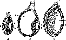
Relationship of the tunics, testicle, and epididymis (schematically): A-normal testicle; B-hydrocele of the testicle; C-orchitis; 1-tunica propria of the testicle, 2-testicle, 3-epididymis, 4-hydrocele.
levations with mumps. Orthophoria is described in cases of joint rheumatism, gout, influenza, croupous pneumonia, malaria, typhoid fever, and smallpox. The fourth form of orthophoria, primary acute inflammation of the testicles in children, occurs most frequently at ages 6-15 years. The etiological factor is not always clear. There are indications of a connection between the disease and masturbation. Ombredanne, out of 7 cases of primary acute orthophoria in children, found twisting of the testicle in 4 cases. Pathological anatomy. In acute inflammation, the testicle is enlarged (see figure), has an elastic or even firm consistency, the tunica albuginea is thickened, the interstitial tissue and seminiferous tubules are filled with serous-purulent content. When the tubules are damaged, hyperplasia of Sertoli cells occurs fairly quickly. Leydig cells increase in number, lose fat, and acquire phagocytic properties (Morgenstern). Subsequently, small foci of pus form, gradually merging with each other; the process ends either with resorption and scar formation, or the abscess becomes encapsulated, and its contents turn into a mass rich in cholesterol. An abscess following its natural course may rupture into the cavity of the tunics and outward through the skin, forming a fistula, around the opening of which granulation and even fungoid growths (fungus benignus testis) form. The clinical picture of acute orthophoria depends on the etiological factor. In traumatic forms, if there is no hemorrhage, the swelling is usually moderate, the pain is insignificant, and the course is rapid. In metastatic diseases, the process begins acutely, temperature rises sharply, pain appears, and a tumor develops in the corresponding half of the scrotum. In urethral orthophoria, the process develops more slowly; the scrotal skin is hyperemic, the testicle is enlarged in size. Fever and chills gradually intensify and reach their maximum by the second week, then slowly subside. The pain is localized in the scrotum, in the inguinal region, from where it radiates to the sacrum, the lumbar region, and sometimes to the legs. Primary acute orthophoria in children usually begins suddenly, proceeds with severe pain in the corresponding half of the scrotum and with a rapidly increasing high temperature. In differential diagnosis, the possibility of epididymitis, hydrocele, and incarcerated hernia should be considered. External examination of the scrotal organs in connection with palpation allows differentiation of orthophoria from epididymitis. In orthophoria, the anterior surface of the scrotum presents an oval, smooth, uniform, tightly stretched surface, to which the unchanged epididymis is attached from behind. It is somewhat more difficult to distinguish orthophoria from hydrocele, the characteristic signs of which are fluctuation and translucency. In doubtful cases, puncture of the testicular tunics should be performed. The difference between orthophoria and 'elastic' incarcerated hernia is established based on the history and the presence of gastrointestinal symptoms (vomiting, constipation, etc.) in intestinal incarceration. In the vast majority of cases, orthophoria ends in complete recovery, more rarely - in atrophy of the testicle as a result of proliferation of connective tissue elements. When an abscess has formed, the process can spread along the spermatic cord into the pelvic cavity. Treatment consists in therapy of the underlying condition, in keeping the patient in bed; the scrotum is placed in a suspensory and heat is applied to it. In orthophoria based on mumps, Zoller recommends injecting convalescent serum. When an abscess appears, an incision of the tunics and testicular tissue should be made as early as possible. With extensive destruction or gangrene of the testicle, castration should be resorted to. Chronic orthophoria. In addition to specific orthophoria based on tuberculosis and syphilis (see Testicle), chronic inflammatory orthophorias caused by ordinary flora are observed. They develop either from acute forms through gradual subsidence of the process, or from the very beginning the inflammation proceeds sluggishly, torpidly. According to König, two forms of chronic orthophoria should be distinguished: orthophoria circumscripta with abscess formation and orthophoria chronica diffusa with severe connective tissue changes. In orthophoria circumscripta, the tunics usually adhere to the skin in places where the suppuration approaches the surface, the testicle is slightly enlarged, and the consistency in the affected areas is firm. The process ends with the formation of a fistulous tract. The course of the process is slow; the testicle is almost painless, with shooting pains in the lower back, sacrum, or abdomen. In differential diagnosis, tuberculous and syphilitic orthophoria should be kept in mind. In tuberculosis, painful sensations are usually absent, the process is combined with lesions of the seminal vesicles, prostate, and mainly the epididymis. In differentiation from syphilitic lesions, the history, other signs of syphilis, and finally the results of antisyphilitic treatment should be taken into account. Treatment consists in eliminating the causal factors maintaining the inflammatory process in the testicles. When an abscess forms, an incision should be made, and with extensive necrosis of the testicle, castration should not be delayed. Orthophoria chronica diffusa is characterized by abundant proliferation of connective tissue elements and is designated as fibrosis testis. In mild cases, there is formation of connective tissue strands or separate areas of callus tissue. In more severe cases, almost all glandular tissue is replaced by a dense mass of fibrous nature. Microscopic examination reveals destruction of the epithelium, thickening of the tubule walls, and in places complete obliteration of the lumens. The process sometimes lasts for many years without special complaints from the patient. In differential diagnosis, tuberculosis, syphilis, tumor of the testicle, as well as hemorrhagic periorchitis should be taken into account. In the latter, the boundaries of the epididymis are usually blurred, the consistency is uneven, firm in places, soft in others; puncture reveals bloody disintegration. Therapy consists in removal of the testicle and the administration of iodine preparations internally.
Related articles
Mentioned in
Cite this page
“Orthophoria.” Soviet Medical Encyclopedia. English translation of Bolshaya Meditsinskaya Entsiklopediya, 1st ed. (Moscow, 1928–1936), ed. N. A. Semashko. https://sovietmedicalencyclopedia.pages.dev/article/orthophoria/