Binocular Vision
Historical document, translated for reference. It reflects medical knowledge of the 1920s–30s and is not medical advice.
Summary
Binocular vision is the process where images from both eyes combine into a single unified picture. This visual ability allows for depth perception and distance estimation through the coordination of eye muscles and the convergence of visual axes.
Encyclopedia article (1928–1936)
BINOCULAR VISION, vision with two eyes, in which the images received on the retina of the right and left eyes are combined into one common picture. Both eyes have such innervation of the muscles that fixation by one eye of any point in space causes fixation by the other eye of that same point, with the corresponding point being depicted in a specific location of the yellow spot of both eyes. The direction in which a given fixed point is seen corresponds to the direction in which a single eye, placed in the sagittal plane of the skull, would fix that point
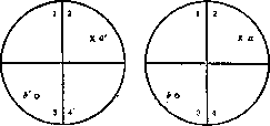
Figure 1.
between our two eyes (cyclopean eye). If we imagine that the extracted and maintaining their shape retinas of both eyes are superimposed one on the other so that the right part of one retina coincides with the right part of the other eye and the left part of one with the left of the other, with the yellow spot of one eye coinciding with the yellow spot of the other eye, then the overlapping points
„, of both retinas are called
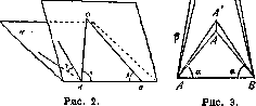
corresponding points. In the center is placed the yellow spot, and with crosses aa' (in quadrants 2) and circles ЪЪ' (in quadrants 3) are depicted corresponding points. If the eye fixes any particular point object and, consequently, its image is depicted on the yellow spot, then points in space that give images on corresponding points of the retina are perceived as single points. Those objects that are not depicted on corresponding points of the retina give double images. The meaning of B. v. can be easily understood if we imagine the following: let us imagine that we have two points A and B (see figure 2), representing our two eyes, and let us assume that we wish to determine the position of some point in space-O. The position of this point will be determined if we know two angles « and β and if
at the same time know the angle γ of the inclination of the plane AOB to the horizon H; if at the same time the length of the line AB is known, then all points in space can be determined using the corresponding angles α, β and γ, which are coordinates characterizing the position of points in space. When, with the help of B. v., fixation of different points in space is performed, then, in essence, the angles α, β γ are determined by the tension of the contracting eye muscles, and this allows determining the position of point O in space. Thus, eye movements and B. v. are a means of evaluating distances, which are necessary for animals to orient themselves in space. The more the angles α and β approach being straight, the more inaccurate the determination of the distance of point O becomes, because with this a small change in angle causes a huge change in the distances of the point from the eyes, as can be seen in Fig. 3, where the changes in distances of points A, A' and B, B' in space are given for identical changes in angles A and B by the amount α. The greater the distance AB, the more accurately one can determine, all other conditions being equal, the distances of points lying before us, and thus it can be concluded that for small animals, for example, mice and rabbits, the determination of distances exists in a less precise form than for large animals, for example, elephants, in which the distance between the eyes is more significant.
P. Lazarev.
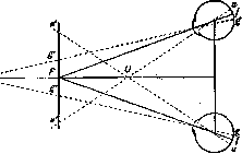
B. field of vision. The ability to combine images received on the retinas of both eyes gives volumetric, depth images of objects. The perception of depth is created due to the muscular act of bringing together the eye axes (convergence), when setting the eyes on more closely located objects. The degree of tension of convergence serves for judging the depth of objects, for evaluating distance. The very fusion of the images received by the right and left eyes already occurs in the visual centers of the brain. It can occur only in cases when images of objects fall on corresponding associated points of the retinal shell of the eyes (see Eye Association). The ability of B. v. allows us to consider both eyes as one combined apparatus. The sum of all points in space, the images of which, with a given position of the eyes, fall on associated points of the retina, is called the horopter (see). All objects located closer or farther than the fixed point no longer fall on associated points of the retina and therefore are seen double. At the same time, the images of objects located closer to the fixation point seem crossed to us, and the images of objects located farther than this point seem spread apart, as can be seen in Fig. 4. This phenomenon is called physiological doubling, in contrast to pathological doubling, which occurs with an incorrect relative position of the eyes, i.e., when the images being fixed fall on non-associated points of the retina. Such pathological doubling is encountered in clinical practice in cases of strabismus. Complaints of doubling are noted, however, only in recent cases
«-
Fig. 4. Physiological doubling with normal binocular vision: F - the fixed point, the images of which fall on corresponding points of the retina (//). U and d-points lying closer and farther than the fixation point. Their images fall on non-associated points of the retina (u and u d d) and therefore are seen double. strabismus (in recent cases of paralysis of the eye muscles). Subsequently, there is an unconscious exclusion of the images of one eye, and doubling ceases to bother patients.
Related articles
Mentioned in
Cite this page
“Binocular Vision.” Soviet Medical Encyclopedia. English translation of Bolshaya Meditsinskaya Entsiklopediya, 1st ed. (Moscow, 1928–1936), ed. N. A. Semashko. https://sovietmedicalencyclopedia.pages.dev/article/binocular-vision/