Field of Vision
Historical document, translated for reference. It reflects medical knowledge of the 1920s–30s and is not medical advice.
Summary
This article from the 1928–1936 Great Medical Encyclopedia discusses the definition and methods of examining the field of vision, including the Donders control method and campimetry. It details the technical procedures, clinical applications, and geometric limitations of these examination techniques in ophthalmology.
Encyclopedia article (1928–1936)
FIELD OF VISION represents the space, all points of which are simultaneously visible with a fixed gaze. During monocular fixation, the image of the fixed object is formed in the region of the macula lutea, whereas the images of objects located at some distance from the fixed object are formed not in the region of the macula lutea, but on the more peripheral parts of the retina. Therefore, they are seen unclearly. Examination of the field of vision is performed using perimetry (see); in addition, there are 2 more methods for examining the field of vision, each of which has its own practical application, although in a more limited form than examination using a perimeter. The first of them was proposed by Donders and is usually known under the name of the control method, because it is based on a kind of control with the investigator's own eye. Its methodology consists of the following. The subject sits with their back to the light, closes one eye, e.g., the right, with the palm or a light bandage, and with the other continuously fixes the opposing eye of the investigator, who is positioned opposite at approximately a distance of 1.5 m and also covers one eye, but already the opposite one, i.e., in this case the left; conversely, if it is necessary to know the state of the field of vision of the patient's right eye, the latter closes his left, and the physician his right eye. The study of the function of the peripheral parts of the retina by the control method in principle reduces to an approximate determination of the outer boundaries of the patient's field of vision by comparing it with the field of vision of the investigator, which of course is assumed to be normal. Practically, this is carried out in such a way that the investigator, carefully watching the patient's opposing eye, slowly moves the ends of the fingers of his hand or, even better, a small square of white paper of 1 cm2 fastened to the end of a thin stick in space between himself and the patient from the periphery to the center until the subject notices the movement of this object. If, when moving it from various sides (at least from 4), these moments occur simultaneously for both the physician and the patient, the patient's field of vision is considered normal. Conversely, the absence of such a coincidence indicates disorders on the part of the patient's peripheral vision and in general terms even gives the investigator an idea of the very nature of these disorders, thus indicating the need to subject the patient to a more thorough and subtle examination. For all its primitiveness, the described method has fairly wide application, being indispensable for preliminary orientation at outpatient admissions, when visiting patients at home, and generally in all cases where it is impossible to use special instruments, as for example in patients with a severe general condition requiring a fixed position in bed, etc. On the other hand, having in principle a purely subjective and only comparative character, the control method gives no objective representation of the state of peripheral vision in either the subject or the investigator himself; in other words, it does not provide the possibility of any graphic representation, let alone measurement of the field of vision, which, however, is extremely necessary. In its simplest form, these requirements are met by the projection of the field of vision onto a plane; the second of the mentioned methods, proposed in 1855 by Graefe and named campimetry, is built on this principle. Any smooth black surface (in the form of a wall, blackboard, piece of fabric stretched on a frame, etc.) can serve as an instrument for this purpose (a campimeter), in front of which the subject is placed at a distance of 15-25 cm, and this distance must be kept unchanged throughout the entire study; one eye is bandaged, and the other fixes some mark, e.g., a white cross drawn opposite to it on the campimeter. Having seated the patient in this manner, the investigator, with small oscillating movements, moves some white object against the black background of the campimeter, e.g., a small piece of chalk or another suitable object on the end of a thin black rod (in the form of an ivory ball or a square of white paper), etc., and the object is slowly moved from the periphery toward the fixation point until the subject notices its approach; the place on the campimeter opposite which the object first appears in the patient's field of vision is marked with the same chalk. A similar manipulation is performed in at least 4 directions, namely in the vertical, horizontal, and two intermediate ones, as a result of which 8 points are marked at the ends of the indicated meridians, connecting which we obtain a clear image of the field of vision in its projection onto the plane, and at the same time an idea of its shape and size. The simplest way to determine the latter consists in linear measurements of the distances from the center to the outer boundaries in each of the indicated meridians. However, in view of the fact that the dimensions of the outline itself change depending on the change in the distance between the eye and the campimeter, and with an increase in distance the projection of the field of vision also increases, for this purpose it is more appropriate to use a constant value, namely the tangent of the angle formed by the visual line directed to the fixation point and the line connecting the boundary of the found field of vision with the optical center of the eye. Practically, for this, one can use a special table for converting angular values into linear ones, calculated in centimeters and corresponding on the campimeter to the tangents of certain angles. The described method, despite its simplicity and ability within certain limits to give fairly accurate results, is not devoid of certain shortcomings, stemming mainly from the fact that it is based on the projection of the spherical surface of the retina onto a plane. The most important of them are as follows. 1. As measurements on a degree grid show, the boundaries of the field of vision projected onto a plane on average extend inward from the center by 60°, upward by 65°, downward by 75°, and outward by 90°. From this it follows that the determination of the outer boundary of the field of vision on the campimeter is impossible, because the tangent of an angle of 90° is equal to infinity and consequently the line directed to the outer boundary of the field of vision will be parallel to the plane of the campimeter. Thus, the figure of the field of vision on the campimeter must be a parabola open outward. 2. In view of the fact that linear values on a plane as they distance from the center increase faster than the corresponding values on the spherical surface of the retina, the projection of various kinds of fundus changes on the campimeter will give incorrect ideas about their actual dimensions and specifically in the direction of exaggeration. Therefore, campimetry as a rule has a limited scope of application, mainly for examining the central parts of the field of vision with a radius of approximately up to 30°, namely for determining paracentral scotomas, enlargement of the blind spot (see Bjerrum's method), etc. 3. The campimetric method does not provide the ability to monitor the correct fixation of the center by the tested eye if the study is performed at too close a distance (15-25 cm); lengthening the distance, on the other hand, entails...
a drop in accuracy.
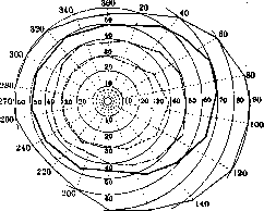
Boundaries of the field of vision. [...] This condition in turn creates inconveniences in that an eye with a sharp drop in visual acuity is not able to distinguish the movement of the object on the peripheral parts of the campimeter, which are too far removed from the center. All these listed shortcomings are completely eliminated only when examining peripheral vision on a perimeter, which is why this method is the most commonly used. The normal field of vision has definite boundaries, which, however, depend to a significant extent on the research methodology. The boundaries of the field of vision are widest for white color; for red, green, yellow, and blue colors, they are considerably narrower (see figure). But the boundaries of the field of vision for white color are also subject to considerable fluctuations depending on the structure of the facial parts surrounding the eye. For example, sharply protruding superciliary arches or a deep sinking of the eye can significantly narrow the field of vision. In addition, the boundaries of the field of vision depend to a large extent on the lighting conditions during the study and on the intellect of the patient. All this, taken together, leads to the fact that the boundaries given by various authors for the field of vision for white color show a rather large discrepancy, as can be seen from the following table, borrowed from Landolt. Boundaries of the f[ield] o[f] v[ision] for a white mark of 10 sq. mm. [...] Even if such contradictory data are obtained when determining the boundaries for white color, an even greater discrepancy exists regarding the boundaries for colors. The fact is that most colors on the periphery of the field of vision change their color hue, which makes the determination of boundaries largely inaccurate. Those boundaries that are marked in printed diagrams for clinical studies as normal boundaries of the field of vision for white color and colors represent only the average boundaries of the normal field of vision. Therefore, when evaluating perimeter data, great caution should be exercised, especially in cases where these deviations are not sharply expressed. In general, reliable results in any doubtful cases can be achieved only through repeated studies. The binocular field of vision differs in some features from the monocular field of vision. When looking with both eyes, the field of vision of one eye is superimposed on the field of vision of the other in such a way that the nasal half of the field of vision of the right eye moves onto the temporal half of the field of vision of the left eye and vice versa. Since the boundaries of the field of vision from the temporal side are wider than from the nasal side (by approximately 30°), the following ratios are obtained: the middle part of the resulting field of vision is indeed served by both eyes, while on both temporal sides there are sickle-shaped areas served only by the eye of the corresponding side. These areas are called temporal sickles (temporaler Sichel) of the field of vision. As anatomical and clinical studies show, they are supplied by special bundles of nerve fibers, which, starting from the chiasm and all the way to the cortical visual centers, run in an isolated bundle and have a completely definite position. Changes in the field of vision can be caused by both organic and functional diseases. The first group includes changes in the field of vision in lesions of the vascular membrane, retina, and visual pathways; the second group includes changes in neuroses. By their nature, all changes in the field of vision can be divided into two groups: narrowing of the field of vision and scotomas. Narrowing is understood as deviations from the boundaries of the field of vision. Scotomas represent limited, isolated defects of the field of vision; they have the appearance of islets of various outlines located in different parts of the field of vision. Narrowing of the field of vision can be expressed in the form of more or less regular concentric narrowing, loss of individual sectors, entire quadrants, and sometimes even entire halves of the field of vision. The latter type of defects of the field of vision is called hemianopia (see). The diagnostic significance of these forms of narrowing of the field of vision varies. In lesions of the vascular membrane and retina, mainly various varieties of concentric narrowing are observed, while in diseases of the visual pathways, all the above forms are encountered. In general, in diseases of the vascular and retinal membranes, the study of the field of vision only complements the data obtained by other methods of examining the eye, since in the overwhelming majority of cases the diagnosis is made by ophthalmoscopy. Completely different ratios are observed in lesions of the visual pathways. Here, changes in the fundus oculi are far from always present, and in a number of clinical forms (retrobulbar neuritis, diseases of the chiasm, lesions of Gratiolet's bundle and cortical visual centers), the diagnosis can be made only on the basis of perimetric data. Regarding the lesion of the visual pathways, of particular importance is the circumstance that the shape of the defects of the field of vision depends on the localization of the pathological focus. Lesions of the optic nerves from the eyeball to the chiasm give various forms of concentric narrowing of the field of vision and sometimes loss of individual sectors. In cases where both optic nerves are affected, the defects of the field of vision in both eyes, with rare exceptions, do not have a symmetrical character. Starting from the chiasm, the character of the defects of the field of vision changes sharply; they acquire a symmetrical character and are expressed in the form of complete or incomplete hemianopias (see) (see volume VI, art. 499-504, figures 1-5). In complete hemianopias, the entire corresponding half of the field of vision drops out; in incomplete ones, only a more or less sharp narrowing of the field of vision in the corresponding areas is observed. At the same time, depending on whether the conductivity of these bundles of nerve fibers is completely interrupted or only partially disrupted, hemianopia for white color or only hemianopia for colors is observed. Such a relationship is observed not only in hemianopias, but also in other forms of narrowing of the field of vision, as well as in scotomas (see). First of all, it must be noted that already in the normal field of vision there is a physiological paracentral scotoma, which is the blind spot. Of the pathological paracentral scotomas, Björrum's scotomas are of great diagnostic significance. These are arcuate scotomas originating from the blind spot and surrounding the fixation point. They represent one of the characteristic signs of changes in the field of vision in glaucoma. Enlargement of the blind spot is also sometimes observed in lesions of the optic nerves (see nervus opticus). Of the functional disorders of the field of vision, concentric narrowing in severe forms of hysteria is of particular importance. In many cases, it can reach very significant degrees, and then only through repeated studies and observations is it possible to distinguish it from concentric narrowing on the basis of an organic lesion of the visual pathways. In cases of sharply expressed fatigue, the so-called spiral field of vision is obtained, the essence of which is as follows: if after determining the boundaries of the field of vision in various meridians, one again determines the boundary in the first of the studied meridians, it turns out to be significantly closer to the fixation point than during the first perimetry of the same meridian. (Field of vision in microscopic technique—see Microscope.) The significance of the field of vision for professional selection. The study of the field of vision is important not only from the point of view of diagnosing diseases of the visual pathways and retina. In a number of cases, the state of the field of vision must also be taken into account to judge professional suitability. When evaluating the professional significance of certain defects of the field of vision, it should be remembered that the binocular field of vision is for the most part served by both eyes. Therefore, even blindness of one of the eyes with a good state of the field of vision in the other eye gives only a small narrowing of the binocular field of vision from the side of the blind eye. Professional suitability is most reduced by a sharply expressed concentric narrowing of the field of vision in both eyes, observed, for example, in retinitis pigmentosa and certain forms of optic atrophy. In these cases, even with satisfactory central visual acuity, the patient moves only with extreme uncertainty. This is explained by the fact that the patient, although seeing well directly in front of himself, completely fails to notice objects surrounding him. Hemianopias also strongly reduce professional suitability. In this regard, heteronymous (bitemporal and binasal) and homonymous (right-sided and left-sided) hemianopias have different significance. Heteronymous hemianopias have little effect on professional suitability, because even in sharply expressed degrees they narrow the binocular field of vision very little. For example, in bitemporal hemianopia, the nasal half of the field of vision of the left eye replaces a large part of the temporal half of the field of vision of the left eye and vice versa. The situation is completely different in homonymous hemianopia. Here, the nasal part of the field of vision on one eye and the temporal part on the other always drop out simultaneously, and the dropped out part of the field of vision is therefore no longer compensated for by anything. Central scotomas are important only if they sharply reduce visual acuity. The latter circumstance is always observed in absolute scotomas for white color.
Central scotomas for colors, especially relative ones, can occur even without a sharp drop in vision. The state of the field of vision is especially important for those professions in which work takes place in factory and industrial premises. When moving among operating machinery, it is especially important, in addition to central visual acuity, to also have a good field of vision, since defects in the field of vision can be a cause of injuries. This applies equally to skilled workers operating machinery directly, as well as to laborers. From this point of view, it is important to take into account not only the profession, but also the detailed working conditions. For example, a cleaning woman with a severely narrowed field of vision can work in school and office premises and at the same time be completely unsuitable for work in factory and industrial premises. The state of the field of vision is also of great importance for transport work. The highest requirements must be set for driving professions (for example, city tram drivers, chauffeurs, railway engineers). Of all types of transport, the state of the field of vision is most important for aviation. When determining occupational suitability, there is no need for such meticulous perimetry as usually takes place in clinical examination for diagnostic purposes. In most cases, testing for white color is quite sufficient. Perimetry is especially important in cases where there are changes in the fundus with relatively good visual acuity, since defects in the field of vision are particularly common here and are decisive in terms of determining occupational suitability. All of the above should be taken into account not only in vocational selection and vocational counseling, but also in expertise from the point of view of disability.
E. Tron.
Related articles
Mentioned in
Cite this page
“Field of Vision.” Soviet Medical Encyclopedia. English translation of Bolshaya Meditsinskaya Entsiklopediya, 1st ed. (Moscow, 1928–1936), ed. N. A. Semashko. https://sovietmedicalencyclopedia.pages.dev/article/field-of-vision/