Ophthalmoplegia
Historical document, translated for reference. It reflects medical knowledge of the 1920s–30s and is not medical advice.
Summary
This article from the 1928–1936 Soviet medical encyclopedia defines ophthalmoplegia as the simultaneous paralysis of several eye muscles innervated by different nerves. It details the classification into complete and partial forms, the symptoms including the characteristic 'Hutchinson's face,' and the various causes ranging from organic nerve damage to infections, tumors, and toxic substances.
Encyclopedia article (1928–1936)
OPHTHALMOPLEGIA (from Greek ophthalmos- eye and plesso - I strike), simultaneous paralysis of several eye muscles innervated by different nerves (nn. oculomotorius, trochlearis, abducens). More often, O. refers to the disease of the above character in both eyes; however, combined paralysis of two or three nerves of one eye also fits this name. O. is divided into complete and partial. In complete O., both the external muscles of the eye (straight, oblique, and the levator of the upper eyelid) and the internal ones (sphincter of the iris and ciliary) are affected. Partial O. is of two kinds: external O., when only the external eye muscles are affected, and internal O., when the internal muscle of the eye is paralyzed. In the vast majority of cases, O. depends on organic damage to the peripheral neurons of the above three nerves (more often their nuclei, less often their roots and the nerves themselves).
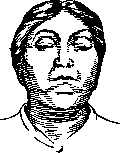
Figure 1. Complete ophthalmoplegia. Hutchinson's face. Bilateral ptosis.
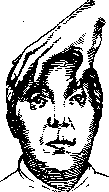
Figure 2. Complete ophthalmoplegia. Hutchinson's face. Upon artificial elevation of both eyelids, the eyeballs are found to be immobile.
The symptomatology of pronounced O. is reduced to the drooping of both eyelids and immobility of the eyeballs - the so-called 'Hutchinson's face' (Fig. 1 and 2); in internal O., the pupils are dilated and do not react to light nor to convergence; accommodation is often impaired due to paralysis of the ciliary muscle. In less severe cases, paralysis is observed in some muscles, while others show only paresis. Diagnosis of complete external or internal O. presents no difficulties - complete absence of eye mobility, ptosis in external O., and absence of pupil movement and accommodation in internal O. are easily determined. In incomplete O. with paralysis or paresis of individual muscles, diagnosis requires examination of diplopia, the field of vision, etc., as is done in determining paralytic strabismus (see Diplopia, Strabismus). According to course, acute and chronic O. are distinguished. The latter usually has a progressive character. Acute O. is often unilateral as a result of rapidly developing necrosis of vascular origin, being then combined with a number of other symptoms indicating damage to nearby systems (see Alternating syndromes). More often, acute O. is bilateral. It occurs under the influence of various intoxications and infections. Among chemical substances, alcohol is first, then lead, ergot, and sulfur derivatives. Methyl alcohol causes isolated acute internal O. O. is often due to poisoning with meat, fish, sausages; in such cases, it is sometimes difficult to decide whether the matter is poisoning with chemical substances contained in these products or with infection entering the organism together with them.
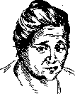
Figure 3. Ptosis of the left eyelid, abduction of both eyes to the right, dilatation of the left pupil, constriction of the right pupil.
Acute O. occupies a prominent place in the symptomatology of a number of infectious diseases of the brain and its membranes (epidemic encephalitis, tuberculous meningitis, acute hemorrhagic poliomyelitis, etc.). Sometimes acute O. occurs after fracture of the base of the skull with subsequent hemorrhage. Subacute O. develops sometimes in tumors of the brain stem, brain base, and cerebellum; it is observed much more often in syphilitic diseases of the brain (Fig. 3). Chronic O. very rarely represents an independent disease; in the vast majority of cases it develops against the background of an already existing disease of the nervous system or precedes it (sometimes long before). Of these diseases, tabes and progressive paralysis are first, then myasthenia, funiculomyelosis, multiple sclerosis, chronic poliomyelitis, even rarer Basedow's disease, cysticercosis, and sarcoma of the meninges. Isolated internal O. can be the only manifestation of neurosis for many years. Chronic external O. usually has a progressive character, and as a rule, in its further development it does not pass into complete, i.e., does not spread to the internal muscle of the eye, which speaks in favor of its nuclear localization. The pathological essence of chronic O. usually comes down to an inflammatory process at the bottom of the third ventricle and the aqueduct of Sylvius, which ultimately leads to the death of the nuclei of the III, IV, and VI nerves.
Ophthalmoplegia occupies a peculiar place in migraine (see Hemicrania). In rare cases, 'migraine ophthalmoplegique' O. occurs on the side of the pain. It either disappears after a migraine attack or leaves residual paresis, which intensify with a new migraine attack and pass into chronic O. Congenital O., depending on underdevelopment of the nuclei of the III, IV, and VI nerves, stands apart. This O., as a rule, is external, and the eyelid elevators are especially affected, while the ability to converge is usually preserved. Double images do not occur with this O. Sometimes this form of O. is familial; in essence, it belongs to aplastic or abiotrophic processes and can be considered as a partial atavism: in a number of lowly organized animals, the eyes sit immovably in the orbits. In individual cases of pseudobulbar paralysis, external O. was observed, depending on interruption of central paths from the cortex to the nuclei of the III, IV, and VI nerves. For this form of O., the absence or limitation of voluntary eye movements is characteristic with preservation of reflex movements. This form was named by Wernicke pseudophthalmoplegia. Cases where it is due to primary damage not of the nervous apparatus but of the muscle can be referred to false O. Thus is O. in scleroderma. Finally, hysterical O. occurs. In it, the internal muscle of the eye is not affected, automatic eye movements are preserved, and the O. itself sometimes disappears when the patient's attention is distracted.
Related articles
Mentioned in
Cite this page
“Ophthalmoplegia.” Soviet Medical Encyclopedia. English translation of Bolshaya Meditsinskaya Entsiklopediya, 1st ed. (Moscow, 1928–1936), ed. N. A. Semashko. https://sovietmedicalencyclopedia.pages.dev/article/ophthalmoplegia/