Urethroscopy
Historical document, translated for reference. It reflects medical knowledge of the 1920s–30s and is not medical advice.
Summary
Urethroscopy is a method for examining the urethra using special instruments called urethroscopes. The article describes different types of urethroscopes, their components, and the procedures for both dry and irrigation urethroscopy.
Encyclopedia article (1928–1936)
URETHROSCOPY, URETHROSCOPES. Urethroscopy is a method for examining the urethra by means of special instruments called urethroscopes. Two systems of urethroscopes are distinguished: for dry and irrigation urethroscopy. The dry urethroscope (Valentin or Lewis) consists of a grip (light carrier), a set of tubes, and electrical cords. The grip consists of two metal plates insulated from each other. The upper plate consists of two parts separated from each other by insulation. On the upper plate there is a contact slider for turning on the current. The lower plate at the front end has three holes on the underside: one for screwing in a bulb located on a long rod, and two for fixing the tube. At the rear end, each plate has recesses for electrical wires. Nickel-plated tubes with diameters from No. 21 to No. 29 (according to Charrière), straight, 15 cm long (Fig. 1). One of them, longer, is curved like a beak at the central end and has a notch for viewing the seminal hillock. The straight tubes at the central end are cut at an angle and the edges are somewhat concave. To prevent injury to the urethral mucosa when inserting the tube, they are provided with obturators (mandrins) ending conically. On the upper plate there is an attachment for fixing a lens or Kaufmann's magnifying optical instrument. The latter in the viewing urethroscope almost directly adjoins the grip, while in the urethroscope adapted for endourethral interventions, it is set 5-6 cm from the grip (Fig. 2).-The irrigation urethroscope of Goldschmidt consists of a straight metal tube closed at the end with two oval holes on the sides. For posterior urethroscopy, the catheter is longer, with a beak at the bladder end and with a notch on its posterior side (Fig. 3). For posterior urethroscopy, the tube brought to the external sphincter is lowered by advancing the funnel, thereby overcoming the spasm of the external sphincter. Then the tube is advanced forward by 11/2-2 cm to the seminal hillock. To advance it, the peripheral end of the tube must be tilted so that its longitudinal axis coincides with the longitudinal axis of the subject's body. Fixing the tube with the left hand in this position, the mandrin is removed with the right hand, the field of vision is dried with cotton swabs, and the illuminating instrument is introduced.

Figure 1. Metal tubes with obturator for illuminating the urethra.
The examination of the urethra, both in anterior and posterior urethroscopy, is performed from back to front, gradually withdrawing the tube and repeatedly drying the field of vision. For orientation, it is necessary that the "central figure" be in the field of view of the urethroscope, i.e., the lumen of the urethra, where the radially arranged folds of the mucous membrane converge. The "central figure" in the retrocollicular part of the urethra appears in the form of a slightly gaping semicircle. The "central figure" in the area of the seminal hillock retains a semicircular shape, with the folds of the mucous membrane of the anterior wall of the urethra directly adjoining the apex of the seminal hillock, the shape of which determines the appearance of the central figure of this area. On the sides of the seminal hillock, two openings of the seminal ducts can be seen, and in the center there is a depression leading into the cavity of the utriculi masculini (see Urethra). The "central figure" of the membranous and pendulous parts of the canal has a dot-like appearance. In the area of the bulb and glans, it appears as a vertical slit. The color of the mucous membrane changes depending on the condition of the body as a whole and, in particular, the vascular system. It is usually pinkish in the area of the glans, redder in the pendulous part, and in the bulbar part the mucosa is more hyperemic. Toward the front, the coloring weakens and in the area of the navicular fossa the color is yellowish-white. The surface of a normal mucous membrane is moist, shiny, and smooth. In the radially arranged folds, vessels passing under the epithelium, the openings of Morgagni's sinuses, and the excretory ducts of Litre's glands are visible.
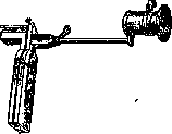
Figure 3. Irrigation urethroscope: Obj-objective; W-faucet for admitting liquid; at-sleeve with optics.
Indications for dry urethroscopy are: 1) chronic inflammatory processes in the urethra, as well as establishing recovery from urethritis; 2) suspicion of foreign bodies, diverticula of the urethra, ulcers, tumors, or cysts. If the latter are found, a puncture is made with a urethral probe shaped like a stiletto by Colman, which is a thin metal probe 12 cm long, pointed at the end. Urethroscopy is contraindicated in acute inflammatory processes in the urethra. Irrigation urethroscopy is performed with the patient in the same position as for cystoscopy. The urethroscope tube with the mandrin inserted is introduced into the bladder and connected to an Esmarch's syringe filled with indifferent liquid. After removing the obturator, the flow of liquid is turned on and at this time the optical system is inserted into the tube. The left hand regulates the flow of liquid by squeezing and releasing the driving rubber tube, providing sufficient stretching of the urethral walls. After turning on the flow, the internal sphincter of the bladder is found. Gradually withdrawing the instrument, the posterior urethra, the prostatic fossa, and the seminal hillock are examined. At the end of urethroscopy, the optical system is removed, the obturator is inserted, and only then is the urethroscope removed. Anterior irrigation urethroscopy is performed with a straight tube. The mucosa appears paler than with dry urethroscopy. The indication for irrigation urethroscopy is suspicion of the presence in the urethra, especially in the area of the seminal hillock, of tumors, polyps, growths, or ulcerative processes.
Related articles
Mentioned in
Cite this page
“Urethroscopy.” Soviet Medical Encyclopedia. English translation of Bolshaya Meditsinskaya Entsiklopediya, 1st ed. (Moscow, 1928–1936), ed. N. A. Semashko. https://sovietmedicalencyclopedia.pages.dev/article/urethroscopy/