Rectoscopy
Historical document, translated for reference. It reflects medical knowledge of the 1920s–30s and is not medical advice.
Summary
Rectoscopy is a method for examining the lumen of the rectum, using a tubular instrument with a light source. It is used to diagnose and treat conditions such as hemorrhoids, polyps, and tumors, and to perform minor surgical procedures.
Encyclopedia article (1928–1936)
RECTOSCOPY (syn. proctoscopy), rectoscopes (from Latin rectum - rectum and Greek scopeo - I look). Rectoscopy is a method of examination used for inspecting the lumen of the rectum. It was already used in ancient times with various devices that dilated the sphincter ani. Scientific development of R. was obtained in 1895 thanks to the works of Kelly and Fyodorov, who proposed a series of metal tubes with removable obturators, equipped with a man-dren. The principle of these tubes is preserved in modern instruments, which have only technical improvements and a light source. The application of R. significantly expanded after the works of Fyodorov (1896), who proved through numerous studies on corpses that anatomical conditions fully allow the introduction of rigid tubes into the rectum and even into the beginning of the flex sigmoi-deae (recto-romanoscopy) at a distance of 30-40 cm from the anus. The works of Kelly and Fyodorov led to wider application of R. A number of rectoscopes appeared on the market, gradually improving in their design (Fig. 1--5). Especially great, one could say decisive significance in the further development of the method played the use of a light source - an electric bulb - during rectoscopy, first in the form of a pan-electroscope by Casper, then a small incandescent electric bulb (Minyon), attached at the end of the tube on a special rod serving simultaneously as a conductor of electrical energy. Of the numerous types of rectoscopes existing on the market and differing from each other only by insignificant technical modifications, the most convenient and technically improved can be considered the Straus rectoscope (Fig. 5), manufactured by various German firms. It consists of a metal tube (e) 20-30 cm long (usually sets of three tubes of 20, 25 and 30 cm are manufactured). The diameter of the tube for adults is 17a-2 cm, for children 1 cm. The tube is marked with divisions indicating the length of the part introduced into the rectum, starting from its peripheral end. The tube is screwed onto a short cylinder a, to which a narrow rod is attached, entering the tube and almost equal in length to it; at the end of the rod an electric lamp is screwed on. A handle b is screwed to the cylinder from below, through which an electrical conductor passes from the rheostat. The current is switched on by moving the contact g. Before introducing the rectoscope into the rectum, a rod-man-dren d is inserted into it on a long rod zh. After introducing the rectoscope into the rectum, the man-dren is removed and the opening in the cylinder is closed by a small ocular z or a magnifying and bringing the image closer lens i. The cylinder has a valve on the side for pumping air into the rectum. For the performance of R. it is necessary to thoroughly clean the intestines, achieved by prescribing a laxative the day before the examination, and in the morning of the day of examination one or two enemas. Rectoscopy is usually performed in the knee-elbow position; in weak patients in the supine or lateral position (better on the right). The end of the tube and man-dren are abundantly smeared with vaseline, and the rectoscope is introduced into the rectum by rotary movements without violence. To overcome the resistance of the sphincter during its spasm (for example, during fissures) or in nervous subjects prone to fear, it is necessary to apply local anesthesia, but usually, with careful introduction of the rectoscope, the moment of passing it through the sphincter is not very painful. After introducing the rectoscope into the intestine, the man-dren is removed and, having attached the optical system, the further advancement of the rectoscope is continued under eye control, with the light on. At this moment, the opening of the tube appears tightly closed by the adherent mucous membrane. Carefully pumping air with a balloon, the folds of the mucosa are straightened and the recto-scope is advanced into the opened lumen of the intestine. Acting in this way under eye control, it is usually easy to introduce the rectoscope for the entire length of the rectum, and in calm and patient patients to introduce it 5-10 cm into the lumen of the S-Romani (recto-romanoscopy). Special caution is required when advancing the rectoscope past the promontorium, for which it is best to hold the posterior wall of the rectum, carefully deflecting the handle of the rectoscope forward and slightly to the left. One should advance all the time slowly, stopping at the slightest complaint of the patient and carefully continuing to inflate the folds of the mucosa. The examination of the rectum is continued and when the rectoscope is withdrawn, especially well the area of the internal and external sphincters can be examined. The picture that opens upon introduction of the rectoscope appears (as stated above) in the form of folds of the mucosa, sometimes even invading the lumen of the rectoscope. The mucosa is light-red in color, smooth on the surface, shiny, and through the mucosa, especially in young subjects, blood vessels under the mucosa are visible. In older subjects, the color of the mucosa is paler, the surface of the mucosa is completely smooth. Indications for R.: hemorrhoids, polyps, ulcers of the rectum, strictures, new growths, proctitis, sigmoiditis, foreign bodies, fistulas, bleeding of unknown origin. With the help of a rectoscope, under eye control, certain operations are performed - cauterization of the mucosa, incision of strictures, removal of polyps with a thermocauter, removal of pieces of tumors for microscopic examination, etc. For this purpose, a number of special long instruments in the form of scissors, spoons, galvanocauter, etc., are provided for rectoscopes. : With insufficiently careful introduction of the rectoscope, accompanied by violence, damage to the wall of the intestine can occur, especially in the area of the promontorium. Straus describes a case of perforation of the intestine during R. Insufficiently careful contact of the tube with a tumor can cause severe intestinal bleeding, therefore, during R., as during any other endoscopy, the basic rule is careful, gentle introduction of the instrument without any violence.
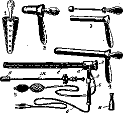
Figures 1-5.
H. Blumental. J REC.TOSELE, bulging of the posterior wall of the vagina with the formation of a diverticulum of the anterior wall of the rectum (see figure). The bulging can be variously expressed, being detected primarily during abdominal pressure strains. The studies of Halban and Tandler show that with R. from the side of the rectal muscle no defects are noted. According to Halban, Tandler and Martin (E. Martin), the leading significance in the origin of R. is the insufficiency of that perineal wedge which is located between the rectum and the vagina. Indeed, the height of the perineal wedge under ordinary conditions is 3-4 cm, and with R., according to Halban and Tandler, is not more than 1/4-2г'я cm. Thus, with R. there is always a defect in the tissues located between the rectum and the vagina. Clarifying the question of the origin of R., Martin attaches exceptional significance to the weakening of the fasciae of the rectum and vagina. Such a violation of the stability of the fasciae is possible not only with ruptures of the perineum, but also with the integrity of the external tissues, when
<г
Ш/г
^--^
------'""^
stability is a predisposing moment for the formation of R., the development of which is favored by stagnation of fecal masses in the lower part of the rectum, accumulation of gases. R., forming as a result of a violation of the stability of the perineal wedge, can be observed without displacement and prolapse of the uterus. However, R. is encountered much more often in conjunction with prolapse and prolapse of the uterus, since damage to the perineal wedge is rarely observed without a violation of the stability of the pelvic floor. In connection with this, the symptoms observed during rectocele are usually lost in the general picture of the disease characteristic for prolapse of the uterus, vagina (see.). With the isolated existence of R., patients note a bulging from the genital slit of the posterior wall of the vagina, which they often mistakenly take for prolapse of the uterus. The existing gaping of the genital slit favors irritation of the vagina (see.). With a significantly expressed R., accumulation of fecal masses in the formed diverticulum of the rectum is often observed, which can lead to diagnostic errors (tumor from the posterior wall of the vagina).- Diagnosis of R. is not difficult, especially if a study through the rectum is added to the usual examination. Prevention of R. consists in careful layered suturing of perineal ruptures; it is necessary to ensure that in the postpartum period there is no accumulation of fecal masses in the lower part of the rectum. Treatment of R. is only surgical and should be directed towards restoring and strengthening the fasciae of the rectum and vagina.
Related articles
Mentioned in
Cite this page
“Rectoscopy.” Soviet Medical Encyclopedia. English translation of Bolshaya Meditsinskaya Entsiklopediya, 1st ed. (Moscow, 1928–1936), ed. N. A. Semashko. https://sovietmedicalencyclopedia.pages.dev/article/rectoscopy/