Rectal Prolapse
Historical document, translated for reference. It reflects medical knowledge of the 1920s–30s and is not medical advice.
Summary
This article from the 1928–1936 Soviet medical encyclopedia discusses the etiology and anatomical theories behind rectal prolapse. It details the roles of the pelvic floor, the coccyx, and the Douglas pouch in the development of this condition.
Encyclopedia article (1928–1936)
RECTAL PROLAPSE (prolapsus recti), the temporary or permanent presence of the rectum (or a part of it) outside the anal orifice. The maintenance of the rectum in its natural, normal position depends on the equilibrium of its fixation apparatus and intra-abdominal pressure. The fixation apparatus generally consists of: a) the muscular part of the pelvic floor (levator ani), b) connective tissue bundles of the fascial coverings connecting the rectum with the prostate gland, urinary bladder, diaphragm (muscular floor) of the pelvis, and the coccyx, c) the musculature of the rectal sphincter (sphincter ani). Etiology. There are many theories to explain the etiology of rectal prolapse, the most important of which are the following: Esmarch and Fischl see the cause of rectal prolapse in the weakening of the attachments of the lower part of the rectum, depending on an inflammatory process in the mucosa, which causes edema of the submucosal tissue, from which the mucosa detaches, descending downwards and subsequently dragging the other layers of the rectum with it. The inflammatory process, in turn, reflexively causes a spasm of the sphincter, which leads to increased work of the abdominal press to overcome this spasm during defecation—a vicious circle, leading ultimately to rectal prolapse. This point of view is valid for certain types of cases, as patients often point to prolonged catarrh of the colon and rectum as the cause of rectal prolapse. Some authors believe that the presence of hemorrhoids leads to an inflammatory state of the mucosa, as a result of which conditions favoring rectal prolapse may be created. Having performed a number of histological studies of the wall in rectal prolapse, Ludloff found changes in it consisting of thinning of the mucosal and muscular layers as a probable consequence of the inflammatory process. Hoffmann views rectal prolapse as a consequence of insufficiency of the pelvic floor, which has lost its normal elasticity and stretches under the influence of the tension of the abdominal press. In the pathological process, in all probability, not only the muscular part of the pelvic floor (sphincters, mm. levator ani, coccygeus, recto-urethralis, transv. perinei) with the pelvic fascia takes part, but also the bony skeleton of the pelvis. Of particular importance in this is the coccyx, which in a normal adult has an arcuate shape—concave anteriorly (see Figure 1). In subjects suffering from rectal prolapse, and in children, the coccyx has a vertical direction, which is physiological in the fetus (see Figure 2). Regarding the latter...
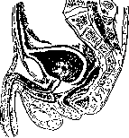
Figure 1. Sagittal section through the pelvis of an adult male.
anomaly, Waldeyer, Ludloff, and Bereznegovsky, who examined a large number of corpses of children suffering from rectal prolapse, also pointed it out. Due to the absence of this normal depression (excavatio recto-coccygealis), in which the ampulla of the rectum is normally located, conditions are created for the development of rectal prolapse. Ludloff and Napalkov, having microscopically examined the tissue of the m. levator ani in patients with rectal prolapse, found sharp degeneration and atrophy of it. The muscular tissue was to a significant extent replaced by connective tissue. The theory of the deep position of the Douglas pouch (see), proposed by Waldeyer and Zuckerkandl, who viewed rectal prolapse as a perineal hernia, and confirmed by interesting studies of Napalkov and Bereznegovsky on corpses, has significantly more adherents. The mechanism of the origin and development of rectal prolapse, according to this theory, consists in the fact that with a low position of the Douglas pouch, the pressure of the abdominal press is transmitted from the rectum not to the sacrum (as is normal), but to the movable coccyx, which yields to this pressure. In this case, the anterior wall of the rectum is pushed into its own lumen, as a result of which a gradual development of rectal prolapse follows. This theory is confirmed by the frequency of rectal prolapse in children, in whom the Douglas fold is normally situated low—approximately at the level of the upper edge of the prostate gland, while in a normal adult subject it reaches only the level of the upper edge of the seminal vesicles. According to the third theory (Jeannel), the cause of rectal prolapse lies in the insufficiency of the apparatus suspending the rectum. Fixed on a weak, incapable of stretching, suspension apparatus, the rectum, following its own weight, exerts pressure on the m. levator ani, under the influence of which the latter undergoes atrophy, which leads to rectal prolapse. Jeannel, as well as Verneuil, see confirmation of this theory in the fact that even extensive destruction of the sphincter
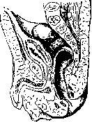
Figure 2. Sagittal section through the pelvis of a newborn (after K. Peter). At various operations (anal fistulas, rectal cancer), with a normally functioning suspension apparatus, do not lead to prolapse; consequently, the muscular floor of the pelvis itself does not play a role in the mechanism of rectal prolapse, and its insufficiency is secondary. Janel wittily compares the rectum to a prisoner sitting on a chain. The anus is the door of the prison. With a strong chain, the prisoner cannot leave even through open doors, but as soon as the chain weakens or lengthens, the prisoner will leave at the first opening of the doors, and the doors (anus), in the expression of Verneuil, "open at every defecation" ("elle s'ouvre a chaque defecation"). The correctness of this theory is disputed by Ludloff, Lennormant, and other authors. During the famine years in the Volga region, Schotter and Lehnbach observed rectal prolapse in adults and children severely emaciated by prolonged starvation. Rectal prolapse in children is often a consequence of dysentery or prolonged diarrhea. The habit often encountered in children of sitting on the potty for a long time is, according to the observation of pediatricians, a factor also contributing to rectal prolapse. Sometimes rectal prolapse is observed in subjects severely emaciated due to debilitating diseases (often in cancer of internal organs). Often rectal prolapse is combined with other developmental defects: bladder exstrophy, congenital hernias, splanchnoptosis. Terebinsky, who performed X-rays of the sacrum in all cases of rectal prolapse he encountered, found in almost all cases non-fusion of the arches of the sacral vertebrae - a symptom indicating underdevelopment. Experience and research accumulated in numerous works on the subject of the etiology of this disease do not provide sufficient grounds to give preference to any one of the three main theories. Thus, rectal prolapse should be considered as a consequence of a whole series of factors, partly of congenital, partly of acquired character. There is no one single factor that is causal for all cases of rectal prolapse. Among other factors predisposing to prolapse and causing it, one should note bronchitis, whooping cough, and phimosis in children, chronic constipation, multiple births with perineal tears leading to relaxation of the perirectal tissue, and getting up too early after childbirth. Rectal prolapse is predominantly observed in workers of heavy physical labor. Hereditary transmission of this disease is observed rarely (according to Napalkov - 4 in 96, according to Lennormant - 4 in 273), but in the anamnesis of patients, one often finds an indication of hernias in parents or close relatives of the patient. Course of the disease. In the initial stages, patients do not experience particular inconveniences or suffering, since the intestine usually prolapses only during defecation and is very easily reduced by the patient himself. With the passage of time, the size of the prolapsed part of the rectum increases, prolapse occurs even from slight straining (lifting a slight weight, coughing, sneezing), reduction becomes increasingly difficult and succeeds only in a certain position of the patient (on the back with legs pressed to the abdomen, on the side, etc.), after an enema and abundant lubrication of the prolapsed intestine with vaseline and other measures, sometimes prolonged and painful. Constant stretching of the sphincter by the prolapsed intestine leads to its insufficiency, which is often manifested by incontinence of gas, and then of feces. The patient becomes unbearable in society. The mucosa of the prolapsed intestine becomes edematous, ulcerates, and bleeds at the slightest contact with underwear, and especially with passing fecal masses. Ulcers become infected, secondary lymphadenitis develops in regional glands (inguinal region, mesenteric glands). Extensive retroperitoneal abscesses have been described, for which the ulcerated mucosa of the prolapsed rectum served as the entry gate. Cases of cancer that developed on the ulcerated surface of the prolapsed rectum are known. A moment comes when the edematous intestine, not amenable to reduction, becomes incarcerated in the anal orifice and becomes necrotic, which can lead to peritonitis. Incarceration of only the prolapsed rectal mucosa, of course, is not such a dangerous complication. Blumenthal observed a six-month-old child in whom a prolapsed cylinder of rectal mucosa 10 cm long, which had prolapsed after prolonged diarrhea, became necrotic. The child recovered. After 4 months, only a slight narrowing of the lower segment of the rectum could be noted. Clinically, one distinguishes prolapse of the rectal mucosa (prolapsus ani - of German authors) and prolapse of all layers of the rectum (prolapsus recti et ani - according to German authors). The prolapsed intestine has a very characteristic shape, resembling a rosette, consisting of concentrically arranged folds of the rectal mucosa (see Figure 3). In the center of the prolapsed intestine, in the depth of the rosette, a narrow opening of the intestinal lumen is visible. The intestine usually prolapses easily under the influence of straining, as a result of which, for the diagnosis of the disease and determination of the degree and type of prolapse, the patient is made to squat and strain as if for defecation. (Since defecation often occurs during this, this examination is recommended to be performed over some vessel.) The size of the prolapsed segment of the intestine varies: from a small fold of mucosa, prolapsing only during very strong straining, to a tumor-like mass, sometimes reaching enormous sizes. In prolapse of all layers of the rectum, the prolapsed part consists of two cylindrical segments of the intestine inserted into each other (see Figures 4 and 5). The outer tube is lined
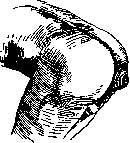
Figure 3.
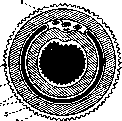
Figure 4. Diagram of a cross-section of the prolapsed part of the rectum: 1 - mesocolon; 2 - mucosa of the inner ring; 3 - muscular coat of the inner ring; 4 - peritoneum; 5 - muscular coat of the outer ring; 6 - mucosa of the outer ring (after Pels-Leusden).
with mucosa on the outside, the inner one - on the inside. The cavity formed between the inner and outer cylinder, lined with peritoneum, is called rectocele or hedrocele. Rectal prolapse occurs in men and women equally often. In all statistics (Ludloff, Napalkov, Lennormant), the vast majority of cases of rectal prolapse fall on the age under 10 years; for example, out of 96 cases collected by Napalkov, 70 relate to patients under 10 years of age.

Treatment of rectal prolapse. Upon the appearance of the first symptoms of rectal prolapse - complete prohibition of physical labor and appropriate treatment. With a clear etiology - elimination of causal factors: treatment of dysentery, colitis, elimination of constipation, bronchitis, whooping cough, phimosis, and other causes increasing intra-abdominal pressure. Elimination of factors hindering defecation (cold toilet, uncomfortable position during defecation), treatment of anal fissures and fistulas, hemorrhoids, rectal polyps. These measures in the majority of cases of rectal prolapse in children lead to a complete cure. Prophylaxis of rectal prolapse should proceed in the same direction. Thure Brandt at one time proposed a special method of rectal massage, using which some authors (Ludloff and others) achieved a good result. The method consists of percussion of the sacral region and other special manipulations on the muscles and the suspension apparatus of the rectum. At present, this method is little used. To strengthen the sphincter, Karewski injected paraffin into the perianal region, Roux - alcohol, Schotter and Lehnbach - milk. It is hardly possible to recommend such injections into the rectal region, which is abundantly supplied with vessels (danger of thrombosis) and inhabited by a rich bacterial flora (danger of developing paraproctitis). Among conservative methods, one should mention pelottes attached with adhesive plaster bandages, and devices of the hernia truss type (see Figure 6), proposed by Esmarch, Bauer, and others and used, mainly, in cases of rectal prolapse depending on temporary causes (e.g., bronchitis, whooping cough, phimosis). In
Figure 5. Hedrocele (after Ludloff).
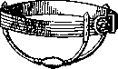
many cases, however, conservative therapy does not give the desired results, and one has to resort to one or another of the very numerous operations proposed for the treatment of rectal prolapse. Analogous to the three main theories cited above to explain the etiology of rectal prolapse, operations can be reduced to the following groups: 1) operations on the anal orifice, 2) operations to strengthen the pelvic floor (pelvic floor plasty), 3) operations on the suspension apparatus and the rectum, 4) combined operations. Velpeau, Dupuytren, Dieffenbach, and then
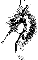
Roberts performed an excision of a wedge-shaped piece of the sphincter followed by suturing. The operation was very popular, but quite often resulted in complications in the form of incontinence of gas and even feces, or narrowing of the anal orifice. In 1891, the German surgeon Thiersch proposed inserting a wire ring under the skin of the transitional fold to strengthen the sphincter. The technique of the operation is simple (see Figure 7); the operation is performed under local anesthesia. Besides narrowing the anal orifice with the ring itself, the latter, being a foreign body, causes the formation of scars around the anal orifice, which remain even after the removal of the wire ring. The Thiersch method is especially convenient in children with prolapse of only the mucous membrane of the rectum due to dysentery, etc., as it is a very minor intervention. After recovery (cessation of prolapse), the wire ring can be removed. Sometimes it has to be removed earlier due to pain during defecation. Not infrequently, the ring cuts through soon after the operation, and then it has to be renewed. To avoid the danger of obstruction, Vreden does not advise tightening the ring too much. A number of authors (Raug, Kirschner, Brunn, and Vakulenko) have proposed replacing the wire ring with a strip of broad fascia rolled into a tube. Although the technique of this modification is slightly more complex than the Thiersch method, it lacks the negative aspects of the latter: the rigidity of the ring, pain, and other phenomena caused by a foreign body. Bereznegovsky strengthens the sphincter with muscle flaps formed from the gluteal muscles. The operation is complex in its technique, very bloody, and has not received
Figure 7. wide distribution due to the easy possibility of infection, which nullifies most plastic operations in the region of the anal orifice. Gersuny and Vreden proposed, after separating the lower section of the rectum, to rotate it 180° around its axis, after which the mucosa is sutured to the skin in the new position, which achieves shortening of the rectum and narrowing of its lower segment. The method is anti-physiological and harbors a number of dangers: necrosis of the rectum, thrombosis, etc.
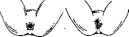
Figure 8.
Figure 9. To eliminate the low position of the Douglas pouch, Napalkov and others proposed a number of very complex operations, which, due to technical difficulty and high mortality, also did not receive wide distribution. To strengthen the pelvic floor, the Hoffman operation is most often used, which is very similar to the Lawson-Tait operation for strengthening the perineum. An incision is made at the border of the skin and mucosa around the posterior circumference of the anal orifice (see Figure 8); the rectum is exposed (for a length of 4-5 cm) and pulled anteriorly, which creates a deep funnel-shaped wound, at the bottom of which
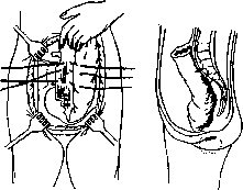
Figure 10.
FIG. 11. a series of sutures are placed in the transverse direction. Skin sutures are placed in the same direction (see Figure 9). In severe cases, colopexy is added (see below). Recently, operations that strengthen the rectum from the abdominal cavity side—colopexia or rectopexia—have become most widespread. Most often, Kummell's colopexy is used: laparotomy in the Trendelenburg position; the rectum is pulled ad maximum out of the small pelvis and sutured with several silk sutures to the sacrum (to the ligamentum longitudinale anter. et poster., see Figures 10 and 11). At the XVII Congress of Russian Surgeons, Kuzmin reported good results obtained by this method; Ratner and Nikitin, who published a number of operations according to Kummell, consider it the operation of choice. The addition of a Thiersch wire ring or a fascial ring further improves the results. Lenormant, Rotter, and others suture the pulled-out rectum to the anterior abdominal wall; Friedrich and Herzen, having placed an anastomosis between the descending colon and the pulled-out rectum (see Figure 12), suture the loop formed in this way to the anterior abdominal wall. Hagen-Torn proposed a very complex operation consisting of a combination of pelvic floor plastic surgery (perineorrhaphy) and strengthening of the rectum (rectopexy). In severe, neglected cases of rectal prolapse that do not yield to treatment with plastic surgery, Mikulicz in 1889 proposed
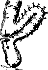
Figure 12.
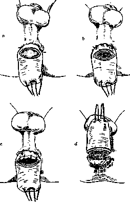
Figure 13.
to resect the prolapsed part of the rectum. In 120 cases, he obtained a good result. The technique of the method is clear from Figure 13. At present, the operation is used relatively rarely, only in the most severe cases, and results in a mortality rate (peritonitis) of up to 12% (Ludloff, Lenormant). Delorme, and then Rehn, proposed resecting

only the mucous membrane of the prolapsed part. The large number of proposed operations testifies to the fact that there is still no sure method for all cases. Recurrences are observed with all methods. When choosing an operation, one must strictly individualize each case of rectal prolapse, trying to find the most suitable method through a carefully collected anamnesis and detailed examination. Most often, it is necessary to perform combined operations (e.g., colopexy + pelvic floor plastic surgery or colopexy + wire or fascial ring). It is better to start with the simplest operations (Thiersch), which sometimes give an effect even with large prolapses, and in case of failure, resort to more complex ones.
Related articles
Mentioned in
Cite this page
“Rectal Prolapse.” Soviet Medical Encyclopedia. English translation of Bolshaya Meditsinskaya Entsiklopediya, 1st ed. (Moscow, 1928–1936), ed. N. A. Semashko. https://sovietmedicalencyclopedia.pages.dev/article/rectal-prolapse/