Intussusception (a1159)
Historical document, translated for reference. It reflects medical knowledge of the 1920s–30s and is not medical advice.
Summary
This article from the 1928–1936 Soviet Great Medical Encyclopedia defines intussusception as the telescoping of one part of the intestine into another. It details the clinical forms, mechanical and spastic causes, and the pathological anatomy associated with this condition, particularly in infants.
Encyclopedia article (1928–1936)
INTUSSUSCEPTION (invaginatio, intussusceptio), an obstruction that has developed as a result of the insertion of one part of the intestine into another. A distinction is made between invaginatio ileo-caecalis (80%) (Figs. 1 and 2), enterica (Fig. 3), and colica (Fig. 4). Clinically, there are 2 forms of intussusception. The first—the most frequent and relatively easily recognizable—is acute intussusception in infants; the other form—rare and difficult to diagnose—is subacute, chronic intussusception at a much later age. The acute form is of practical importance due to its frequency, danger to life, and the relative ease of diagnosis. Intussusception is observed much more often in boys than in girls (on average, there are 3 girls for every 10 boys). Up to 80% of all intussusceptions are observed in the first year of life. The highest frequency falls on the 5th–9th months (up to 60%). In the second year of life, intussusception is significantly rarer, and in the future, the number of cases progressively falls with age. In the origin of intussusception, mechanical
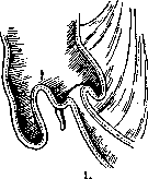
Fig.
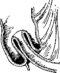
Figure 2.
is observed; subsequently, the number
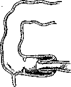
Figure 3.
causes play an undoubted role: tumors, polyps, inflammatory changes, infiltration of the wall, weakness of the sphincter in invaginatio ileo-caecalis, enlargement of glands, as well as hemorrhages into the intestinal wall, etc. In addition to these causes of a mechanical nature, spastic phenomena can also play a known role. In both cases, antiperistaltic movements, which exist in the large intestine as a physiological phenomenon, are of great importance. Predisposing moments for the occurrence of intussusception include a long mesentery poor in adipose tissue, a mobile caecum, a relatively large lumen of the ileocecal valve, and also a strong development of Peyer's patches. Among the precipitating factors of intussusception, one must point to polyps protruding into the intestinal lumen, submucosal lipomas, angiomas and fibromas, Meckel's diverticulum, local inflammatory processes of the intestinal wall, as well as spasms and pareses. Usually, the causes are multiple. In the case of the presence of mechanical factors, intussusception is easily explained by the insertion of the central section of the intestine into the peripheral one due to the movement of fecal masses. In the absence of precipitating mechanical factors, the occurrence of intussusception is much more difficult to explain. The experiments of Nothnagel and Propping significantly reinforced the spastic theory, pushing the paralytic theory into the background. These authors induced local spasm of the intestine by electrical stimulation or injection of spasmodic agents and obtained experimental intussusception in this way. According to Nothnagel, the insertion occurs due to the contraction of the longitudinal musculature of the intestinal segment bordering the spastically contracted section of the intestine, thanks to which the spastically contracted section is drawn into the intestine. According to Propping, the matter consists in the contraction not of the longitudinal, but of the circular musculature. The first impulse for intussusception is given by the simultaneous contraction of the circular musculature and the lengthening of the intestinal loop. Peristaltic waves then insert the contracted section of the intestine further. However, experimentally, Propping was not able to obtain long-lasting intussusceptions. From what has been said, it is clear that the mechanism of intussusception is not yet fully clear. With short insertions, the patency and
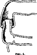
nutrition of the intestine are not affected at all or suffer relatively insignificantly. When a longer segment of the intestine is inserted, as a rule, there occurs a delay of intestinal contents, disturbances of blood circulation, and nutrition of the wall and mesentery of the inserted segment. Intussusception can often be observed as a banal phenomenon during autopsies of young children who died from any cause. This intussusception develops during agony. It is easily recognized due to the fact that stretching the intestine in length easily eliminates it—in these cases, the intussusception is not fixed. Such intussusceptions, of course, have no clinical significance. Usually, single and descending intussusceptions are observed, i.e., thanks to peristalsis, the intestine is inserted more and more in the direction of the anus; but there can be double intussusceptions, etc. (Figs. 5 and 6). Due to the disturbance of blood circulation from the compression of the mesentery during acute strangulation, gangrene of the inserted
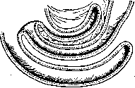
Figure 5.
intestine develops, with its subsequent detachment. In chronic intussusception, a gradual necrosis of the inserted intestine is usually observed. In both cases, a diffuse perforated peritonitis can easily develop. In the most frequent ileocecal form, the ileum, together with the Bauhin valve, is inserted into the large intestine, carrying with it the cecum and the vermiform appendix. In rarer forms, the ileum is inserted without the participation of the Bauhin valve. An even rarer form is that in which the insertion of the small intestine into the small intestine occurs, and a complete rarity is the intussusception of the large intestine into the small intestine. The pathological-anatomical picture is extremely different depending on the time and duration of the intussusception. The length of the intussusceptum also plays a role. With a short intussusceptum, the changes in the intestine consist of mild edema and punctate hemorrhages on the serosa and mucosa, which are discovered only after disinvagination. With a longer intussusceptum with the insertion of the corresponding
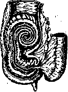
Figure 6.
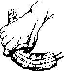
Figure 7. In the section of the mesentery, the phenomena of circulatory disturbance in the form of venous congestion, edema, hemorrhages, and thrombosis appear more sharply. However, in such cases, the patency of the intestinal canal may still be preserved. With continuing intussusception, all these phenomena intensify, ending in gangrene of the affected area. With a long intussusceptum, the indicated changes occur faster and more intensely. If intussusception develops slowly, then the slowly developing gangrene manages to become demarcated, adhesion of the intestine surrounding the intussusceptum with the ring of invagination occurs, and sometimes the intussusceptum becomes necrotic and can be expelled naturally through the intestinal lumen. This spontaneous cure is observed, however, very rarely and is possible only in chronic intussusception, mainly in adults. Usually, however, local or general peritonitis develops. Depending on the indicated conditions, the appearance of the intussusceptum during an autopsy of the abdominal cavity is different, ranging from barely definable changes to a picture of severe gangrene of the intestine. The symptomatology of intussusception is not complex and consists of phenomena of obstruction and the discharge of blood with mucus from the rectum. Pains in the form of very strong colic manifest as sharp cries, changes in the face, general appearance, and condition of the patient; vomiting is added to them. It is accepted to think that vomiting in intussusception of the small intestine occurs very early, and in intussusception of the large intestine—much later. Attacks of pain continue all the time with small intervals of calm. The passage of gas through the rectum ceases completely, and along with this, there is a discharge of blood with mucus. The temperature usually rises, but can also remain normal. Continuing vomiting sometimes acquires a fecal odor. Left to itself, the child dies on the 4th-6th day. —The most essential thing is early diagnosis. In acute cases, it is not difficult. Usually, amidst complete health, sharp pains in the abdomen suddenly occur, accompanied by cries, vomiting, urinary retention, and mucous-bloody stools; these data are sufficient for diagnosis and immediate intervention. Palpation of a sausage-shaped tumor is already a relatively late sign, and one should not wait for its presence. Peritoneal phenomena in acute intussusception develop with great rapidity, and even early operations within 12-24 hours still yield 20% mortality, which after 24 hours progressively rises to 75% and higher. In very rare cases, the invaginated intestine becomes necrotic and separates into the intestinal lumen, and previously formed adhesions prevent the development of perforated peritonitis. Palpation of the abdomen usually does not make it possible to determine intussusception. In other cases, and often already in the first days, a soft, cylindrical, somewhat curved tumor, moving from place to place, can be palpated in the abdominal cavity. Examination per rectum reveals blood in a greater or lesser amount. Per rectum, it is often easy to palpate the tumor of the intussusceptum. Radiography data do not help the diagnosis, and one should not resort to X-rays in acute cases. Thus, the most important signs of acute intussusception are sudden sharp pains, vomiting, mucous-bloody stools, absence of gas and stool, as well as the presence of a palpable tumor. In the differential diagnosis of acute intussusception, one should keep in mind acute intestinal catarrhs, as well as congenital strictures and atresias. The latter, however, are very rare. In subacute or chronically developing intussusceptions, the main symptom of more or less pronounced phenomena of chronic obstruction is the palpation of a sausage-shaped tumor, sometimes soft, sometimes harder and clearly demarcated. Therapeutic treatment, although yielding an effect in some cases, must be completely discarded (enemas under high pressure, under general anesthesia, with digital disimpaction of the tumor). According to Kocher's statistics, such treatment yielded incomplete reduction of intussusception in 15 cases, with intestinal perforation in one case, and severe collapse with a fatal outcome in three cases. Thus, attempts to reduce intussusception under anesthesia are dangerous, and one should not lose precious time, which is more valuable here than even in strangulated hernia. Surgical intervention is, of course, performed under general anesthesia (predominantly chloroform). The incision is below the navel, along the midline or pararectal—depending on the suspected site of intussusception. The most rational method is disinvagination, performed according to the method depicted in Figure 7. When reduction of intussusception has succeeded, it is best, in order to avoid its recurrence, to fix the invaginated part of the intestine to the parietal peritoneum. A minimum of time should be spent on these manipulations. Closure of the abdominal cavity must be performed with special care and securely, keeping in mind the tension of the abdominal wall after the operation, which is common when a child cries. It is best to pull the abdominal wall together with wide strips of adhesive plaster. Such disinvagination is the best method of treating intussusception. In 309 cases at the London Hospital, 78% recoveries were obtained with this method (the statistics cover the time from 1903 to 1920). If surgical intervention is applied late (after 24-36 hours) and disinvagination does not succeed, one has to resort to the operation of creating an anus with or without resection of the affected area, or to resection of the affected section with the establishment of intestinal patency. All these methods yield a huge percentage of mortality. The creation of an anus is especially hopeless. Resection with subsequent suturing of the intestine yields a somewhat better result. Such high mortality is caused mainly by the late time of operating and depends on intoxication, peritonitis, and surgical shock. Attempts to reduce the severity of the operation by resecting only the invaginated section of the intestine after simple incision of the invaginated intestine are, of course, simpler than total resection with anastomosis (Mansell-Moullin, Delore). The results of this intervention are so far doubtful. Thus, if disinvagination cannot be performed, then all other interventions appear in the vast majority of cases to be hopeless. In general, it can be said that for the first 24 hours, success can be expected from the intervention. In the following days, the possibility of cure drops sharply and progressively. Chronic intussusception is observed relatively rarely, usually at a later childhood age and mainly in adults. The cause in the vast majority of cases is benign tumors and polyps. Diagnosis comes down to the presence of a sausage-shaped tumor, phenomena of chronic obstruction, diarrhea, often with mucous content, sometimes with bloody content. The X-ray picture can give, unlike acute obstruction, a typical picture of intussusception, but only in intussusception of the large intestine. Treatment of chronic intussusception is surgical and consists of wide resection of the entire affected section. To reduce the time of the operation and reduce surgical shock, the Delore method can be used—resection of only the invaginated intestine through a longitudinal incision of the invaginating intestine. Lit.: Dieterichs M., On the theory of intussusception, Khirurgicheskiy Arkhiv Velyaminova, vol. XXX, book 1, 1914; Poteenko V., Chronic intussusception of the large intestine, Khirurgiya, vol. XXXI, 1912; Rufanov I., On the invagination of intestines, Ibid., vol. XXXV, 1914; Edington G., Intussusception, Brit. med. journal, v. II, 1921; Perrin W. a. Lindsay I., Intussusception, a monograph based on 400 cases, Brit. journ. of surgery, v. IX, 1921; Propping R., Uber den Mechanismus der Darminvagination, Mitteil. a. d. Grenzgeb. d. Medizin u. Chirurgie, B. XXI, 1920; Thompson W., Acute intussusception in children, Brit. med. journ., v. I, 1924.
Related articles
Cite this page
“Intussusception (a1159).” Soviet Medical Encyclopedia. English translation of Bolshaya Meditsinskaya Entsiklopediya, 1st ed. (Moscow, 1928–1936), ed. N. A. Semashko. https://sovietmedicalencyclopedia.pages.dev/article/intussusception-2/