Hirschsprung Disease
Historical document, translated for reference. It reflects medical knowledge of the 1920s–30s and is not medical advice.
Summary
Hirschsprung disease is a rare congenital condition characterized by idiopathic dilation of the colon with hypertrophy of the walls and persistent constipation. The disease was first identified as a distinct entity by Hirschsprung in 1888 and primarily affects children, with males being more commonly affected.
Encyclopedia article (1928–1936)
HIRSCHSPRUNG DISEASE (more correctly - Hirschsprung, Hirschsprung), megasigmoid, megacolon congenitum, idiopathic congenital dilation of a greater or lesser segment or even the entire large intestine, with hypertrophy of the walls and persistent constipation. Individual cases of this disease were occasionally described since the 17th century; in 1888, Hirschsprung identified it as an independent nosological unit. The dilation affects the colon, more often the sigmoid, rarely the rectum and cecum, and it is impossible to detect the presence of any mechanical obstacles, which distinguishes Hirschsprung disease from secondary dilatation of the large intestine in connection with some acquired obstacle (adhesions, scars, etc.). The disease is rare, most often observed in childhood (of 137 cases collected by Bogolyubov, in 91 cases it was observed before the age of 15). The pathogenesis has not yet been sufficiently clarified. Some (Hirschsprung and his supporters) consider the dilation and hypertrophy to be primary and classify H. d. as congenital developmental defects in the sense of partial gigantism, similar to pyloric hypertrophy; according to others, the dilation and hypertrophy are secondary - a consequence of congenital abnormalities in position, abnormal length of the descending colon or sigmoid colon, causing bends, folds and thereby stagnation of contents and dilation of the intestines (Marfan, Konjetzny, Neter, etc.). Explanations were sought in spasm of the anal sphincter (Fenwick) or spasm at the transition of the sigmoid to the rectum, in aplasia of the muscular layer of a certain segment of the large intestine with subsequent dilation in this place (Concetti). The dilation and hypertrophy are also explained by a violation of the innervation of the large intestine, leading to a kind of partial giant growth of it (Bruning - trauma to the sympathetic nerve, Ishikawa - violation of autonomic innervation of the longitudinal muscles, facilitating defecation). Cases of unmistakable complete intrauterine development of the disease without the presence of any mechanical causes, as well as combinations with other congenital defects - for example, absence of the uterus and vagina (Hartley), defect of the abdominal wall in a newborn (de Blasi), etc., have been observed. Cases of familial megacolon in a mother, three sons and a grandson (Romani), in two sisters (Bruening), in twins are known. In more pronounced cases, the disease reaches significant development by birth and manifests in the first days and months; in other cases, there is apparently only a predisposition to the disease in the form of excessive length, width or abnormal position of the large intestine, but without violation of functions (the disease develops imperceptibly, not manifesting itself in any way, and begins to manifest only after reaching significant development, when the formation of bends is possible due to the large size of the loop, twists, stagnation due to violation of compensation, etc.). Cases that do not manifest themselves until deep
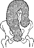
Figure 1.
old age are known. Excessive length (glossosolen) or abnormal position do not yet serve as an unconditional cause for the development of Hirschsprung disease. The dilated part of the intestine is often longer than normal (see Figure 1) and can reach enormous sizes: 16-20 cm in circumference in small children and up to 50-76 cm in adults. The walls are thickened to 0.25-0.5 and 1 cm in adults (normal - 0.15 cm) mainly due to the circular muscle layer; the longitudinal muscles are weak or completely unnoticeable, more rarely expressed in the form of a thin continuous layer; haustra are not expressed. Thinned (secondary atrophy) sections of the intestine may be found among hypertrophied ones. There is not always a sharp boundary between the dilated and healthy, properly functioning part of the intestine. The walls of the intestines are sometimes quite fragile, the mucous membrane is thickened, in a state of chronic inflammation, in long-lasting cases atrophic, with defects of the epithelium, pigmentation (traces of hemorrhages); ulcers, abscesses in the submucosal layer may be present on it. By the end of the disease, there may be perforations of ulcers with local or diffuse peritonitis. On microscopic examination, thickening of muscularis mucosae and especially the circular muscle with hypertrophy of individual fibers, inflammatory infiltration, dilation of vessels, edema, connective tissue growth of the submucosal layer were found. The dilated intestine contains huge sometimes (e.g. 14 kg in a 15-year-old girl) quantities of fecal masses of varying density; voluminous fecal concretions are common. The enlarged loop occupies a large part of the abdominal cavity, pushing backward-upward and compressing all abdominal organs and venous vessels, elevating the diaphragm, thereby displacing the lungs and heart (see Figure 2), which leads to increased respiration, tachycardia, occasionally - slowing of the pulse, cyanosis, edema. The lower opening of the chest cavity is dilated, the line of the costal margin is directed across. The disease affects the male sex more (78-80%). The disease manifests more often after birth (according to Lowenstein, in 55 out of 88 cases) or in the first years of life with persistent sluggishness of the intestine, gradual increase and
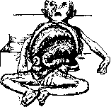
Figure 2. Megacolon congenitum (five-month-old child). (According to Pfaundler-Schlossmann.) protrusion of the abdomen due to meteorism and accumulation of fecal masses. The skin of the abdomen is thin, pale, with a developed venous collateral network, sometimes edematous. Through the abdominal coverings, the contours of the dilated intestine can be seen during peristalsis, and even peristalsis can be felt by hand. The increase in the abdomen reaches significant sizes (see Figures 3 and 4); in an adult, an abdominal circumference of 220 cm was observed. The stool is once
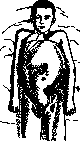
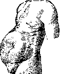
Figure 3.
Figure 4.
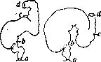
in 6-10 days, sometimes once every few weeks, even in 2-3-4 months. Cases of prolonged, up to 4 months, delay of stool without visible change in the general condition of patients have been described. The action of the intestine may remain periodically satisfactory, constipation is sometimes replaced by diarrhea. The intestinal loop at the site of enlargement and dilation forms rather sharp bends, more often at the border of the mobile sigmoid colon and the beginning of the immobile rectum (see Figures 5 and 6). The folds and valve-like folds of the mucous membrane formed by the intestinal loop can cause retention of gases released when the patient changes position. In children, a deeply inserted finger into the rectum can lift such a fold protruding into the lumen of the intestine, and the finger enters a large cavity, causing subsequent abundant laxation and exit of gases. Fissures of the anus with spasmodic contraction of the sphincter are observed, but very often the sphincter is freely passable. With prolonged retention of feces and gases in the final stage, there is vomiting, more often with bile, sometimes fecal (observed in a 14-day-old child). Appetite is impaired, especially at the end of the disease. In urine - indican, sometimes protein, cylinders. At first, children develop satisfactorily on breast feeding; with the development of the disease, growth slows down; nutrition falls, the skin gets a yellowish-white color, the limbs are delicate, thin, the eyes and cheeks sink in, the head is small. The general condition is depressed, headaches, at the end of the disease - apathy, drowsiness, cachexia, cerebral phenomena, nausea and other dyspeptic phenomena, pain in the abdomen. Abundant emptyings
Figures 5 and 6. a-b-rectum; b-c - II. sigmoidea; c-d - colon desc. (According to Perthes.)
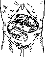
of the intestine temporarily improve the child's condition and general state. Patients die either from intoxication or from perforation or from aggravated obstruction. In small children, the course of the disease is usually more severe than in cases of later development of the disease in adolescents and adults. In typical cases, diagnosis is not difficult, but in atypical cases, correct diagnosis is not always easy. In small children, Hirschsprung disease is confused with the results of dietary errors, stenoses, atresias. Later, in the presence of fecal concretions, it is confused with various tumors of the abdominal cavity. In cases of development of the disease in adults, the history sometimes indicates sluggishness of the intestine in childhood, which increased with age, with a simultaneous increase in the abdomen and signs of chronic intoxication. Diagnosis is facilitated by: distension of the intestine, X-ray examination, proctoscopy, during which the proctoscope is inserted into the intestine without resistance, is mobile in all directions, as if in a large pot, and can be palpated through the abdominal wall (see Figure 7); the possibility of introducing an unusually large amount of water during an enema, sometimes it does not flow back; abundant laxatives. - The prognosis is unfavorable with rapid development of the disease in early childhood, more favorable with slow development and at a later age. - Conservative treatment usually does not eliminate the expansion and hypertrophy of the intestines, it only maintains relative well-being without significant harm to the development and nutrition of the body. It consists of symptomatic measures and diet. Children are recommended to be breastfed longer, as switching to artificial feeding intensifies the manifestations of the disease. Later, a light, mixed semi-liquid food that gives the smallest possible residue is prescribed. Thorough regular daily emptying of the intestine with simple, high and siphon enemas. Frequent use of a gas tube. Laxatives are less effective and often even harmful. Sometimes electrization in the form of electric enemas (not to be used in cases of intestinal ulcers and in weakened patients). In persistent constipation - stretching of the sphincter and in extreme cases manual emptying of the intestine. Mortality with conservative treatment is 88-93%. Surgical treatment is more effective - gives more than 50% complete cure. The most appropriate form of surgical treatment - resection of the affected section of the intestine, one-stage and two-stage, depending on the patient's condition and the conditions of the operation. With the development of technique, resection gives more and more success (mortality with one-stage is 28%, with two-stage - 8%, Monnier) and actually cures the patient. Less radical and less recommended (more often as a preliminary operation) is exclusion
Figure 7. Intestine palpated after a 7-week delay in bowel movement. (According to Luria.) of the diseased intestine by creating an anastomosis. In severe, neglected cases in weakened patients with severe intoxication, the creation of an anus praeternaturalis is resorted to for emptying the intestine and restoring the patient's strength, after which a radical operation is performed. Suturing the dilated intestine or its fixation gives no results. Surgical intervention must be timely, while the body has not yet been exhausted by the disease, can tolerate the operation and restore the lost strength.
Related articles
Cite this page
“Hirschsprung Disease.” Soviet Medical Encyclopedia. English translation of Bolshaya Meditsinskaya Entsiklopediya, 1st ed. (Moscow, 1928–1936), ed. N. A. Semashko. https://sovietmedicalencyclopedia.pages.dev/article/hirschsprung-disease/