Ortho
Historical document, translated for reference. It reflects medical knowledge of the 1920s–30s and is not medical advice.
Summary
Ortho is a prefix used in pathology with various meanings, such as in orthostatic albuminuria and orthognathism. Orthodiagraphy is a method for measuring the body by tracing and measuring the contours of its shadow when illuminated by parallel X-rays.
Encyclopedia article (1928–1936)
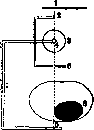
ORTHO (from Greek orthos- straight, correct), a prefix often used in pathology with various meanings. Thus, one speaks of orthostatic albuminuria, referring to the appearance of protein in urine when the body transitions from a lying to a standing position (straight); one speaks of orthognathism, referring to the straight position of teeth; when speaking of orthophoria, one imagines a completely correct (parallel) position of the visual lines of both eyes. See also the articles Orthography, Orthopedics. In organic chemistry, the prefix ortho- has a specific designation - see Organic Chemistry. ORTHO GRAPHY, more correctly - orthodiagraphy (from Greek orthos- straight, correct, dia- through and grapho- I write), a method of measuring the body by drawing and measuring the contours of its shadow obtained when illuminated by parallel X-rays. This is achieved as follows: when X-rays are passed through, the point of impact on the screen of the so-called central ray, i.e., that of the rays emanating from the anticathode which is strictly perpendicular to the plane of the screen, is marked in some way. For this purpose, a cross of thin wire or another object that gives on the screen as small as possible but a distinct shadow can be fixed immovably to the tube in the path of this ray. The tube on a corresponding stand must be movable in a plane strictly parallel to the plane of the immovably fixed screen. Then, on the screen, the shadow of the central ray marker is successively set at various points on the boundary of the shadow of the object being measured, and these points are marked on the screen. If, in this way, the contours of the shadow of the measured body are outlined by the shadow of the central ray marker, marking all points of the contour of the shadow on the screen, then an image of the silhouette of the measured body, obtained when illuminated by parallel rays, is obtained on the screen; this silhouette will in size exactly correspond to the dimensions of the body being studied in a plane parallel to the plane of the screen. By giving the object different positions in relation to the screen and each time drawing its shadow in the same way, this body can be measured in all planes. Instead of drawing the silhouette of the measured body on the screen, the screen can be fixed on the same movable stand that holds the tube, immovably to the tube and movably with it in planes strictly perpendicular to the central ray; on the screen, the point of impact of the central ray is marked in some way, or a small screen, 2-3 cm in diameter, is taken, the center of which exactly corresponds to the central ray. Paper is fixed immovably but in a plane strictly parallel to the plane of movement of the tube and screen on a frame (behind the tube or somewhere nearby), on which the contour of the shadow of the measured object is drawn with the help of a recording device fixed on the same movable frame of the stand to which the tube and screen are attached. This principle was first realized in a special instrument - the orthodiagraph according to Groedel (see Groedel's orthodiagraph scheme: 1-paper; 2-pencil; 3-X-ray tube; 4 and 5-central ray; 6-blinder; 7-stand; 8-heart, fig.). For drawing the contours of the shadow instead of a pen or pencil that pops out when pressing a balloon, one can use, by stretching paper in a frame, a needle (according to Schick) that pierces the paper when squeezing the balloon, or even better an electric spark jumping between two electrodes moving on both sides of the paper (according to Kharakhorkin). With the spark, the paper is burned through with small holes (possibility of recording orthodiagrams in several copies simultaneously). The Groedel orthodiagraph is most widely used. But if a recording device according to the principles just indicated is adapted to a movable stand of the 'clinoscope' type by Veifa or 'universal' by Siemens, then each such stand can be turned into an orthodiagraph that satisfies all requirements. The basic conditions for obtaining correct results with O. are as follows: 1) the plane of movement of the tube (and screen) must be a) strictly parallel to the screen and the plane on which the drawing takes place, and b) perpendicular to the central ray; 2) the patient must be immobilized in the position given to him. Non-compliance with these conditions leads to errors, and the method then not only loses its meaning but also causes harm, creating an illusion of precision where there is none. Orthodiagraphy was first implemented and developed by Moritz after corresponding efforts by Rosenfeld, Payne, and Levy-Dorn. It is used mainly for measuring the size of the heart, less frequently for determining the size of foreign bodies that have entered the body, very rarely for determining the size of the stomach (with it filled with contrast food), liver, spleen. In recent times, special attention is being paid to orthodiagraphic measurement of the aorta. The heart can be orthodiagraphed both in the standing position and in the sitting and lying position of the patient. Each position has its advantages and disadvantages. In the lying position, it is easier to immobilize the patient, but due to the elevation of the diaphragm, the heart changes its shape and its contours become less distinct, especially the apex; moreover, severe cardiac patients cannot always tolerate a prolonged lying position. At present, O. in the sitting position is most common.
To obtain a silhouette of an object that closely approaches its true dimensions, one can also use tele-radiography. This method has over O. the advantage of complete objectivity and does not require the researcher to remain on the rays for a relatively long time (2-3 min.). But for tele-radiography of the heart, powerful equipment, special devices and expenditure of photographic materials are necessary. Moreover, tele-radiography of the heart still gives some increase in its shadow and in oblique positions usually does not give accurate data due to the unclear boundaries of the heart shadow due to its fusion with the shadows of the chest wall. In tele-radiography of the heart in the dorso-ventral position, the boundaries of the cardiac apex are not obtained if they are located below the left dome of the diaphragm, as is often the case with a short chest cavity and high position of the diaphragm. In addition, a great advantage of measuring the size of the heart by O. is that the different arcs of the cardiac shadow can be precisely distinguished from each other by the difference in character, which is necessary for more detailed measurement of the orthodiagraphic silhouette of the heart. Therefore, orthodiagraphy for measuring the size of the heart and for accurately determining its configuration deserves preference in most cases over tele-radiography. It is believed that the errors in drawing the boundary of the cardiac shadow by O. in an experienced researcher do not exceed 3 mm.
Related articles
Mentioned in
Cite this page
“Ortho.” Soviet Medical Encyclopedia. English translation of Bolshaya Meditsinskaya Entsiklopediya, 1st ed. (Moscow, 1928–1936), ed. N. A. Semashko. https://sovietmedicalencyclopedia.pages.dev/article/ortho/