Exophthalmos
Historical document, translated for reference. It reflects medical knowledge of the 1920s–30s and is not medical advice.
Summary
This article defines exophthalmos as the protrusion of the eyeball, noting it as a symptom of various local and systemic conditions. It details diagnostic methods, including the use of exophthalmometers, and discusses the classification of causes ranging from orbital to systemic diseases like Basedow's disease.
Encyclopedia article (1928–1936)
EXOPHTHALMOS, or protrusio bulbi, the protrusion of the eye, or bug-eyedness, is a symptom of both general and local processes. The position of the eye in the orbit is determined by the relationship between it and the orbital cavity with all its contents; normally, the apex of the cornea is situated in the plane of the orbital opening, although prolonged inclination of the head downward produces a slight physiological exophthalmos. It is necessary to keep in mind that sometimes an eye or eyes appear protruding, whereas in reality they are in their proper place—this is false exophthalmos. Such a phenomenon is observed in paralysis of the eyelid muscles (lagophthalmos) and in high degrees of myopia, especially if it is unilateral, as well as in buphthalmos, where, by contrast with the size of the other (normal) eye, the impression of the presence of exophthalmos is created. According to Golovin's classification, all cases of exophthalmos are divided into three large groups: 1) E. depending on extraorbital causes, 2) E. from parietal causes (bony walls of the orbit), and 3) E. from orbital causes.
In true exophthalmos, there is a displacement of the entire eyeball forward—and consequently, of its point of rotation. The degree of forward displacement is quite variable and can reach 2 cm or even more, approaching the picture of a dislocation of the eye. In Figure 1, it can be seen that the edges of the eyelids already lie behind the equator, which is why blinking and voluntary closing of the eyes are impossible—and this is a significant complication.
For the purpose of measuring the degree of protrusion, one can use a ruler. In cases of unilateral exophthalmos, one can measure the distance from the outer edge of the orbit to the apex of the cornea with sufficient accuracy in millimeters.
For measurements, and especially for observing changes in the magnitude of the protrusion, special instruments (Cohn's, Hertel's, Birch-Hirschfeld's, and others), called ophthalmometers or exophthalmometers, are used.
The most convenient and simple of these is the Hertel exophthalmometer, in which the main parts are obliquely placed mirrors, which are applied by their bases to the outer edges of the orbits.
In these mirrors (Figure 2), for especially precise measurements, the photographic apparatus of Birch-Hirschfeld is used.
It is quite natural that in exophthalmos, which is only a symptom, the most thorough examination is necessary not only of the orbit but also of the adjacent air-bearing cavities of the cranial box with its contents, as well as of the entire organism, since the cause of exophthalmos, especially bilateral, often lies in general diseases or poisonings—Basedow's disease (see), diseases of the hematopoietic organs, etc.
When examining the orbit, one tries to penetrate as deeply as possible with the thinnest finger—the little finger—between its walls and the eyeball, at which point the question of the presence of unusual indurations and protrusions on its walls or in the depths is resolved. The presence of pulsation or vibration in this case should compel one to resort to auscultation of the eye, during which the noise of hydatids or blowing, buzzing noises in pulsating exophthalmos may be heard. By applying pressure to the eye through the eyelid, one determines, on the one hand, the presence of tenderness, and on the other, the possibility of a temporary reduction or even disappearance of the exophthalmos.
An increase in the contents of the muscular funnel produces protrusion of the eyes along the axis of the orbit, i.e., forward, outward, and downward (Figure 3); isolated enlargement of only one muscle produces displacement forward and to the side; the same is observed in lesions of the orbital wall or an air-bearing cavity (Figure 4). A uniform reduction of the orbit or a uniform increase of its contents produces direct exophthalmos, etc.
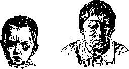
Certain diseases of the nervous system—brain tumors, hydrocephalus, increased intracranial pressure—can cause E., which in this case will be bilateral; tumors of the optic nerve or parts of the brain directly adjacent to the orbit can also cause E., but unilateral, and finally, irritation of the cervical section of the sympathetic system leads to the development of E.; this last fact is used by some authors to explain E. in general diseases: irritation of the sympathetic nerve is transmitted to Müller's muscle, increasing its contraction, which causes the displacement of the eye anteriorly (phenomena opposite to what is observed in enophthalmos).
The prognosis and treatment depend on the etiological factor.
Figure 1. Figure 2. Figure 3. Figure 4.
Related articles
Mentioned in
Cite this page
“Exophthalmos.” Soviet Medical Encyclopedia. English translation of Bolshaya Meditsinskaya Entsiklopediya, 1st ed. (Moscow, 1928–1936), ed. N. A. Semashko. https://sovietmedicalencyclopedia.pages.dev/article/exophthalmus/