FRONTAL BONE
Historical document, translated for reference. It reflects medical knowledge of the 1920s–30s and is not medical advice.
Summary
The frontal bone forms the anterior part of the cranial vault and base, and constitutes the upper wall of the orbit. It consists of the squama, nasal part, and two orbital parts, with frontal sinuses that develop during the first year of life.
Encyclopedia article (1928–1936)
FRONTAL BONE (os frontale) forms the anterior part of the vault of the skull and base, and at the same time constitutes the upper wall of the orbit. In it, the following parts are distinguished: the squama, nasal part, and two orbital parts. The squama (squama frontalis) is a bony plate extending at almost a right angle from the orbital parts and curved in the sagittal and frontal planes. Its uneven, serrated edge (margo parietalis) connects with the parietal bones by the coronal suture (sutura coronalis, s. fronto-parietalis) and in part with the sphenoid bone (sutura spheno-frontalis). The lower boundary is defined by the nasal part and the well-expressed supraorbital margin (margo supraorbitalis), which laterally passes into the zygomatic process (proc. zygomaticus os. front.), connecting by the suture (sutura zygomatico-frontalis) with the frontal process of the zygomatic bone. The anterior, convex surface of the squama (facies frontalis) is divided by a sagittally directed ridge into two parts (fig. 1). This ridge represents the remains of the frontal suture, which is usually closed
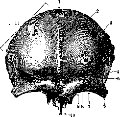
Figure 1. Frontal bone from the front: 1-squama; 2-facies front.; 3-tuber front.; 4-facies temp.; 5-linea temp.; 6-processus zygomat.; 7-margo supraorbit.; 8-foramen supraorbit.; 9-incisura front.; 10-spina front.; 12-margo parietalis. (After Spalteholz.)
at the second year of life, but in some cases remains open until 40-50 years, when all cranial sutures close. On either side of the ridge there is a smooth, protruding forward tubercle (tuber frontale), which is separated from the well-expressed ridge of the supraorbital arch (arcus superciliaris), located above the orbital margin, by a small depression. Between the inner
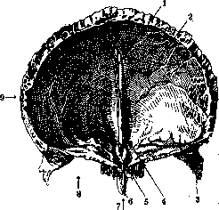
Figure 2. Frontal bone from behind: 1-sulcus sagitt.; 2-fies cerebralis; 3-proc. zygomat.; 4-crista front.; 5-for. caecum; 6-spina front.; 7-pars nasal.; 8-pars orbit.; 9-squama. (After Spalteholz.)
ends of these ridges there is a depressed area - the glabella (glabella, see); the outer ends extend laterally and slightly upward and gradually smooth out. The supraorbital margin at the boundary of the inner and middle thirds has a small notch, and sometimes an opening (incisura frontalis or foramen frontale), through which the vessels and nerve of the frontal region pass, and somewhat laterally - the supraorbital notch or opening (incisura supraorbit. or foramen supraorbit.) for vessels and nerves of the same name (fig. 2). From the outer edge of the zygomatic process, a rough, arched ridge (crista temporalis) extends, which, directing backward, divides into the superior and inferior temporal lines. Behind this line is the temporal surface of the frontal bone (facies temporalis), forming the upper-anterior part of the base of the temporal fossa (see Temporal region). The posterior (fig. 2), concave surface of the squama (facies cerebralis) has depressions from Pacchionian granulations in the form of fairly deep pits, and in the middle is divided by a groove (sulcus frontalis), which passes into the sagittal groove (sulcus sagittalis), intended
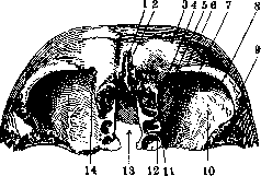
Figure 3. Frontal bone from below: 1-spina front.; 2-glabella; 3-arcus supercil.; 4-incisura front.; 5-spina trochl.; 6-for. supraorbit.; 7-margo supraorbit.; 8-fossa glandulae lacr.; 9-proc. zygomat.; 10-facies orbit.; 11-12-for. ethmoid, post, et ant.; 13-incisura ethmoid.; 14-fovea trochl. (After Spalteholz.)
for the superior sagittal sinus. In the lower part of the edge of this groove, rising, it forms a ridge (crista frontalis) for the process of the falx cerebri. This ridge reaches the ethmoidal notch and together with the alar processes of the ethmoid bone forms the blind foramen (foramen caecum), limiting it in front. The orbital parts (partes orbitales) (fig. 3) are divided by a deep notch (incisura ethmoidalis) and have an almost triangular shape, with their inner sides located parallel, and the lateral sides converging medially and backward. In front they are bounded by the supraorbital margin, on the sides they pass into the zygomatic processes, behind by a serrated edge they connect with the small wings of the sphenoid bone, and more laterally - with the frontal margin of the greater wings (sutura spheno-frontalis). The ethmoidal notch is filled by the ethmoid bone, the cribriform plate of which connects at the edge of the notch by the suture (sutura fronto-ethmoidalis), supplemented in front by the connection of the posterior free edge of the nasal part of the frontal bone with the cribriform plate. In the outer part of the edge of the notch there is a projection to which the lacrimal bone adheres, behind connecting with the lamina papyracea of the ethmoid bone. The grooves of the edge, combining with the grooves of the labyrinth, form two openings on each side - anterior and posterior (for. ethmoidale ant. et post.), for the anterior and posterior arteries, veins, and nerves of the ethmoid region and orbital branches of the spheno-palatine ganglion. The lower, orbital surface (facies orbitalis) is evenly concave, on the inner-anterior angle it has a depression (fovea trochlearis) and next to it a small projection (spina trochlearis), in which the trochlea of the superior oblique muscle of the eye is located. In the outer part, in the area of the zygomatic process, there is a depression for the lacrimal gland (fossa glandulae lacrimalis), which in front is bounded by the supraorbital margin. The nasal part (pars nasalis) is located between the orbital parts. Its lower serrated edge (margo nasalis) connects with the nasal bones and with the frontal process of the maxilla; in the middle part it has a projection (spina nasalis ossis frontalis), the anterior surface of which adheres to the nasal bones and in part to the frontal processes of the maxillae, and the posterior surface - to the rooster's comb (crista galli) and to the perpendicular plate of the ethmoid bone. By its posterior free edge, the nasal part participates in the formation of the ethmoidal notch, supplementing it in front (see above). Near the spina frontalis there are on each side openings (aperturae sinuum frontalis), leading into the paired frontal sinuses (sinus frontalis). The latter are located between the plates of the orbital part symmetrically in the area of both supraorbital arches. They are separated by a thin partition located in the sagittal direction and usually asymmetrically, as a result of which the right and left sinuses are expressed differently. On average, their capacity does not exceed 5 cm3; sometimes they have a honeycomb appearance, consisting of separate chambers communicating with each other and opening by a common opening (infundibulum) into the middle nasal passage. The shape of the sinuses and the limits of their extension vary greatly: laterally they can reach the zygomatic bone, and posteriorly - to the small wings of the sphenoid bone, being separated from the ethmoid cells only by a very thin bony plate. Their cavity is lined with mucous membrane, weakly fused with the bone, of the same structure as the mucous membrane of the nasal cavity, but thinner, smoother, and poorer in nerves and blood vessels. In its development, the frontal bone belongs to the membrane bones and represents a paired formation, and even in a newborn it consists of two parts, which fuse at the 2nd year of life, but sometimes remain unfused for a long period (see above); then a suture remains between them (sutura frontalis, s. metopica). Ossification occurs from two symmetric points appearing at the end of the 2nd month or at the beginning of the 3rd month of intrauterine life, located near the supraorbital margin; in addition there are accessory ossification points in the proc. zygomaticus. The frontal sinuses are formed at the 1st year of life.
Related articles
Mentioned in
Cite this page
“FRONTAL BONE.” Soviet Medical Encyclopedia. English translation of Bolshaya Meditsinskaya Entsiklopediya, 1st ed. (Moscow, 1928–1936), ed. N. A. Semashko. https://sovietmedicalencyclopedia.pages.dev/article/frontal-bone/