Dural Sinuses
Historical document, translated for reference. It reflects medical knowledge of the 1920s–30s and is not medical advice.
Summary
This article from the 1928–1936 Soviet medical encyclopedia describes the anatomy, connections, and clinical significance of the dural venous sinuses, including the superior sagittal sinus, cavernous sinus, and transverse sinus.
Encyclopedia article (1928–1936)
DURAL SINUSES (sinus durae matris), or venous sinuses, represent non-collapsible reservoirs, devoid of valves, and in most cases triangular in cross-section. They contain partitions, especially pronounced in the so-called cavernous sinus. The walls of the D. are formed by leaves of the dura mater's processes (falx), the inner surface of which is covered by intima, formed by a layer of elastic fibers lined with endothelium. Venous blood flows into the D. through the superficial and deep veins of the brain and meninges, through the veins of the skull bones (vv. diploicae, s. diploeticae) and emissary veins, through the veins of the orbit, the external coverings of the head and face. It is considered that blood from the sinuses drains via: 1) v. jugularis int., 2) emissary veins, 3) vv. diploicae and 4) vv. vertebrales. All D. are interconnected. The largest of them is the s. sagittalis, located between the leaves of the dura mater according to its attachment along the sagittal suture of the skull. Slightly deviating from the suture line to the right and widening posteriorly, the sinus extends from the foramen caecum to the protuberantia occipitalis interna, where it drains into the confluens sinuum (Fig. 1). Along its course there are bulges, the so-called lacunae laterales, the size of which varies considerably: from 1.1 to 3.3 cm. The lacunae are most pronounced in the posterior (occipital) part of the D. and especially in brachycephals, in whom the D. has a form that earned it the name "lacunar" (Walker). Lobular outgrowths of the arachnoid membrane, the so-called granulationes arachnoideales Pacchioni, project into the lacunae, sometimes almost completely filling them. The width of the D. also varies within the limits of 1.0 to 3.0 cm.
Usually, the more developed the s. sagittalis, the less pronounced the other venous D. In the area of the protuberantia occipitalis interna, the longitudinal D. drains into the transverse sinus, which is especially wide (mainly on the right side). The site of the D. fusion is called the confluens sinuum, s. torcular Herophili. The sinus rectus and sinus occipitalis also drain here. The former receives the v. magna Galeni (Fig. 2), located along the line of attachment of the falx cerebri to the tentorium cerebelli and connects with the sinus sagittalis inf., which runs along the free edge of the falx cerebri facing the corpus callosum. The sinus occipitalis, collecting around the circumference of the foramen magnum, then proceeds towards the confluens sinuum along the line of attachment of the falx cerebelli, s. processus falciformis minor. The sinus transversus on each side, from the area of the protuberantia occipit. int. (confluens sinuum), proceeds towards the asterion according to the attachment of the tentorium cerebelli along the transverse sulcus of the occipital bone. In the area of the asterion, in the mastoid part of the temporal bone, the sinus lies in the sigmoid sulcus and, changing direction, passes into the first dilated part of the v. jugularis int. (bulbus). The angle at which the sinus transversus passes into the sinus sigmoideus differs in different skull types: in brachycephals it approaches a straight line, in dolichocephals it is more than straight. The right sinus transversus is usually more developed than the left and the s. sagittalis drains entirely into it; into the left sinus transversus drain the sinus rectus and sinus occipitalis. The direction and position of the transverse sinus externally correspond to the sup. nuchal line and is quite constant along its course to the asterion; however, from the asterion to the for. jugulare (sinus sigmoideus) its position is very variable. These variations have significant practical importance in operations on the mastoid process, when in cases of the so-called sinus prehension there is a danger of injuring it. The degree of sinus prehension can be judged by the thickness of the mastoid process, determined by the position of the mastoid incisure: the thicker the proc. mastoideus, the further the sinus is located, and consequently the less danger of its damage. Along the lateral surfaces of the sella turcica, on both sides, between the superior orbital fissure and the apex of the temporal bone pyramid, lies the cavernous sinus (sinus cavernosus) of considerable width and indeterminate shape. Anteriorly and posteriorly, the cavernous sinuses are connected by transverse anastomoses (sinus intercavernosus anterior and posterior) and generally form a circular D. - sinus circularis (Ridley). Into it drain the v. ophtalmica (anastomoses with the plex. venosus pterygoideus), sinus alae parvae, s. spheno-parietalis, s. vena meningea media - anteriorly, and the sinus petrosus superior et inferior and veins coming from the plex. basilaris - posteriorly. Inside the D., tightly wrapped by it, lies the a. carotis int.; externally it is adjacent to the n. abducens, medially from which in the thickness of the sinus wall lies the n. trochlearis and even more medially the n. oculomotorius; in close proximity to it are the Gasserian nodes, the first branch (n. ophthalmicus) lying in immediate proximity; above, covering the sinus intercavernosus anterior, lies the optic chiasm. The presence of well-expressed anastomoses between the venous network of the head and face, on the one hand, and the sinuses, on the other, explains the ease of infection spreading to the D. This especially concerns the cavernous sinus, which is abundantly connected by anastomoses with the veins of the orbit (vv. ophthalmicae) and the face (v. facialis - v. angularis and plex. pterygoideus) (Fig. 3). Carbuncles of the upper lip are distinguished by the severity of the course of the process and high mortality precisely due to the proximity of the D., first of all the cavernous sinus, through the v. angularis - v. ophthalmica, connected with the v. facialis, by which the infection, spreading rapidly, leads to thrombosis of the D. and meningitis. To prevent the spread of infection in this case Roeder advises ligating the v. angularis. Thromboses and thrombophlebitis of the sinuses are rarely primary. In these cases they develop as a marantic thrombophlebitis on the ground of cardiac insufficiency.
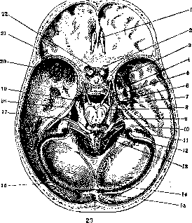
Figure 1. Topography of the sinuses on the base of the skull: 1-bulbus olfactorius; 2-n. opticus; 3-n. ophthalmicus; 4-n. maxillaris; 5-n. oculomotorius; 6-n. mandibularis; 7-n. trigeminus; 8-n. trochlearis; 9-n. acusticus; 10-n. glossopharyngeus; 11-n. vagus; 12-n. accessorius; 13-n. hypoglossus; 14-sinus occipitalis; 15-confluens sinuum; 16-sinus transversus; 17-sinus petrosus inf.; 18-sinus petrosus sup.; 19-n. abducens; 20-sinus cavernosus; 21-sinus alae parvae; 22-sinus intercavernosus ant.; 23-sinus sagittalis sup.
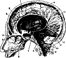
Figure 2. Sinuses on the sagittal section: 1-sinus sagittalis sup.; 2-sinus sagittalis inf.; 3-falx cerebri; 4-v. ethmoidalis post.; 5-v. ethmoidalis ant.; 6-branches of v. spheno-palatina; 7-plexus basilaris; 8-sinus petrosus inf.; 9-plexus venosi vertebrates interni; 10-sinus occipitalis; 11-sinus transversus; 12-sinus rectus; 13-v. cerebri magna (Galeni); 14-vv. meningeae mediae.
and even more medially the n. oculomotorius; in close proximity to it are the Gasserian nodes, the first branch (n. ophthalmicus) lying in immediate proximity; above, covering the sinus intercavernosus anterior, lies the optic chiasm. The presence of well-expressed anastomoses between the venous network of the head and face, on the one hand, and the sinuses, on the other, explains the ease of infection spreading to the D. This especially concerns the cavernous sinus, which is abundantly connected by anastomoses with the veins of the orbit (vv. ophthalmicae) and the face (v. facialis - v. angularis and plex. pterygoideus) (Fig. 3). Carbuncles of the upper lip are distinguished by the severity of the course of the process and high mortality precisely due to the proximity of the D., first of all the cavernous sinus, through the v. angularis - v. ophthalmica, connected with the v. facialis, by which the infection, spreading rapidly, leads to thrombosis of the D. and meningitis. To prevent the spread of infection in this case Roeder advises ligating the v. angularis. Thromboses and thrombophlebitis of the sinuses are rarely primary. In these cases they develop as a marantic thrombophlebitis on the ground of cardiac insufficiency.
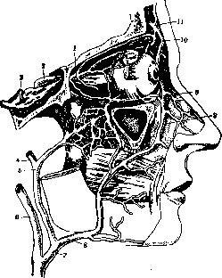
Figure 3. Anastomoses of the veins of the orbit, face and neck (partly according to Henle): 1-v. ophtalmica; 2-n. opticus; 3-sinus cavernosus; 4-v. temporalis superficialis; 5-v. facialis post.; 6-v. jugularis int.; 7-v. facialis communis; 8-v. facialis ant.; 9-anastomosis of v. ophtalmica inf. with plexus pterygoideus; 10-v. angularis; 11-v. naso-frontalis.
(see Brain, special pathology). Secondary thrombosis is more often observed as a consequence of bone disease during purulent processes of the middle and internal ear—thrombosis of the sigmoid sinus and jugular vein; in most cases the process is complicated by extradural suppuration, limited meningitis, abscesses in the temporal lobe of the brain or in the cerebellum. Otogenic thrombosis of the sinus can develop during acute and chronic suppuration of the middle ear, when pus, after destruction of the bony substance of the mastoid process, penetrates to the posterior cranial fossa and reaches the outer wall of the sigmoid sinus. As an exception, thrombi may form in small bone veins (osteophlebitis of Celsus) and from there spread to the sinus. A thrombus in the dural sinuses is usually wall-like and only upon further enlargement reaches the opposite wall and closes the lumen of the sinus (occluding thrombus). Usually, due to the ingress of infection from the primary focus, the thrombus undergoes purulent decomposition. The spread of the thrombus is possible both in the direction of the heart and towards the brain. Reaching the bulb of the jugular vein, the thrombus can penetrate to the subclavian vein. Spreading in the opposite direction and occluding the superior and inferior petrosal sinuses, the thrombus grows further into the cavernous sinus, and from there can infect the ophthalmic vein and the cavernous sinus of the other side (circular sinus); spread is also possible along the transverse sinus, where the thrombus reaches the torcular Herophili and can cross to the other side. In addition, transition of infection is possible from the floor of the tympanic cavity to the bulb of the jugular vein, from the anterior wall of the tympanic cavity to the carotic plexus, from the labyrinth to the superior and inferior petrosal sinuses and the bulb of the jugular vein, and from there into the sigmoid sinus. As a result of the decomposition of infectious thrombi, pyemia and metastases are observed in the lungs, joints, muscles, subcutaneous tissue, eye, brain, kidneys, and heart valves. Infected thrombosis of the dural sinuses proceeds clinically with general signs of sepsis. Among local symptoms of sigmoid sinus thrombosis, Gerhardt's symptom (see) and Grissinger's symptom (see) are observed; if the thrombus penetrates into the internal jugular vein, a painful cord is palpable at the inner edge of the sternocleidomastoid muscle; during thrombosis of the bulb of the jugular vein, sometimes due to compression in the jugular fossa (foramen lacerum posterius) phenomena from the vagus, glossopharyngeal and accessory nerves are observed. During cavernous sinus thrombosis, swelling of the eyelids, orbital cellulitis, optic neuritis are noted, and sometimes the abducens, oculomotor, trochlear nerves and the first branch of the trigeminal nerve are involved with corresponding manifestations. Treatment of dural sinus thromboses is only operative and consists in opening the sinus and removing the thrombus; with the spread of the process the prognosis is very unfavorable. Operative technique has been developed and is successfully carried out during thrombosis of the sigmoid sinus (methods of Grunert, Vass, Piffle, Ivanov, etc.). Intervention on the cavernous sinus appears extremely difficult from a technical standpoint. Vass's attempt to open the thrombosed cavernous sinus remained unsuccessful.
Related articles
Mentioned in
Cite this page
“Dural Sinuses.” Soviet Medical Encyclopedia. English translation of Bolshaya Meditsinskaya Entsiklopediya, 1st ed. (Moscow, 1928–1936), ed. N. A. Semashko. https://sovietmedicalencyclopedia.pages.dev/article/dural-sinuses/