Mastoid Process
Historical document, translated for reference. It reflects medical knowledge of the 1920s–30s and is not medical advice.
Summary
The mastoid process is a part of the temporal bone located behind the external auditory canal. It contains air-filled cells that develop in early childhood and varies between pneumatic and sclerotic types in adults.
Encyclopedia article (1928–1936)
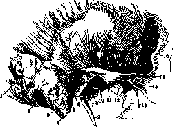
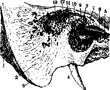
Mastoid process, processus mastoideus, part of the temporal bone, located behind the external auditory canal, behind the squamo-mastoid suture, and connecting with the squamous and tympanic parts of the temporal bone. The upper edge of the mastoid process connects with the parietal bone, the posterior edge with the occipital bone (fig. 1). The outer surface of the mastoid process is convex and rough, as five muscles attach to it here: mm. splenius capitis, sternocleidomastoideus, occipitalis, auricularis post., complexus min. The inner surface is concave and smooth. Here there is a wide curved groove for the sigmoid sinus. On the outer surface there are several small openings (foramina mastoidea), the number, shape, and direction of which are variable. They open on the inner surface in the groove of the sinus. These are emissaries through which the veins of the outer part of the skull communicate with the intracranial veins and sinus. From the medial part, the mastoid process is surrounded by a deep incision (incisura mastoidea), where the posterior belly of the digastric muscle begins and passes. Slightly behind the incisura mastoidea and parallel to it is the groove for the occipital artery. Between the mastoid process and the squamous part of the temporal bone is the parietal incisure, into which the mastoid part of the occipital bone extends for its connection with the temporal bone. The apex of the mastoid process is directed downward. The mastoid process appears only in the first years of life. From the end of the first year of life, air-filled bone cells (cellulae mastoideae) appear and develop in it, parallel with the formation of
Figure 1. External view of the temporal bone and mastoid process: 1-occipito-mastoid suture. 2-groove for m. biventer; 3-cells of the mastoid process; 4-mastoid process; 5-tympanic cavity; 6-spina suprameatum; 7-cavum tympani; 8 and 9-Eustachian tube; 10-styloid process; 11-fossa mandibularis; 12-nasopharyngeal opening of the Eustachian tube; 13-lateral plate of the pterygoid process; 14-articular tubercle; 15-proc. zygomaticus; 16-greater wing of the sphenoid bone.
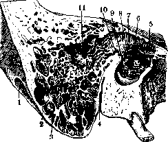
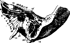
Figure 2. Pneumatic type of mastoid process: 1-groove for m. biventer; 2-wall of the mastoid process; 3-cellulae mastoideae; 4-canal for n. facialis; 5-m. cochleariformis; 6-oval window; 7 and 8-canal of n. facialis; 9-pyramid; 10-aditus ad antrum; 11-antrum.
several small openings (foramina mastoidea), the number, shape, and direction of which are variable. They open on the inner surface in the groove of the sinus. These are emissaries through which the veins of the outer part of the skull communicate with the intracranial veins and sinus. From the medial part, the mastoid process is surrounded by a deep incision (incisura mastoidea), where the posterior belly of the digastric muscle begins and passes. Slightly behind the incisura mastoidea and parallel to it is the groove for the occipital artery. Between the mastoid process and the squamous part of the temporal bone is the parietal incisure, into which the mastoid part of the occipital bone extends for its connection with the temporal bone. The apex of the mastoid process is directed downward. The mastoid process appears only in the first years of life. From the end of the first year of life, air-filled bone cells (cellulae mastoideae) appear and develop in it, parallel with the formation of
Figure 3. Transitional pneumatic type of mastoid process: 1-groove for m. digastricus; 2-wall of the mastoid process; 3 and 6-diploe (spongy bone tissue); 4-canal for n. facialis; 5-proc. styloideus; 7-Eustachian tube; 8-promontorium; 9-cochleariform process; 10 and 11-canal of facial nerve; 12-pyramid; 13-entrance to the antrum and prominence of the lateral semicircular canal; 14-antrum.
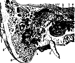
the process are filled with air bone cells (cellulae mastoideae). In adults, two types of mastoid process are distinguished: pneumatic and sclerotic (fig. 2-4). The first is rich in cells, lined with mucous membrane of the same structure as the mucous membrane of the tympanic cavity. The cells communicate with each other, and one of them, somewhat larger, located forward and upward (antrum mastoideum, antrum tympanicum), communicates with the epitympanic recess of the tympanic cavity through a wide opening (aditus ad antrum tympanicum). The sclerotic (senile) type usually does not contain cells and consists of compact bone mass. The cells of the mastoid process are arranged in a fan around the tympanic antrum; most of them are located below and outside. While the mastoid antrum is already present at birth, the mastoid cells begin to develop only at the end of the first year of life, so it is quite correct to believe that the mastoid antrum really represents a part of the tympanic cavity, and the mastoid cells are only an appendage of the tympanic cavity. The cells lie very close to the transverse sinus and to the canal of the facial nerve. - It is important for the otologist and practical surgeon to remember that during trepanation of the mastoid process, one must chisel the bone only within the limits of the triangle of Schiff, the sides of which are formed from above by the temporal line, continuation of the root of the zygomatic process,
Figure 4. Transitional sclerotic type of mastoid process: 1-groove for m. digastricus; 2-wall of the mastoid process; 3-diploe; 4-cellulae; 5-styloid process; 6-Eustachian tube; 7-proc. cochleariformis; 8-promontorium; 9-oval window; 10 and 11-canal of n. facialis; 12-pyramid; 13-entrance to the antrum and prominence of the lateral semicircular canal; 14-antrum.
from behind by a perpendicular dropped from this line to the apex of the mastoid process (this line often coincides with the mastoid crest), and from front by a line connecting the apex of the process and the spina suprameatum (see Mastoiditis). Going upward from the temporal line, one can damage the dura mater and enter the middle cranial fossa, from behind-damage the sinus and at the same time cause significant bleeding, and subsequently-phlebitis of the sinus, thrombophlebitis, general
Figure 5. Sclerotic type of mastoid process: 1-groove for m. digastricus; 2-sclerotic tissue; 3-wall of the mastoid process; 4-styloid process; 5-Eustachian tube; 6 and 7-proc. cochleariformis; 8 and 9-canal of n. facialis; 10 and 11-entrance to the antrum; 12-pyramid; 13-antrum.
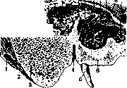
sepsis and very rapid death of the patient; from front-damage to the facial nerve with subsequent paralysis of the muscles of the corresponding half of the face. It is exceptionally important for the surgeon to know in advance what type of mastoid process he has de- Figure 8. Relationship between the tympanic antrum and the transverse sinus, protruding far laterally and occupying almost the entire anterior part of the mastoid part of the temporal bone, and with complete absence of mastoid cells: 1-occipito-mastoid suture; 2-sinus transversus; 3-groove for m. biventer; 4-mastoid process; 5-bony layer 1 mm thick; 6-external auditory opening; 7-spina suprameatum; 8-antrum tympani; 9-parieto-mastoid suture; 10-bony layer 1 mm thick, lo. The danger of injuring the sinus is especially great in case of its protrusion. The degree of protrusion can be judged by the thickness of the mastoid process, determined by the position of the incisura mastoidea: the
Figure 9. Relationship between the mastoid cells and tympanic antrum on one side and the transverse sinus on the other, with strong development of mastoid cells and moderate depth of sulcus transv. The mastoid cells are exposed by removal of the lateral wall: 1-occipito-mastoid suture; 2-projection of sinus transversi; 3-groove for m. biventer; 4-mastoid cells; 5-external auditory opening; 6-spina suprameatum; 7-antrum tympanicum; 8-mastoid-parietal suture. thicker the mastoid process, the farther the sinus is located, the less, consequently, the danger of its damage. The canal of the facial nerve is bent or
6
Figure 10. Mastoid part of the right temporal bone with strongly developed antrum tympanicum and cellulae mastoideae. The entire mastoid part is occupied by cells, leaving only a thin, paper-like plate; the cells are separated from the sinus transversus only by a very thin bony layer: 1-entrance to the tympanic cavity.
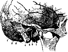
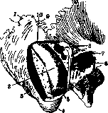
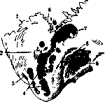
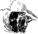
at a large angle or at an angle approaching a right angle. In the first case, when the canal has a more oblique course, its wall is very thin and easily traumatized. The shape of the triangle of Schiff somewhat guides: a triangle with a small height corresponds to the cellular type of pneumatization, and the angle of the facial nerve canal approaches a right angle all the more, the smaller the height of the triangle and the larger its base. The posterior side of the triangle corresponds to the position of the sinus, the upper side to the temporal lobe of the brain, the anterior side to the facial nerve canal (fig. 4-10). So-called 'dangerous' temporal bones are known. They are observed on the right side significantly more often than on the left, and the mastoid process in these cases is poorly developed ('infantile'). Since in children the sigmoid sinus is usually separated from the outer surface only by a thin bone layer, such cases during operations require the greatest caution, as the chisel can easily pierce the lateral wall of the sinus and open it. The blood supply to the mastoid process occurs from the a. meningea med. and a. stylo-mastoidea. The veins flow into the sinus petrosus superior and the sinus sigmoideus. The innervation of the mastoid process consists exclusively of sensory nerves and sympathetic nerves of the same origin as the nerves of the tympanic cavity (see Middle ear). The lymphatic vessels are little studied. Operations in the area of the mastoid process: 1) mastoidectomy - antrumoperation, 2) radical operation of Schwarz-Stucke, 3) opening of the sinus throughout the entire length of bulbi jugularis.
p. shuf'yan.
Related articles
Mentioned in
Cite this page
“Mastoid Process.” Soviet Medical Encyclopedia. English translation of Bolshaya Meditsinskaya Entsiklopediya, 1st ed. (Moscow, 1928–1936), ed. N. A. Semashko. https://sovietmedicalencyclopedia.pages.dev/article/mastoid-process/