Mastoiditis
Historical document, translated for reference. It reflects medical knowledge of the 1920s–30s and is not medical advice.
Summary
Mastoiditis is a complication of purulent otitis media, characterized by the spread of inflammation to the mastoid process with pus accumulation in its cells, often accompanied by bone destruction. The article discusses etiology, pathologic anatomy, and clinical manifestations of this condition.
Encyclopedia article (1928–1936)
MASTOIDITIS (mastoiditis), a complication of purulent otitis media, caused by the spread of the inflammatory process to the mastoid process and consisting of the accumulation of pus in its cells, and often accompanied by the destruction of the bone itself. Since the cells of the mastoid process are lined with mucous membrane, which represents a direct continuation of the mucous membrane of the middle ear cavity, with any inflammatory disease of the middle ear, the cells of the mastoid process are involved to a greater or lesser degree in the process. This involvement of the cells in the process is already manifested in the early stages of otitis in the form of tenderness of the mastoid process on pressure. Therefore, one can speak of M. only from the moment when symptoms appear indicating that the inflammation of these cells has developed into an independent disease. Etiology. The causative agents of the inflammatory process found in M. in the mastoid process are the same as in acute otitis media. At present, there is a tendency to believe that the type of causative agent is not important for the occurrence of M., but affects its further course and complications. A number of studies on the causative agents of M. have given rather uniform results. If one takes the average of the figures obtained by different researchers, it turns out that about 80% of all acute otitis media are caused by various types of streptococcus; the most virulent appears to be Streptococcus mucosus. The main factors that determine that in some cases the accompanying otitis inflammation of the mucous membrane of the mastoid process cells passes without a trace, while in others it leads to a clearly expressed independent disease, can be divided into general and local. Among the general conditions, constitutional peculiarities of the organism and decreased general resistance of tissues (after infectious diseases, in diabetes, etc.) play the main role. A large role in the occurrence of M. also belongs to the primary disease, of which otitis is a complication, which in turn led to M. Thus, it has been noted that otitis after measles, scarlet fever are particularly often complicated by mastoiditis. Among local conditions, the structure of the mastoid process is of primary importance, namely the degree of its pneumatization. The factor put forward in recent years by Wittmaack's theory of the local constitution of the middle ear mucous membrane also plays a large role here. According to this theory, the biological properties of the middle ear of each individual are determined by the course of the pneumatization process (the formation of air-containing cells of the mastoid process in the very first periods of extra-uterine life). Any delay or disruption of the normal course of this process deeply affects the future resistance of the middle ear mucous membrane, which in turn plays a primary role in the etiology of all pathological processes occurring here throughout later life. Pathological anatomy. Already at the very beginning of acute otitis, exudate accumulates in the cells of the mastoid process. Since this secretion does not have sufficient drainage through the narrow, obstructed by swollen mucous membrane communication of these cells with antrum, in each cell it will be under more or less high pressure, i.e., each cell will represent a small "empyema". This stage, which can be called exudative (Levin), is characterized by the fact that the process is concentrated in the mucoperiosteal covering of the cells; no noticeable changes in the bone are observed either in the walls of the cells or in the bone marrow cavities (empyema; Levin). In the vast majority of cases, the exudative stage passes without a trace, and the process in the mastoid process heals. In the presence of the above-mentioned unfavorable etiological moments, phenomena of bone destruction occur, usually combined with the process of "apposition" (new formation of bone); since the latter lags far behind the former, whole groups of cells merge with each other due to the decay of the partitions between them, forming a "bone abscess". The melting of the bone gradually reaches the cortical layer, which under the pressure of pus can rupture, forming a bone fistula, through which pus spreads under the periosteum. Such ruptures of the bone with the formation of pus collections between the bone and periosteum (subperiosteal abscess) can occur at any point on the surface of the mastoid process. However, depending on local conditions, ruptures occur much more frequently in certain places. Such a point is first of all the fossa mastoidea, corresponding to the location of the largest reservoir filled with pus (i.e., antrum mast.) and penetrated by many vascular openings. The second, although not so frequent, but practically very important place of rupture is the inner wall of the apex of the mastoid process, where for rupture there are also relatively favorable conditions in the form of the thinness of this wall and the close proximity of large ("terminal") cells. The pus that has ruptured flows down into the neck area and accumulates under the muscles located here (sterno-cleido-mastoideus, splenius and longissimus capitis), forming an infiltrate, always very hard to the touch and not giving fluctuation ("Bezold's mastoiditis"). Since rupture can occur wherever there are pneumatic cells, and since the latter, as is known, can extend far beyond the limits of the mastoid process (e.g., upward and forward into the root of proc. zygomat.), subperiosteal abscesses can also be observed in these places (zygomatitis). - There is no definite period between the beginning of acute otitis and M. In some cases, M. 87' occurs almost simultaneously with acute otitis, in others - very late, months after the beginning of otitis, when all its symptoms have already passed. Apparently these extremes are explained by the peculiarities of epidemics and the causative agent. The most common time for the onset of M. should still be considered the 3-5th week from the beginning of otitis.
The main points determining that in some cases the inflammation of the mastoid process cells accompanying otitis passes without a trace, while in others it leads to a clearly expressed independent disease, can be divided into general and local. Among the general conditions, constitutional peculiarities of the organism and decreased general resistance of tissues (after infectious diseases, in diabetes, etc.) play the main role. A large role in the occurrence of M. also belongs to the primary disease, of which otitis is a complication, which in turn led to M. Thus, it has been noted that otitis after measles, scarlet fever are particularly often complicated by mastoiditis. Among local conditions, the structure of the mastoid process is of primary importance, namely the degree of its pneumatization. The factor put forward in recent years by Wittmaack's theory of the local constitution of the middle ear mucous membrane also plays a large role here. According to this theory, the biological properties of the middle ear of each individual are determined by the course of the pneumatization process (the formation of air-containing cells of the mastoid process in the very first periods of extra-uterine life). Any delay or disruption of the normal course of this process deeply affects the future resistance of the middle ear mucous membrane, which in turn plays a primary role in the etiology of all pathological processes occurring here throughout later life. Pathological anatomy. Already at the very beginning of acute otitis, exudate accumulates in the cells of the mastoid process. Since this secretion does not have sufficient drainage through the narrow, obstructed by swollen mucous membrane communication of these cells with antrum, in each cell it will be under more or less high pressure, i.e., each cell will represent a small "empyema". This stage, which can be called exudative (Levin), is characterized by the fact that the process is concentrated in the mucoperiosteal covering of the cells; no noticeable changes in the bone are observed either in the walls of the cells or in the bone marrow cavities (empyema; Levin). In the vast majority of cases, the exudative stage passes without a trace, and the process in the mastoid process heals. In the presence of the above-mentioned unfavorable etiological moments, phenomena of bone destruction occur, usually combined with the process of "apposition" (new formation of bone); since the latter lags far behind the former, whole groups of cells merge with each other due to the decay of the partitions between them, forming a "bone abscess". The melting of the bone gradually reaches the cortical layer, which under the pressure of pus can rupture, forming a bone fistula, through which pus spreads under the periosteum. Such ruptures of the bone with the formation of pus collections between the bone and periosteum (subperiosteal abscess) can occur at any point on the surface of the mastoid process. However, depending on local conditions, ruptures occur much more frequently in certain places. Such a point is first of all the fossa mastoidea, corresponding to the location of the largest reservoir filled with pus (i.e., antrum mast.) and penetrated by many vascular openings. The second, although not so frequent, but practically very important place of rupture is the inner wall of the apex of the mastoid process, where for rupture there are also relatively favorable conditions in the form of the thinness of this wall and the close proximity of large ("terminal") cells. The pus that has ruptured flows down into the neck area and accumulates under the muscles located here (sterno-cleido-mastoideus, splenius and longissimus capitis), forming an infiltrate, always very hard to the touch and not giving fluctuation ("Bezold's mastoiditis"). Since rupture can occur wherever there are pneumatic cells, and since the latter, as is known, can extend far beyond the limits of the mastoid process (e.g., upward and forward into the root of proc. zygomat.), subperiosteal abscesses can also be observed in these places (zygomatitis). - There is no definite period between the beginning of acute otitis and M. In some cases, M. occurs almost simultaneously with acute otitis, in others - very late, months after the beginning of otitis, when all its symptoms have already passed. Apparently these extremes are explained by the peculiarities of epidemics and the causative agent. The most common time for the onset of M. should still be considered the 3-5th week from the beginning of otitis.
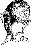
Symptomatology. I. Objective symptoms. 1. The subperiosteal abscess should be placed first in diagnostic value. It appears as a greater or lesser, often occupying the entire surface of the mastoid process, swelling, fluctuating and often covered with red shiny skin. In markedly expressed cases, the subperiosteal abscess pushes the auricle forward and downward (Fig. 1), causing it to stand more or less far from the mastoid process. In less marked cases, this is manifested as a smoothing of the line of attachment of the auricle. This symptom leaves no doubt as to the presence of M. 2. More or less sharply expressed swelling and infiltration of the outer coverings of the mastoid process without fluctuation, serving as an expression of periostitis, by itself is not so conclusive for M., as the subperiosteal abscess, because such periostitis is possible without the presence of M., and as an independent stage in the course of acute otitis. Therefore, this symptom acquires diagnostic value only in combination with other symptoms or when conservative treatment for 2-3 days gives no improvement. The same sign takes on a different meaning when the swelling and infiltration are located on the posterior-superior wall of the auditory canal, causing a slit-like narrowing of the latter. This sign is considered pathognomonic for M.-Of the other objective signs of M., it is necessary to point to the long and persistently profuse purulent discharge from the tympanic cavity. 3. Fever is an important, but not constant symptom of M. An increase in t°, continuing for some time after paracentesis or spontaneous perforation of the eardrum, should give serious suspicion of a complication from the mastoid process. Usually t° in M. remains in adults within 38-38.5°, in small children it can reach up to 40°. However, it must be remembered that M. often occurs without an increase in t°. II. Subjective symptoms. 1. Pain in the mastoid process on percussion and pressure has significance only when they are constant, gradually increase and are felt on pressure over the entire mastoid process, and not only over its apex. Spontaneous pains have much greater significance, already indicating a significant development of the process. They can reach a high degree, causing serious suffering to the patient and causing a noticeable deterioration of his general condition. 2. Special significance is attached to the general condition of the patient. The general poor appearance, loss of appetite, general malaise, drowsiness, etc., beyond any other symptoms, should arouse suspicion of a complication of otitis. Diagnosis. In cases with typical and clearly expressed signs, recognition of M. presents no difficulty, but there are many cases where the clinical symptoms are not typical and partly entirely absent, partly expressed extremely weakly. The diagnosis here can long remain doubtful even for an experienced otiatrist. The presence of a fluctuating subperiosteal abscess makes the diagnosis of M. unquestionable. Similarly, cases where there is free drainage of pus with an increase in t° and general malaise, and the patient complains of pain in the mastoid process, increasing on pressure, where the coverings of the mastoid process are more or less swollen and infiltrated, and finally where all measures of conservative treatment give no improvement, should not cause special difficulties. The diagnosis of M. is made immediately and in the presence of a slit-like narrowing of the auditory canal due to swelling of its posterior-superior wall. In doubtful cases, percussion tone auscultation on the diseased mastoid process and comparison with the tone on the corresponding place of the healthy side can help: on the diseased side, absolute dullness is often obtained, or the patient himself gets a subjective sensation of dullness of the tone on the diseased side. Important auxiliary significance also belongs to X-ray examination of the mastoid process, especially if upon its repeated production it is possible to establish an increase in changes on the X-ray. Prevention. Since M. is always a complication of acute otitis, preventive measures of both social and individual character should be the same as against the main disease of the middle ear. There are a number of factors contributing to the occurrence of M. in the presence of acute otitis, against which struggle is powerless. It is difficult, for example, to influence the general or local decreased resistance of the organism, on the peculiarities of this or that epidemic, on unfavorable anatomical conditions in a given individual, etc. There is no doubt, however, that in a whole series of cases it is possible to prevent the occurrence of acute M., provided that all clinical and bacteriological peculiarities of the given case are taken into account, and the main role is played by the correct treatment of acute otitis (timely performance of paracentesis, etc.). Treatment. 1. Conservative treatment is permissible only in the initial stages of M. and in doubtful cases where the diagnosis has not yet been finally established. Besides the correct treatment of acute otitis, it consists in strict bed rest and in eliminating any harmful influences. Locally-continuous application of ice to the mastoid process. 2. Operative treatment. There are cases where indications for operation can be established immediately, without any hesitation, namely: 1. Cases where during acute otitis symptoms appear that give serious suspicion of complications from the cranial cavities or general infection. Here the operation must be performed immediately. 2. Cases with a clearly expressed fluctuating subperiosteal abscess. In all such cases, indications for operation should be considered absolute. But even more numerous are those cases of M. where indications for operation are not so indisputable and where among otiatrists there are still disagreements. These indications can be called relative. Operation is considered indicated in principle in all cases where the diagnosis of M. is established with greater or less certainty, especially when symptoms appear approximately on the 3rd-4th week of acute otitis, when there are all reasons to believe that the bone process inside the mastoid process has gone so far that there is little chance of spontaneous healing. But operation may also be indicated where, although there is no actual M., the symptoms are so violent and sharp that they indicate a lightning-fast infection threatening more serious complications. If, for example, even in the first days of otitis, exhausting pains of the patient appear, if the general condition is unsatisfactory, if t° reaches 39-40°, then the question of whether M. is already present or not recedes into the background. The greatest doubts and fluctuations are caused by those cases where the diagnosis of M. is doubtful, where symptoms are indefinite, the course of otitis is unusual or protracted. In such cases, it is better to operate, risking that the operation may turn out to be superfluous, than not to operate, risking waiting for an irreparable complication. In recent times, the question of when to operate-the question of early and late operations-has been raised with special force. Some (Scheibe, Beck, Kumpf and others) sharply speak out against early (1st-2nd week of otitis) operation. Others (Bruhl, Voss, Neumann and others) with the same decisiveness defend the advantages of early operation. The latter point of view is based on the fact that, since complications (up to fatal ones) can occur already in the very first days of otitis and since the operation for M. is in general safe, the best way to prevent complications is such an early prophylactic operation as possible. The shortcomings of early operation are pointed out as follows: 1) although rarely, it is still possible in a number of cases to achieve healing of M. by expectant management, 2) in early operations there is not yet a clear demarcation of diseased bone from healthy, so that it is always necessary to remove much healthy bone, expose the sinus and dura mater, which significantly complicates and difficulties the entire technique of the operation. Thus, both early and late operations are relative concepts; neither should be elevated to a principle. The question can only be about a timely operation. The latter is determined by correct indications, the only purpose of which should be the prevention of fatal complications. Technique of operation. Anesthesia, where possible, is applied locally. The incision immediately through the soft tissues to the bone is made with a slight arc backward from the level of the upper end of the concha to the apex of the mastoid process, retreating from the line of attachment of the concha approximately by 1/2-1 cm.
After stopping the bleeding, the soft parts are pushed back with a periosteal elevator to the posterior edge of the process, and forward to the spina suprameatum, after which self-retaining hooks are inserted into the upper and lower angles of the wound, which are spread apart sufficiently so that the operative field lies well exposed (fig. 2).-For the operation, chisels (preferably grooved ones) of several different sizes are needed, as well as a hammer, forceps, and sharp curettes (fig. 3). In
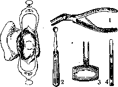
Figure 2.
Figure 3.
Figure 2. The operative field is exposed, spina and fossa suprameatum are visible, the trepanation borders are marked with a chisel (arcs). Figure 3. 1-bone forceps; 2-sharp curette; 3-wooden hammer; i-grooved chisel. To avoid concussion of the skull, it is necessary, wherever possible, to replace the work with chisels with forceps or curettes. The operation in all cases begins with wide opening of the antrum (antrotomy). In cases where there is a fistula, the operation begins at the site of the latter. To determine the place of access to the antrum, the following landmarks are used
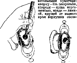
Figure 4.
Figure 5.
Figure 4. Opening of the antrum: the chisel is deepened from behind and above, forward and downward. Figure 5. Completed operation: 1-spina suprameatum; g-antrum mastoideum. of the prominent process perpendicular to the temporal line. Within these borders, the corticalis is removed in the form of a circle with blows of the chisel, within which it is necessary to reach the antrum so that the chisel always has direction from behind and above forward and downward (fig. 4). At a depth of about 1/2 cm, an opening leading into the antrum is usually encountered. The opening is expanded as much as possible (fig. 5), and the antrum is curetted with a sharp curette.
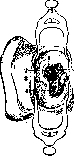
After antrotomy, they proceed to mastoidotomy, or rather to "mastoidectomy," since the modern operation for M. consists in the most complete removal of the entire cellular system of the mastoid process up to the lamina vitrea. For this, a sufficient knowledge of the topographic anatomy of these cellular groups, well developed by More and Beyer (Moret, Beyer), is required. Following the opening of the antrum, the cellulae zygomaticae are removed-the cellular group covered by the root of the mastoid process. Further, the cells covering the sigmoid sinus are removed, up to the exposure of the wall of the sigmoid sulcus or, if the latter is affected, the sinus itself (fig. 6). The next step is the removal of the cells lying in front of the sinus and the cells lying behind the descending branch of the facial nerve, the so-called "retrofacial" cells. Next comes the removal of the cells of the apex or, if necessary, the entire apex. Finally, the cells along the posterior and posterosuperior periphery of the process remain. Here they begin with the cells behind the sinus ("marginal"), then remove the cells in the area of the emissarium and from here, ascending further upward, the so-called "angular cells" lying at the upper bend of the sinus. The operation is completed by inserting a small strip of gauze loosely into the antrum and the auditory canal, and a similar loose packing of the entire wound cavity. Sutures are applied to the upper and lower angles of the wound, but only to the extent that the remaining unsealed opening is sufficient for inspection of the wound cavity during postoperative treatment. The latter aims at gradual filling of the bone defect with healthy granulations, their final transformation into strong scar tissue, and healing of the skin opening with a satisfactory cosmetic result. This is achieved by daily or every other day systematic packing of the wound with iodoform gauze. Communication with the antrum is maintained until complete cessation of purulent discharge from the auditory canal, after which it is allowed to close gradually with granulations. L. Levin.
Mastoiditis in children. Mastoiditis in early childhood runs somewhat differently from mastoiditis in adults. Subperiosteal abscesses in mastoiditis in early childhood are always located higher than in older children (always think of tuberculosis!). Pus from the antrum can easily penetrate into the cranial cavity due to the non-closure of the petro-squamous fissure (this explains the frequency of purulent meningitis in early childhood). Finally, most frequently (in hypotrophics and atrophics) are observed mastoiditis that run under the guise of oto-antritis. In the mastoid process of children under one year, there are usually few cells; there is a well-developed antrum, equal in size to the antrum of adults, and one or two cells lying around the antrum. In addition, the eardrum is thicker (due to the stratum cutaneum and stratum mucosum) than in an adult (perforation therefore does not occur so quickly); the connection of the antrum with the tympanic cavity is narrow and crossed by abundant strands of connective tissue, which become so swollen during inflammation that both cavities (antrum and cavum tympani) are separated; in such cases, otitis can turn into central mastoiditis. Such central M. can run 1) under the guise of toxic dyspepsia: vomiting, diarrhea, not stopping from diet, rapid loss in weight; 2) in other cases, phenomena of bronchitis or pneumonia dominate, with t° not falling when the phenomena in the lung are already small; often central M. give a picture of meningism, i.e., rigidity of the neck (in general, a frequent symptom in inflammation of the middle ear in early childhood), Kernig's symptom, a soporous state, convulsions of the eyes and general (eyes look up and to the side; upper extremities in flexion, lower in extension). All these phenomena disappear (if there is no true meningitis) after paracentesis or after trepanation. At the same time, there are often no particularly noticeable phenomena from the mastoid process. Still, sometimes one can note pain in the bone in the area of the antri (posterosuperior edge of the auditory canal), some change in color and contour of the entire eardrum (absence of the light reflex of the eardrum), sometimes partial bulging of the membrana tympani, loss of reflex, temperature. In older children, the petro-squamous fissure closes; complete pneumatization occurs by 3-5 years; therefore, M. in them runs almost as in an adult, with the difference that the rupture of pus occurs faster than in an adult. M. is most frequently observed with influenza, but it also occurs with other infections, e.g., with measles and scarlet fever; less frequently with diphtheria, whooping cough, etc. It develops the more frequently the earlier otitis begins in these diseases. The most severe is the scarlatinal M., often accompanied by the formation of large sequestra, the occurrence of which must be attributed not only to the severe lesion of the mucous cells but also to the thrombosis of the vessels feeding the bone. In measles and scarlatinal M., more frequently than in M. with other etiology, a) paralysis of the facial nerve (caries of the canal, sometimes simple compression by granulations), b) paralysis of the abducent nerve (Gradenigo), always speaking for a limited leptomeningitis and indicating the seriousness of the process, can be observed.-Complications in M. (thrombosis of sinuses, sepsis, brain abscesses, labyrinthine diseases, intoxication and diseases of the auditory nerve) occur in children more frequently than in adults. In the differential diagnosis of M., one should keep in mind: 1) furuncle, especially in the presence of otorrhea; in a furuncle, the puffiness is located somewhat lower than in M.: pressure in the area of the antri is painless, t° gives smaller fluctuations; a radiogram can help in diagnosis. 2) Lymphadenitis, especially in the presence of a furuncle or lesion of the external auditory canal or the hairy part of the head corresponding to the diseased ear; the diagnosis can present difficulty only in early childhood, since adenopathy up to 1 year is much more frequent than in older children: the lymphatic network around the tympanicum is more developed in children due to numerous excretory ducts through open fissures. These fissures in an adult are impassable. If there is no otitis, then these adenopathies are explained by severe rhinopharyngitis. Prognosis: 1) complicated mastoiditis (thrombosis of sinuses, meningitis, sepsis, brain abscess) sharply worsen the prognosis; 2) in necrotic forms with the formation of sequestra, the prognosis is very serious with respect to life and hearing. In uncomplicated M., the prognosis is good for both life and hearing.-Treatment.-Antrotomy. Indications: 1) inflammation of the periosteum with sagging of the posterosuperior wall of the auditory canal; 2) sudden cessation of purulent discharge from the ear, if at the same time the pain behind the ear increases and granulations close the perforation; 3) appearance of an infiltrate under the apex of the mastoid process (rupture through the apex); 4) formation of an abscess under the periosteum; 5) presence of symptoms indicating rupture of pus into the middle or posterior cranial fossa or transition of the process to the sinus; 6) persistent headaches, constantly increasing; 7) large fluctuations of t°, lasting more than 10-14 days; 8) prolonged purulent discharge (to prevent deafness, and in very small children-deaf-mutism); 9) meningism, if paracentesis is not effective; 10) paralysis of the n. facialis or abducentis.-Operation. Anesthesia local: injection of approximately 8-10 cm3 of a 1% solution of novocaine (general anesthesia can also be given). In the presence of a subperiosteal abscess, some authors recommend a two-stage operation (not necessary): 1) incision (according to Wilde) parallel to the attachment of the auricle to remove pus, then tamponade for 2-3 days; 2) opening of the antri (landmark - the posterosuperior angle of the auditory canal). In the absence of a subperiosteal abscess, a ONE-STAGE opening of the antri is performed.
Related articles
Mentioned in
Cite this page
“Mastoiditis.” Soviet Medical Encyclopedia. English translation of Bolshaya Meditsinskaya Entsiklopediya, 1st ed. (Moscow, 1928–1936), ed. N. A. Semashko. https://sovietmedicalencyclopedia.pages.dev/article/mastoiditis/