Rhinitis
Historical document, translated for reference. It reflects medical knowledge of the 1920s–30s and is not medical advice.
Summary
Rhinitis is an inflammation of the nasal mucosa characterized by increased mucus secretion and nasal congestion. This article from the 1930s Soviet encyclopedia classifies rhinitis into acute and chronic forms, describes symptoms, causes, and treatments of the era.
Encyclopedia article (1928–1936)
Rhinitis (rhinitis, coryza), a disease of the nasal mucosa, characterized by increased secretion of mucus and impaired nasal patency (difficulty in nasal breathing), one of the constant symptoms of which is "nose blowing," from which the name of the disease itself is derived. In clinical practice, the term "rhinitis" is most often used, under which various types of inflammatory processes of the nasal mucosa are understood. Rhinitides are divided into acute and chronic; chronic rhinitides in turn are divided into simple chronic catarrhal rhinitis, hypertrophic and atrophic. A special group of rhinitides, which does not constitute a separate nosological unit, but still has its own peculiarities in development and course, should include professional rhinitides. Acute R. (rhinitis acuta) is one of the most frequent diseases of the nose and upper respiratory tract. It appears either idopathically as an independent disease or symptomatically as a phenomenon accompanying acute infectious diseases. In most cases, there is undoubtedly an infection that has entered the nasal cavity either from the outside or by the hematogenous route; more rarely, mechanical or chemical causes lead to the inflammatory disease of the nasal mucosa. Temperature fluctuations in the form of so-called colds, which were previously given a large role in the etiology of R., are merely a predisposing factor for the action of microorganisms on the mucosa. Among acute infections accompanied by acute R. are influenza, in which the nasal cavity often serves simultaneously as the main or primary focus of the disease, then measles, scarlet fever, diphtheria, erysipelas, in which the nose may be affected almost simultaneously with the place of primary localization or becomes ill subsequently, as a complication, gonorrhea with atypical localization in the nose, and finally infections that cause any specific process in the nose, often accompanied by acute irritation of the mucosa, e.g., glanders. Rhinitis from professional causes may occur in various industries where dust, smoke, and irritating gases can also be the cause of acute rhinitis (more often these harmful factors lead to chronic diseases). Acute catarrh of the nose always affects both of its halves at once. The disease begins without significant disturbance of the general condition with sneezing, burning or pricking in the throat, especially in the nasopharynx, and sometimes also with hoarseness; in persons prone to fever, an increase in temperature to 37° with tenths is observed. In the first stage, a great dryness in the nose is subjectively felt, the mucosa is red, swollen, and has a dry appearance. After several hours (and sometimes after two to three days), the picture changes, the swelling of the mucosa decreases, it itself becomes moist, and a very large amount of secretion begins to be separated (rhinorrhea, rhinoblenorrhea), transparent as water; at the same time, the very unpleasant feeling of tension for the patient decreases—the second stage of inflammation. Then the secretions become mucous, take on a gray color from the admixture of desquamating epithelium, and become purulent from the admixture of lymph cells (third stage). The amount of secretion decreases, the inflammatory changes in the mucosa begin to pass quickly, and after 1-2-3 weeks complete recovery occurs. Ordinary catarrhal rhinitis in the first stage causes a feeling of heaviness in the head, as a result of which any mental work and concentration of thought on one subject become difficult. There is a change in the timbre of the voice in the form of nasalness and a decrease in the sense of smell from stuffiness of the nose or from the spread of the inflammatory process to the regio olfactoria. Later, pains often appear in the forehead, above the eyebrows, or in the region of the orbits. Discharges from the nose macerate the skin at the nasal entrance, which becomes red, slightly swollen, and often covered with cracks. Conjunctivitis often develops due to the spread of the process through the nasolacrimal duct, as well as diseases of the middle ear (spread of the process through the Eustachian tube). In the further course of acute R., the catarrh spreads to the fauces, larynx, trachea, and bronchi. Acute R. can have very serious significance in infants. The nasal passages in children in the first years of life are very narrow, and even slight swelling of the mucosa leads to obstruction of the nose, as a result of which not only is nasal breathing impaired, but sucking is greatly hindered, the child becomes restless, loses sleep, and even a decline in nutrition sets in. In posterior rhinoscopy, the mucosa of the nasopharynx is found to be swollen, red, covered with secretions that flow down the posterior wall of the pharynx and are visible behind the soft palate when the mouth is opened.—As a peculiarity of acute R. in influenza, it should be pointed out that at the beginning of the disease the nose is patent, but there is immediately abundant discharge of mucus. R. lasts longer and often complicates diseases of the accessory sinuses of the nose, accompanied by neuralgic pains in the forehead and region of the face. The diagnosis of acute rhinitis is not difficult to make even without rhinoscopy on the basis of the well-known subjective and objective symptoms. In children, however, it must be borne in mind that R. often occurs in the initial stage of acute eruptive infections, especially in measles. In R. in small children that does not yield to ordinary treatment for a long time, it is necessary to consider the possibility of a gonorrheal or syphilitic process, as well as to remember about diphtheria of the nose, occurring without general symptoms.—Prognosis in general favorable.—Treatment should be directed first of all, especially in persons enervated and predisposed to rhinitis, to the preventive hardening of the body against the influence of weather. One should avoid staying in overheated rooms and care for the purity of the air in them; constant and prolonged stay in the open air in any weather, wearing light clothing, and hydrotherapy procedures in the form of cold rubdowns, douches, and wraps are beneficial. Bed rest and staying in a room are necessary only in the presence of elevated temperature, with more pronounced bronchitis, especially in the elderly and children. Since anomalies in the nasal cavity promote the development of catarrh, it is necessary prophylactically to care for the normal patency of the nasal cavity. Existing obstacles in the form of hyperplasia, deviations, growths, neoplasms, and adenoid vegetations should be removed surgically. With acute R. having begun, one can attempt to arrest it by diaphoretic procedures. Sometimes it is possible to stop R. by prescribing antipyretics (phenacetin, aspirin 0.5-1.0 pro dosi). Symptomatic treatment consists in eliminating the stuffiness of the nose, if only for a limited time, which is accompanied by temporary improvement and disappearance of the associated disorders. Most often for this purpose, cocaine with adrenaline and menthol are used. Cocaini mur. 0.15, Natr. chlor. 0.25, Ac. carbol. 0.1, Aq. destill. 30.0 MDS. Nose drops—10 drops 3-4 times a day. Menthol is insufflated in the form of powder (Mentholi 0.2, Coffeae tostae, Sach. albi aa 5.0) or is used in the form of crystals, dropped into hot water, over which one must breathe through the nose. In acute R. in infants with nasal obstruction, it is useful 10 minutes before feeding to pour into both halves of the nose a few drops of a 1:10,000 solution of adrenaline. Crusts that sometimes exist at the entrance to the nose should be softened with olive oil and removed by wiping. Great caution must be exercised in the use of menthol in small children.—Rules for nose blowing: the patient is advised not to blow the nose too strongly (and with both nostrils), so as not to drive infected nasal mucus into the ear and the accessory sinuses of the nose. In chronic R. there are persistent changes in the form of swelling or atrophy of the nasal mucosa and increased discharge of pathological secretion. The causes leading to chronic R. are very different. In many cases, chronic R. develops under the influence of irritation on the basis of frequent repeated acute inflammations and exacerbations, especially when they appear at such a period of acute catarrh when the phenomena of the preceding disease have not yet had time to disappear. This is especially to be observed in various professions when inhaling air with harmful impurities (smoke, dust, hot air, irritating gases). Many infectious diseases, such as influenza, measles, scarlet fever, diphtheria, syphilis, etc., often serve as the initial moment for the development of chronic inflammations of the upper respiratory tract, including chronic R. All factors that lead to prolonged or frequently recurring hyperemia of the nasal cavity (e.g., alcoholism, chronic constipation, cardiac and renal diseases) can lead to chronic R. Causal factors can also be adenoid vegetations in the nasopharynx due to their inherent tendency to frequent inflammations with spread of the inflammatory process to the nasal cavity, as well as empyemas of the accessory sinuses of the nose due to constant irritation from the secretion entering the nasal cavity. According to a number of authors (Voyachek, Raspopov, etc.), some forms of rhinitides (hypertrophic and atrophic forms of them) are inherited according to Mendel's laws.
The number of observations is very small; in addition, the authors have not sufficiently taken into account the influence of socio-domestic factors. In general, chronic rhinitis occurs more frequently in males than in females. Simple chronic catarrhal R. (rhinitis chronica simplex, seu catarrhalis) is characterized by diffuse hyperemia and uniform swelling of the nasal mucosa. Its symptoms basically correspond to the symptoms of acute rhinitis, but are much less intense; the general condition of the patient usually does not suffer. Difficulty in nasal breathing, which in simple catarrh is a temporary phenomenon (rarely constant), is usually more noticeable when lying on the back or side. Usually in such cases there is an influx of blood to the lower parts of the nose; the vessels of the cavernous tissue, being in a relaxed state due to loss of tone, become engorged with blood, which causes nasal obstruction. When moving to the other side, the obstruction shifts to the other side. Discharges from the nose are more or less liquid in character. Complications in chronic catarrh can develop in the form of disturbance of smell-its weakening (hyposmia) or complete loss (anosmia). Another, frequently occurring complication is lesion of the organ of hearing. The thickening of the posterior end of the inferior turbinate, often observed in chronic catarrh, may either directly close the pharyngeal opening of the Eustachian tube or create conditions under which subsequent closure of the tube or even inflammatory diseases of the middle ear may develop. Sometimes the lacrimal apparatus is also affected due to the fact that the hypertrophied anterior end of the inferior turbinate blocks the nearby inferior opening of the nasolacrimal duct and causes tearing, inflammation of the lacrimal sac and conjunctiva. Diagnosis is usually not difficult with the help of anterior and posterior rhinoscopy. To distinguish between simple and hyperplastic forms of chronic rhinitis, a good means is the application of a 1-3% solution of cocaine to the nasal mucosa; almost complete disappearance of the swelling of the mucosa after cocaine application indicates that we are dealing with simple chronic catarrhal R., while little reduction or absence of it is characteristic of hyperplastic forms of chronic R. Palpation of the nasal mucosa with a pug probe also gives an impression of the degree of its swelling. During rhinoscopic examination, attention must be paid to the condition of the nasal septum, since a strong curvature of it toward the obstruction can more hinder nasal breathing than moderate thickening of the nasal mucosa. The prognosis is favorable, since the swelling of the mucous membrane can always be eliminated by conservative or minor surgical measures. Recurrences can be prevented with certainty only in cases where it is possible to eliminate the causes that caused chronic rhinitis. Chronic hypertrophic R. (rhinitis chronica hypertrophica). The formation and growth of connective tissue reaches its highest degree in the hypertrophic form of chronic catarrh. The proliferation of tissue elements does not develop diffusely throughout the mucosa, but mainly in places where cavernous tissue accumulates. Such places are the anterior and posterior ends of the inferior and middle turbinates. Sometimes such thickening extends along the entire lower edge of the inferior turbinate. The hypertrophied areas may have a very different appearance from the surface. Sometimes they appear smooth, but more often they have irregularities on them, especially at the ends of the turbinates, which take the form of bumpy, lobular, or coarse-grained protrusions. The hypertrophied posterior end of the turbinate may protrude into the nasopharyngeal space like a tumor. The surface of the hypertrophied areas depending on the amount of connective tissue already developed and the degree of blood filling may be pale gray-red, or bright red or bluish-purple. Due to the slowness and unevenness of the growth of connective tissue throughout the mucosa, clinically there are transitional forms between the simple and hypertrophic forms of chronic R., the determination of which is very important in order to correctly prescribe treatment. The symptoms of hypertrophic catarrh are the same as those of simple catarrh. Nasal obstruction, depending on more persistent causes-hypertrophy of the mucosa, is more constant and sometimes decreases little even after the nose is treated with cocaine. It does not change, as in simple catarrh, depending on the position of the head and body. The mucus is thicker in character and is difficult to separate. Improper and forceful blowing of the nose often leads to disease of the middle ear. The cause of both simple and hypertrophic chronic catarrh can be acute R., from which chronic rhinitis gradually develops. In addition, from local causes, curvature of the nasal septum, ridges on it, the presence of adenoids, and from general causes-heart defects, emphysema, obesity and a number of other diseases accompanied by disturbance of normal blood circulation are of importance. Constitutional factors also play a role. The working and living conditions of the patient are also of great importance. The prognosis is more or less favorable in terms of the possibility of eliminating swellings in the nasal cavity and restoring nasal breathing. Treatment of chronic rhinitis. Prophylactically, first of all, everything that serves as a cause or contributes to the maintenance of chronic R. should be eliminated, for which it is necessary to pay attention to the general condition of the body (diseases of the heart, kidneys, obesity, etc.) and the hygienic and working conditions of the patient. After clarification and possible elimination of these causes, one can proceed to local treatment with greater chances of success. The latter can be divided into medicinal and surgical. In the treatment of simple chronic R., in many cases one can limit oneself to the use of astringent or cauterizing agents. From astringents, 1-2-3% solutions of protargol or the application of weak solutions of silver nitrate or zinc chloride are used. For smearing the nose with the aim of causing resorption in the mucosa, a 1-1/2% solution of iodoglycerin is more often used.
From surgical methods of treatment, cauterizations are used in the hypertrophic form in the form of diffuse swelling. Cauterization can be performed with substances (chromic, trichloroacetic acid) or a galvanocautery (Fig. 1). It is sufficient to apply 2-3 grooves from back to front along the edge of the turbinate with acid applied to a probe or a heated pointed cautery, so that the subsequent scarring after the slough falls off will lead to a reduction of the swollen mucosa with restoration of tone to the cavernous tissue (Figs. 2 and 3). Of the mentioned agents, silver nitrate produces the most superficial cauterization, trichloroacetic acid is much stronger, and chromic acid acts even more strongly. The strength and depth of action also depend on the duration of exposure and the force with which the chemical substances are pressed against the mucosa. After trichloroacetic acid, a superficial snow-white slough is obtained, with chromic acid a deeper yellowish one, and the reaction in the form of some painfulness and reactive swelling of the mucosa in the first days after cauterization with chromic acid is stronger than
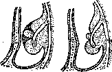
Figure 2.
Figure 3.
Figure 2 and 3. Galvanocautery of the turbinates: fig. 2-before and fig. 3-after cauterization with trichloroacetic acid. In galvanocautery cauterizations of the turbinates, used in far-advanced degenerations of the nasal cavity mucosa, it is necessary to be careful not to cauterize the opposite wall of the nasal septum and not to obtain synechiae later. When removing limited hypertrophy of the anterior and posterior ends and the entire edge of the inferior turbinate, as well as the anterior end of the middle turbinate, it is best to use a cold loop or Beckmann nasal scissors (fig. 4 and 5). A loop corresponding to the size of the hypertrophy is inserted under visual guidance into the nose and applied to the thickening as close to its base as possible, after which the protruding part of the thickening is cut off (fig. 6, 7 and 8). If the hypertrophy occupies the entire lower edge of the turbinate, its removal is performed by means of scissors and a loop (fig. 9,10 and 11). Simple atrophic R. (rhinitis atrophica simplex)-such a chronic diffuse disease of the nasal cavity, which is accompanied by atrophy not only of the mucous membrane, but also of the underlying bony skeleton of the nasal turbinates. The disease is often accompanied by reduced secretion of pathological secretions, which has a tendency to dry and form crusts, but without odor. Regarding the origin of atrophic rhinitis, many different views have been expressed, of which however none explains all cases of the disease. Some authors (Frankel, Schech, Gottstein) adhere to the view that the atrophic form always develops from hyperplastic rhinitis, representing only a later stage of the latter. Indeed, the transition from hyperplasia to atrophy has not been directly observed by anyone, but their connection is considered possible due to the frequently occurring simultaneous coexistence of both processes. In recent years, the literature contains indications of the hereditary origin of atrophic and hypertrophic processes in the nose, of the so-called "dystrophies" of the nasal mucosa; confirmation of this view is the frequent finding of sharply expressed atrophic, less frequently hypertrophic processes in children in the first years of life (Voyachek, Volkovich). Subjective symptoms are often insignificant and almost do not attract the attention of patients. They consist in a distressing feeling of pressure or in pains in the head. In addition, patients often complain of dryness in the nose and throat, which is also often involved in the disease. The most distressing is usually the feeling of nasal obstruction caused by the abundant formation of crusts. The presence of crusts often causes tormenting itching in the nose; to get rid of it, patients often pick their nose with their fingers, thereby causing injuries, especially to the nasal septum, sometimes accompanied by slight bleeding. The spread of atrophy to the regio olfactoria initially causes weakening of the sense of smell, and later its complete loss. On rhinoscopy, the nasal cavity appears very wide due to the atrophy of the mucous membrane and turbinates, but not to the same degree as in ozena. Through the choanae, the nasopharynx can be seen. Sometimes along with the atrophic condition of the inferior turbinate, the middle turbinate is found to be hyperplastically thickened. The diagnosis of rhinitis atrophica is not difficult to make on the basis of rhinoscopic examination and subjective and objective symptoms.-The prognosis in rhinitis atrophica is doubtful. Indeed, it is not difficult to eliminate the most distressing symptoms of atrophy, but it is impossible to fully restore a normally functioning mucous membrane. One cannot expect the return of lost smell in view of the fact that we are dealing with essential anosmia.-Treatment can only be symptomatic, directed against the existing dryness and the crusts forming in the nose. In the initial stages of atrophy, irritating therapy is applied, smearing the nasal mucous membrane daily with a 11/2% solution of iodoglycerin for 2 weeks, in order to enhance the activity of the remaining glands of the mucous membrane. Crusts are well removed from the nose by Gottstein nasal packing with cotton wool, the application of indifferent ointments and nasal douches with physiological or alkaline solutions. It has very unpleasant consequences and strongly worsens the course of atrophic rhinitis an erroneous diagnosis and the application of surgical methods of treatment and galvanocautery in the nose. If one accepts according to the opinion of some authors (Voyachek, Gradenigo, Raspopov and others), that atrophic rhinitis is a hereditary disease, then the fight against it should also be based on eugenic measures. Professional rhinitis do not constitute an independent nosological unit, but


Figure 4.
Figure 5.
Figure 4 and 5. Beckmann scissors for removal of the inferior and middle turbinates.
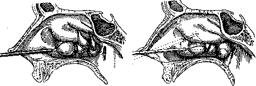
Figure 6.
Figure 7.
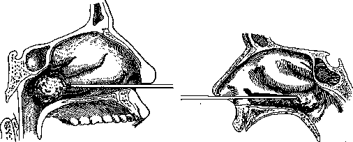
Fig. 8.
Figure 9.
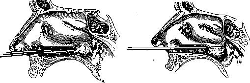
Figure 10.
Figure 11. Figs. 6 and 7. Removal of a polyp from the nose. Figure 8. Removal of the hypertrophied posterior end of the inferior turbinate. Figures 9, 10 and 11. Removal of the inferior turbinate (conchotomy). Nevertheless, the development and course of these conditions have certain peculiarities. The main harmful agents affecting the nasal cavity are dust, gases, and unfavorable meteorological factors. All these harmful agents usually act in combination, but one of them is typically primary. - Dust can be classified according to its effect on the body (according to Kdlsch) as 1) mechanically irritating, 2) chemically acting, 3) infectious, and 4) explosive-destructive. The result of all types of dust is inflammation of the mucous membrane, which in some cases occurs primarily, in others as a secondary infection of the dust-traumatized mucous membrane. The effect of mechanically irritating dust consists in traumatizing the mucous membrane. The degree and location of the trauma depend on the physical properties of the dust: its weight, volume, particle shape, speed of flight, etc. Heavy particles (metallic, mineral dust) mainly affect the initial parts of the nose, while lighter particles (cotton, wool, etc.) affect deeper parts; when particles have a high speed of flight (for example, during grinding of products), the traumatizing effect is intensified. Soft, round dust causes minimal direct trauma, but quickly clogs the excretory ducts of the mucous glands, densely envelops it, and thus promotes drying (chalk, gypsum, flour). Traumatization of the mucous membrane initially causes acute inflammation, often accompanied by nosebleeds. Then infection often sets in, leading to a chronic course. Simple chronic catarrh of the nasal mucosa develops, which sometimes lasts for many years and gradually leads to the proliferation of connective tissue (rhinitis hypertrophica). In some individuals, the stage of hypertrophy does not occur, but rather a rapid drying of the tissue occurs, leading to atrophy of the mucous membrane with subsequent degeneration of the glandular apparatus. Atrophy is also promoted by the scarring of traumatized areas of the mucous membrane. Atrophy of the nasal mucosa occurring in this way differs from genuine forms by the absence of abundant crust formation and subjective complaints. These forms of atrophy rarely lead to thinning of the bony skeleton of the turbinates. The number of such dust-related professional rhinitis is extremely large: according to data from foreign authors from 1908-10, among 300,000 coal miners, 73.57% had respiratory diseases; in porcelain production (according to Temkin's data), 60% had atrophic rhinitis, etc. In addition to inflammatory diseases of the turbinate mucosa, anterior dry rhinitis is often observed in some dusty workshops, which sometimes leads to cartilage perforation (among millers, cement workers, especially under the influence of inhaling chromium dust, etc.); sometimes dust accumulates in large quantities in the middle nasal passage, where concretions and rhinoliths form as a result. - The chemical action of dust always accompanies the traumatic action and consists either in sharp irritation with inflammation of the mucous membrane (inorganic soluble salts) or in cauterizing it. With irritation, catarrhal inflammation phenomena come to the fore, followed by tissue hypertrophy; in the second case, phenomena of scarring, shrinkage, and atrophy occur (chromium dust). - The infectious action of dust, in addition to the traumatic action, causes severe acute rhinitis with subsequent hypertrophy, as well as often inflammation of the paranasal sinuses. The degree of damage to the nasal mucosa when inhaling vapors and gases depends mainly on the degree of solubility of the gas in water. This explains why, for example, ammonia sharply affects the pharynx, while nitrogen oxides and phosgene mainly affect the pulmonary alveoli. Poisoning by certain gases reduces normal resistance to certain types of pathogens (e.g., poisoning by SO2 and H2S with respect to streptococci and staphylococci), and thus leads to inflammation of the nasal mucosa. Clinical manifestations are expressed as irritation, inflammation, ulceration, and necrosis of the mucous membrane, to which infection secondarily joins. In acute cases of gas poisoning, acute rhinitis is more often observed, while in chronic cases, catarrhal inflammation occurs. Often, the olfactory nerve is affected (essential anosmia). - The effect of acids is expressed mainly in that, combining with the protein of cells, they form albuminates. In the protoplasm of cells, homogenization of the substance occurs, the smallest vessels of the mucous membrane become thrombosed, and the entire affected area undergoes necrosis; the mucous membrane heals extremely slowly. If healing occurs, phenomena of shrinkage result, and the mucous membrane becomes particularly resistant to chemical and infectious agents. - Alkalis have less intense cauterizing properties but act very actively on the protoplasm, liquefying it. The inflammatory reaction is less pronounced but has a tendency to spread and ends with significant tissue destruction. - A number of essential oils, petroleum products, and turpentine oil also cause sharp irritation of the mucous membrane, which gradually passes into dry processes; initially there is hyperesthesia, which then turns into anesthesia. - Meteorological influences act reflexively or by affecting the entire circulatory system. As the first symptoms of damage to the nasal mucosa, abundant liquid secretion, edema, and tissue loosening appear. When other harmful factors (dust, gases, and infection) are added, a severe acute inflammatory process develops. This first stage of the disease lasts an extremely long time, and after it, direct drying and atrophy of the mucous membrane often occurs, which develops particularly strongly in those workshops where the air is dry and hot and where, in addition to temperature, dust also acts (stokers, foundry workers, furnace operators). Nervous, vasomotor rhinitis (rhinitis nervosa, vasomotria) is accompanied by abundant, mostly watery discharge, sudden nasal obstruction, redness and swelling of the conjunctiva of the eye, and is often observed in nervous, neurasthenic, and hysterical individuals. Besides mechanical and thermal causes, it can be caused (sometimes quite suddenly) by emotional excitement, sexual arousal, as well as strong olfactory sensations. - Treatment should aim to reduce increased nervous excitability through proper hardening (hydrotherapy) and, if necessary, internal medications (iron, arsenic, quinine, valerian, bromine preparations). Anomalies in the nose should be eliminated surgically. In its manifestations, nervous vasomotor rhinitis resembles hay fever (see).
Related articles
Mentioned in
Cite this page
“Rhinitis.” Soviet Medical Encyclopedia. English translation of Bolshaya Meditsinskaya Entsiklopediya, 1st ed. (Moscow, 1928–1936), ed. N. A. Semashko. https://sovietmedicalencyclopedia.pages.dev/article/rhinitis/