Gastritis
Historical document, translated for reference. It reflects medical knowledge of the 1920s–30s and is not medical advice.
Summary
This article from the 1st edition of the Great Medical Encyclopedia (1928–1936) discusses the etiology and pathological anatomy of gastritis, detailing its acute and chronic forms, catarrhal, phlegmonous, and atrophic changes, as well as various predisposing factors.
Encyclopedia article (1928–1936)
GASTRITIS, gastritis (from Greek gaster - stomach), an inflammatory disease of the stomach, predominantly of a catarrhal character. Older medicine treated the majority of stomach diseases as "indigestion," designating them by various terms such as "indigestion," "apepsia," "bradypepsia," etc. Only since the time of Sauvages and Broussais, i.e., 100 years ago, has the concept of gastritis as an inflammatory disease of the stomach, mainly of its mucous membrane, been established. Etiology. It is not always possible to precisely indicate the immediate cause of gastritis, and it is hardly likely that one and the same specific etiology exists in all cases. Some authors (the Bergman school) advocate the functional theory of the origin of gastritis, according to which a whole series of external factors, individually variable in different people, gradually disrupts the normal working conditions of the stomach, as a result of which the functional ability and resistance of this organ suffer and susceptibility to diseases increases. On this already prepared soil, the inflammatory process then develops. Among such purely functional factors preparing the way for gastritis are: a decrease in secretion, causing a drop in the bactericidal and antitoxic power of gastric juice, irritation of the stomach by toxins, food overload, changes in absorption, and others. According to this theory, the sequence of phenomena is as follows: external factors - functional impairment - increased susceptibility - inflammation. - The main factors usually causing gastritis are the following: 1) disorders of the masticatory apparatus (caries and dental defects); 2) inflammation of the gums and oral cavity, as a result of which the chewing of food and its normal moistening with saliva as the first stage of digestion are disrupted, so that food enters the stomach in a poorly chewed form and causes mechanical irritation; 3) chronic laryngitis, pharyngitis, and especially rhinitis, causing gastric irritation due to the swallowing of inflammatory secretions; 4) very fast eating and eating while working; 5) overloading the stomach with too large quantities of food, especially after preceding fasts and periods of starvation; 6) eating dry food for a prolonged period; 7) thermal irritations from very hot food (tea) or very cold food (ice cream, very cold drinks); 8) chemical irritants, such as: spicy dishes with pepper, mustard, horseradish, vinegar; spices, tea, coffee, and especially alcohol as one of the most frequent causes of gastritis; 9) consumption of low-quality and spoiled products. Some of the above-mentioned factors are often associated with poor general socio-economic conditions, such as: eating in cheap, poor canteens, dormitories, barracks, forced monotony of food, hurried eating during work, etc.; 10) acute and chronic poisonings: with sausage and fish poisons, lead, acids, alkalis, metals, alkaloids, aniline dyes, phosphorus, etc.; occupational poisonings, in which poisons penetrate the organism through the respiratory tract, mouth, or skin; in such cases, the gastric mucosa is affected secondarily as an excretory organ; 11) infections (influenza, typhoid fever, paratyphoid, etc.); 12) Gastritis often arises as a secondary disease in various stomach disorders, for example, in ulcer and cancer, when an inflammatory process develops in the vicinity of the primary focus due to secondary infection. This also includes cases of gastritis on the basis of pyloric stenosis, when food stagnation in the stomach with the formation of a large amount of organic acids leads to catarrhal inflammation of the mucosa. The cause of gastritis can also be pathological functional changes in the stomach, especially secretory disorders and weakening of the neuromuscular apparatus with a decrease in motor function; 13) diseases of the liver and heart leading to circulatory disorders with stagnation in the portal and inferior vena cava systems, as well as azotemic uremias accompanied by gastritis due to the excretion of nitrogenous slag and other pathological metabolic products through the gastric mucosa; 14) severe diseases of the hematopoietic organs, and in particular pernicious anemia or, more precisely, those factors which cause this disease of the hematopoietic apparatus. - Usually, the cause of chronic gastritis is not the isolated action of any single one of the listed factors, but the combined simultaneous influence of several harmful agents. The most frequent causes are fast eating, especially with a poor masticatory apparatus, abuse of spicy dishes and alcohol. One of the important etiological factors is occupational hazards. We may point out the frequent development of gastritis in workers in hot shops (glassblowers), since workers in such professions consume large amounts of cold water. Furthermore, in a number of chemical industries (acid, tinware), gastritis develops on the basis of intoxication, sometimes due to the excretion of these substances by the gastric mucosa.
I. Loriv. Pathological anatomy. The inflammatory process in the stomach affects, mainly, the mucous membrane; in other words, gastritis is predominantly a catarrhal inflammation. - In acute gastritis, the mucous membrane is in one degree or another
Figure 1. Acute gastritis (low magnification): a - gastric glands with preserved mucin-degenerated epithelium; b - glands with complete desquamation of the epithelium and its degeneration (after Henke-Lubarsch).
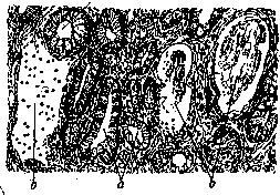
hyperemic, swollen, and covered with an excess amount of mucus; small hemorrhages are frequently observed, which later can give rise to acute erosions and small ulcers; the latter are sometimes located predominantly along the course of the stomach's "track" (Magenstrasse, see Stomach). Microscopic examination in acute gastritis reveals degenerative changes in the epithelium, both surface and gland-lining, with frequent desquamation of epithelial cells, sometimes to the complete denudation of the mucosal surface and gland lumens (see Figure 1). In the fundamental tissue of the mucous membrane and in the submucosal tissue, phenomena of hyperemia, edema, and slight leukocyte infiltration are found. In severe diphtheria in children, croupous gastritis is sometimes observed, manifested by the formation of fibrinous false membranes on the hyperemic mucous membrane; in severe scarlet fever, necrotic gastritis may occur, in which necrosis of the mucous membrane is observed, usually involving the tips of its folds. [It must be admitted, however, that even the normal gastric mucosa during the digestive period can partly simulate the morphological phenomena of gastritis (hyperemia, hypersecretion, desquamation of the epithelium, its degeneration, frequent emigration of leukocytes, e.g., with meat food, etc.).] - Phlegmonous gastritis, manifested by significant thickening of the stomach walls due to their purulent infiltration, is rare and usually occurs as a complication of certain disruptions of the integrity of the mucous membrane. Catarrhal phenomena in this case may recede into the background or be completely absent. - In chronic gastritis, the picture of changes can appear different depending on whether hypertrophic or atrophic changes predominate in the mucous membrane. In hypertrophic chronic gastritis, the mucosa is heavily coated with mucus and unevenly thickened; usually this thickening of the mucosa appears in the form of a large number of protruding elevations giving the mucosal surface a shagreen appearance (etat mamelonné); sometimes, especially in the pyloric part, the formation of multiple small polyps (gastritis polyposa) can be observed. The microscope reveals that these mucosal thickenings are based on the proliferation of glands, often with their cystic expansion, and cellular infiltration of the fundamental interglandular tissue. - Atrophic gastritis, which is frequently the outcome of the hypertrophic period, is characterized by a smooth, shiny appearance of the mucous membrane, its thinning, and a frequent slate-gray tint (due to the deposition, on the basis of blood flushes, of hemosiderin turning into iron sulfide). Upon microscopic examination, a decrease in the number of glands (see Figure 2), their shortening, and simultaneous... [text cuts off in source]
Figure 2. Atrophic gastritis (low magnification). The gastric mucosa is atrophic; the number of glands is greatly reduced; in the depths of the mucosa, small-cell infiltration (after Henke-Lubarsch).

Additionally, the text contains a stray character marker 'ч' from OCR artifacts which is omitted in translation structure.
shrinking of the main tissue; sometimes cystic expansion of individual glands is observed. Atrophy sometimes also affects the muscular layer of the stomach wall, which can contribute to stomach dilation. In some cases, chronic G. involves proliferation and subsequent scarring of connective tissue in all layers of the stomach wall, which can lead to a reduction in the size of the entire stomach (gastr. indurativa, see Gastric Cirrhosis); | in cases when this process mainly affects the pyloric part of the stomach and causes thickening and hardening of its wall, a picture very similar to the fibrous form of cancer (skirrhus) of the stomach is obtained (see Gastric Cirrhosis).-Gastritis nodularis, which is based on hyperplasia of the lymphatic follicles of the stomach wall, is not an inflammation but represents an expression of lymphatism.- Chronic gastritis is also observed in inflammatory processes of a tuberculous and syphilitic nature in the stomach (see Stomach).
a. Abricosov. Clinical symptoms and course. For individual pathological forms of G., it has not yet been possible to establish special clinical pictures, except for atrophic G. with complete disappearance of HCl secretion and enzymes and desquamative G. with characteristic peeling of the epithelium.-Acute G. begins suddenly and is accompanied by a series of violent dyspeptic phenomena: bloating throughout the upper abdomen, salivation, eructation of food, nausea, vomiting, pain under the xiphoid process; appetite is absent, the tongue is coated and dry, temperature is usually slightly elevated, sometimes normal, and in infectious and toxic forms it can give high figures (g. infectiosa, toxica, phlegmonosa); vomit in simple catarrh (g. simplex) is not abundant and contains poorly digested food with a large amount of enveloping mucus and saliva; in severe forms it also contains pus and blood. Microscopically, shed cylindrical epithelium is noted, sometimes entire layers of detached glandular tubules, red blood cells, leukocytes, bacteria. The intestine is almost always irritated, with symptoms of constipation, meteorism, or diarrhea, which plays the role of "drainage" removing the infection. With timely measures and favorable course, simple G. passes in a few days.-Infectious G. proceeds very severely, and phlegmonous and purulent forms-almost always with a fatal outcome.-Chronic G. can develop on the basis of acute. More often, however, it develops gradually, under the influence of prolonged irritation of the stomach by various harmful agents. Dyspeptic phenomena gradually increase. The most characteristic are: bloating after each meal, a feeling of heaviness and fullness even after small food intakes; eructation of food, sometimes sour or rotten; dull pain under the xiphoid process soon after eating, depending on the amount of food and its physical composition (coarse vegetable fiber, connective tissue, etc.), sometimes sharp lancinating pain after eating, tendency to constipation or diarrhea; objective symptoms: moderate abdominal bloating, diffuse tenderness in the epigastric region; from general symptoms-lassitude, fatigue, apathy, tendency to hypochondria. From the side of the secretory apparatus, increased secretion is initially observed due to irritation of the glands (irritative stage, gastritis acida). Later, the secretory apparatus is exhausted, the depressive stage sets in, and the secretion of hydrochloric acid decreases (g. hypacida) or completely disappears (g. ana-cida). Usually, the disappearance of secretion first affects the main cells that secrete hydrochloric acid; enzymes disappear later, and the last to disappear is the rennet enzyme; its absence usually indicates complete atrophy of the glands, which can no longer be restored, leading to permanent disappearance of secretion (g. atro-phicans, anadenia). However, sometimes normal secretory function is preserved (g, normacida). Along with free HCl, high figures of bound hydrochloric acid are often found due to the abundance of mucus. The motor function is usually reduced due to the increased accumulation of mucus, which hinders the mixing and movement of gastric contents. Delayed evacuation often depends on pylorospasm (sometimes even with subsequent stagnant dilation of the stomach) or on the sharp swelling of the mucosa in the pyloric part, leading to relative stenosis of the pylorus ("stenosing Gastritis" of Boas). On the contrary, hypo- and anacidic forms easily lead to weakening of the closing reflex of the pylorus with acceleration of evacuation and subsequent (gastric) diarrhea. In the gastric contents after a test breakfast, with any acidity, there is always mucus characteristic of catarrh: thick, sometimes glassy, falling to the bottom of the vessel (unlike saliva and oral cavity mucus, which float on the surface), and clumps of poorly digested food with a thick layer of enveloping mucus. (However, according to Boas and Noorden, mucus is not a constant sign of G.; on the other hand, there is a picture of so-called "myxorea" on the basis of organic diseases or neurosis of the stomach, but without any presence of G.) The presence of a large amount of mucus in the washing waters on an empty stomach in the form of grayish cloudy flakes is very characteristic. The microscopic picture shows leukocytes, cylindrical and flat epithelium, nuclear grains, myelin cells (Yavorsky spiral cells), goblet cells (see fig. 3); with significant stagnation-remains of food taken the day before, many yeast fungi and sarcina (with preservation of free HCl). The microscopic picture can vary depending on the presence of hydrochloric acid in the stomach: in acid G., cell nuclei, Yavorsky cells, and poorly digested starch grains predominate (see figure 4); in anacidic G.-clumps of leukocytes, cylindrical and flat epithelium with preserved nuclei, goblet cells, and well-digested starch grains (see figure 5). The division of gastritis according to the secretory function of the stomach is fully justified by the fact that the nature of secretion in chronic gastritis gives guiding indications for treatment. Often there is a change in the blood picture in the sense of secondary anemia. The prognosis of acute G. in mild forms is undoubtedly favorable, in severe forms-uncertain. In chronic G., the prognosis is generally favorable in terms of the disappearance of painful symptoms, but complete restoration of health sometimes requires a very long period (months and even years) and constant caution in dietary regimen. In atrophic G., restoration of secretory function is already impossible. The consequences of G. in the form of nutritional decline and anemia can persist for a long time after the disappearance of all main symptoms.-Complications. ^Complete disappearance of gastric secretion, more often in old age or in the presence of alcohol intoxication. 2) Stomach ulcer. According to one theory (Konjetzny and some French authors), chronic G. underlies every ulcer. Undoubtedly in any case that G. can contribute to the formation of ulcers on the mucosa. 3) The most common complication of G.-enterocolitis, which usually occurs due to impaired gastric digestion and irritation of the lower parts of the digestive tract by poorly digested food. 4) Cholecystitis and cholangitis. Whether these diseases develop simultaneously with chronic G. under the influence of the same harmful agent or they are a consequence of G., it is difficult to decide in each individual case. Some authors believe that infection of the bile ducts, apparently, is also facilitated by achylia as one of the frequent consequences of chronic G. In any case, statistics confirm the very frequent incidence of bile duct diseases in chronic G. 5) Anemia, usually of moderate severity, of the chlorosis type; in addition, chronic G. with a decrease in HCl and enzymes is as a rule accompanied by pernicious anemia. 6) Neuroses of the most varied kinds. Gastralgias, diarrhea, and other forms of neuroses of the stomach and intestines are not uncommon. 7) Symptoms of avitaminosis in the form of gingivitis, glossitis, blepharitis, dermatitis, furunculosis, as well as general weakness and joint pains. The cause-prolonged inadequate nutrition and lack of vitamin supply due to too long and strict diet.-Some authors consider chronic gastritis a predisposing factor for stomach cancer, however, in view of the lack of exact data on the origin of stomach cancer, this question must be considered open. The diagnosis of acute gastritis is determined by the above-described complex of symptoms after a preceding acute poisoning, overeating, or infection. Examination of vomit and gastric contents after a test breakfast finally establishes the diagnosis. The diagnosis of chronic gastritis is based on the characteristic complex of symptoms: loss of appetite, unpleasant taste in the mouth, feeling of rapid filling and bloating after eating, heaviness in the epigastric region and nausea, more often on an empty stomach, dull pain after abundant or coarse (in the physical sense) food, eructation of food taste, sometimes vomiting; less characteristic are sharp pains in the epigastric region and heartburn. Morning vomiting in alcoholics, according to the majority of authors, depends on the accompanying chronic G. of pharyngitis. From the objective symptoms, diagnostically important are: the presence of a large amount
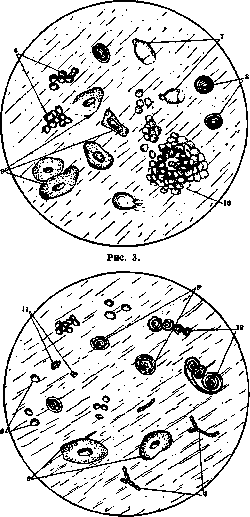
Figure 4.
mucus on an empty stomach in the wash waters and after a test breakfast, abundance of leukocytes and cylindrical epithelial cells in the stomach contents, sometimes-diffuse tenderness in the epigastric region and tension of the abdominal wall of the entire upper abdomen, as well as macro- and microscopic signs of motor insufficiency. A coated tongue in chronic G. by no means has the same diagnostic value as in acute gastritis, and more often depends on insufficient cleaning of the oral cavity and tongue. All these symptoms allow for an accurate diagnosis only when there are certain data in the history, such as, for example, rapid eating, eating dry food, acute gastritis, etc. However, the history and status do not always provide sufficient data for an accurate diagnosis; there are even such (latent) forms that proceed for years without any symptoms. The difficulty of diagnosis is further increased by the fact that almost all properties are characteristic
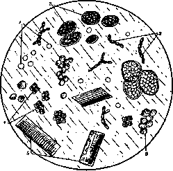
Figure 5.
Figure 3-5. Microscopic picture of gastric contents in gastritis: 1-fat droplets; 2-plant cells; 3-yeast fungi; 4-sardines in G. with motor insufficiency of the stomach; 5-muscle fibers; 6-leukocytes; 7-goblet cells; 8-starch grains; 9-epithelium; 10-glandular tubes; 11-cell nuclei; 12-spiral cells (according to Boas). The chronic G. symptoms (except for the abundant presence of mucus in the stomach) may also depend on another disease.- Anatomical diagnosis in vivo. Some authors (Korczynski, Jaworski, Hayem and others) have long proposed a method for histological examination of pieces of mucosa from washing waters, but, according to modern views (Lubarsch, Boas and others), it does not make it possible to establish individual forms and stages of development of the inflammatory process, for which reason it is little applied in the clinic. Direct examination of the gastric mucosa by means of gastroscopy makes it possible to accurately establish the presence of an inflammatory process in the stomach; unfortunately, due to its complexity, this method is also little used. The newest roentgenoscopic method of Berg (Berg), based on finding swollen folds of the mucosa, allegedly characteristic only for G., has not yet given definite results. In differential diagnosis, one must take into account the possibility of the following diseases. 1) Stomach ulcer gives a characteristic dependence of pains on the time of food intake and on its chemical (and not only physical) composition. In G., for example, acute and salty dishes often do not cause any unpleasant sensations, as is always the case with ulcers. Objectively, in ulcers there is usually a limited painful point, while in G. objective tenderness is either completely absent or has a diffuse character. In the gastric contents there is usually little mucus (if there is not simultaneously a sharply expressed accompanying G.). 2) Cancer of the stomach in the absence of a palpable tumor gives a picture close to atrophic G. with a decline in nutrition. The distinguishing features of cancer are: a more sudden onset, a more rapid development of all symptoms, especially loss of appetite, progressive deterioration, loss of weight, cachexia, despite increased nutrition; in some cases-severe motor insufficiency. In the gastric contents, the characteristic features are the presence of lactic acid (absent in G.), large amounts of combined HCl in the absence of free HCl, and reaction to hidden blood. Failure to obtain a therapeutic effect, despite properly conducted treatment, especially in elderly people and in the presence of achylia and persistent reaction to blood in the stool, should always arouse suspicion of the possibility of development of a malignant tumor. 3) Achilia of functional nature with complete disappearance of gastric secretion is characterized by the fact that subjective and objective symptoms are not constant, do not depend so much on the nature of food, especially its physical composition, as in G., and the secretory function itself is subject to sharp fluctuations (heterochylia). 4) In nervous dyspepsia, the course is less constant, in jumps, deteriorations and improvements do not depend on diet, subjective symptoms predominate over objective ones and depend less on the methods and nature of food than on external influences, mental factors and the general state of the nervous system. Almost always there are also a number of other disorders from the autonomic nervous system and the psychic sphere. 5) In diseases of the bile ducts, even in the absence of typical colic attacks, there is limited palpable tenderness in the area of the gallbladder or the left lobe of the liver. The diagnosis in these cases should be based entirely on objective data (examination of urine and blood for bile pigments, determination of the gallbladder reflex, careful palpation of the liver area, etc.). 6) Chronic enterocolitis can give a picture very similar to chronic G. with all the subjective signs of the latter. The distinguishing symptoms of enterocolitis are: dependence of pains on emptying of the intestine and their colicky character, bloating and splashing in the abdomen, tenderness on palpation of individual parts of the intestine and data from examination of fecal masses. Treatment. Treatment of acute G. consists in emptying the stomach by washing or artificial vomiting, in cleansing the intestine with an enema (and in the absence of vomiting-with a laxative) and in complete deprivation of food for 1-2 days (only tea with lemon is allowed). In case of acute poisoning, an antidote is given. Etiological treatment is exhausted by this and gives way to symptomatic treatment: irritation of the mucous membrane is calmed by solutions of Arg. nitr. (0.1:200.0), anesthesin, orthoform, bismuth; for nausea and vomiting-solution of menthol, validol; for severe pains-injection of atropine with morphine. In the first days, bed rest and warmth on the abdomen are mandatory. Subsequently, gradual expansion of the diet: at first only liquid food, such as mucilaginous soups, weak vegetable broths, liquid tea with lemon or milk, weak black coffee; then semi-liquid (pureed cereals with milk, butter, unsweetened kissels and especially-jellied dishes); later-meat and fish cutlets, boiled lean fish, vegetable purees, white bread. The general principle of the diet is to spare the stomach, avoiding any mechanical and chemical irritations. With a sharp increase in acidity-frequent meals (every 2-3 hours in small portions). The transition to ordinary food should be very gradual, so that the patient for several weeks after the disappearance of all dyspeptic phenomena continues to maintain the diet. In severe forms with continuous vomiting, fever and general intoxication-complete exclusion of nutrition through the mouth, absorption enemas from glucose or physiological solution of salt, and in the presence of severe irritation of the intestine-administration of the above-mentioned solutions under the skin or into the vein. Treatment of chronic gastritis should be directed towards eliminating the harmful etiological factor (correction of the chewing apparatus, treatment of diseases of the gums, nasopharynx, etc.). Dietotherapy is in the forefront, which should pursue a dual purpose. The first period-to spare the affected organ, avoiding sharp stimulants of secretion and irritation of the mucous membrane with coarse food and maintaining rapid evacuation of the stomach. The second period-training and transition to the previous digestive load. In this period of treatment, it is important to fight not only with the remnants of dyspeptic phenomena, but also with the danger of weakening of the body from too strict a diet. One of the main difficulties is the fight against loss of appetite and aversion of the patient to bland food. The necessity to select dishes that are at the same time nutritious and not irritating and sufficiently tasty requires from the doctor, besides knowledge in the field of therapeutic nutrition and therapeutic cuisine, also a great deal of art. For the first period of treatment, besides dishes recommended for acute G., the following are allowed: weak meat and fish broths, vegetable decoctions, milk and pureed vegetable soups, soft-boiled eggs, steamed chopped cutlets from lean meat and fish, pureed cottage cheese with sugar or sweet sour cream, fruit and berry jellies and syrups, stale white bread, better-dry toast and toast. All this food is distributed into 4-5 small meals a day. In the second period of treatment, the main guiding line is the state of gastric secretion. With normal and increased acidity, it is especially important to exclude salty, sour and fried dishes and any sharp seasonings, as well as all strong secretagogues, like strong tea, coffee, strong broths. On the contrary, with reduced acidity, some secretagogue dishes and taste seasonings are indicated, such as broth, fresh caviar, coffee, cocoa, lemon, toast with grated cheese, buttermilk, kefir, ayran, berry syrups, orange and carrot juices. But special care is required in prescribing meat: fatty varieties of meat (goose, duck, pork) are completely forbidden, as well as any meat and fish containing a lot of connective tissue and fat. Throughout the treatment, it is especially important to exclude smoked and fried dishes, products from yeast dough, black bread, coarse vegetable fiber, fresh fruits, as well as alcohol, snacks, canned goods, sharp seasonings and sauces. In prolonged course, decline of strength and weight loss, intensified nutrition is prescribed with abundant content of butter (cream or Provence), pureed greens and fruits, berry and fruit juices and syrups (vitamins). Drug treatment, compared to dietary, plays a secondary role. For sour gastritis, remedies are prescribed to reduce gastric secretion: bitter salts in the amount of 1/1-1/4 teaspoon in hot water or alkaline mixture: Natr. citrici, Magnesiae ustae aa-15.0, Magist. Bismuti 6.0, 1/2 teaspoon 3 times a day, half an hour before meals, or 25% Magn. Perhydrol 0.5, three times a day. For reduced acidity-diluted HCl, Acidol-Pepsin, dog gastric juice in weak solutions. In the presence of severe pains and persistent vomiting-injections of morphine with atropine (Morph. mur. 0.1, Atropin sulfur. 0.01, Aq. destil. 10.0, 1/2 dr 1 syringe several times a day).
In the period of recovery--injections of arsenic and tonics (glycerophosphates, strychnine, iron, Arg. nitr. with belladonna, etc.).-Balneotherapy plays an important role in chronic G. Drinking mineral waters directly affect the diseased gastric mucosa, washing away mucus and desquamated epithelium, due to which the work of the glands is revitalized, the motor function is increased, and all gastric digestion is improved. Waters of sodium chloride are especially indicated. For acid G., Smirnovskaya water, Borjomi, Essentuki No. 4 are successfully used; for decreased acidity-Essentuki Nos. 4, 17 and 18, Izhevsky, Kuyalnitsky and Staro-Russky sources. For abundant mucus content and decreased motor function (especially in desquamative G.), gastric irrigations with mineral waters are indicated. Drinking waters are prescribed in small doses, no more than 3/4-1 glass per dose, 2-3 times a day, 1/2-1 hour before the main meals, necessarily in a heated form. Drinking courses (4-6 weeks) are more beneficial to conduct at a resort, using natural rather than bottled water, however, when choosing a resort, one must consider not only the nature of the source, but also the general climatic conditions and the availability of therapeutic nutrition at this resort. The most indicated resorts are Essentuki, Zheleznovodsk, Staraya Russa, and from Western European ones-Karlovy Vary, Marienbad, Kissingen, Wiesbaden, Ems, Vichy. Prevention. There are few diseases in which prevention would play as important a role as in G., since preventing this disease simultaneously protects from a whole series of others that develop on its soil (enterocolitis, cholecystitis and many others). Personal prevention consists in caring for the hygiene of nutrition: care of teeth, slow and calm eating, eating at set hours without large intervals between meals and without overloading the stomach with large portions, restriction of dishes that are harmful (spicy snacks, hot tea, alcohol, etc.), consumption of only high-quality products and their thorough washing. After transferred diseases, caution is necessary when returning to normal nutrition; at the first signs of G.-strict adherence to diet is necessary. Social prevention is destined to play an extremely important role in preventing chronic G. The beginning has already been laid by Soviet medicine in the form of organizing an expanding network of therapeutic nutrition canteens at hospitals and outpatient clinics and public canteens at schools, factories and plants; such canteens should ensure the quality of products and a rational composition of food. In addition, there is dietary dispensarization, organization of courses in dietetics and therapeutic cooking for doctors, nurses and cooks under the People's Commissariat of Health and provincial health departments, propaganda of the ideas of rational nutrition among the broad masses of the people and among patients through the organization of lecture-conversations and the publication of popular literature.
i. Lorie. Gastritis in children. In children, gastritis is observed in both acute and chronic forms. (For stomach diseases in early childhood, see Dyspepsia and Intoxication.) Acute G. in children over 2 years of age is a fairly common disease and develops more easily the younger the child, which is explained, on the one hand, by the comparative weakness of the digestive apparatus and, on the other hand, by the tendency of children to consume excessive amounts of certain food substances, especially sweets and fruits. The immediate cause of acute G. is the ingestion of generally large amounts of food, even if suitable in composition, as well as hard-to-digest food, especially fatty foods, and the consumption of spoiled products (meat, fish, sausages, canned goods, dairy products). Sometimes it is observed that children cannot tolerate certain food substances, which in themselves cannot be considered harmful. The cause of G. is also poisoning by various poisons of mineral, vegetable, and animal origin. Excessive filling of the stomach, the irritating composition of food, products of its decomposition, especially with prolonged retention in the stomach due to its expansion, and the absorption of these products lead to the picture of G. Symptoms. Sometimes without warning signs, but often after indefinite phenomena in the form of lethargy, pallor, headache, nausea and vomiting occur. Vomiting is very profuse, sometimes with food eaten many hours before the onset of the disease, sometimes single and sometimes repeated and continuing even when there is no food in the stomach, sometimes with an admixture of bile. In many cases, after vomiting, relief occurs and the patient feels recovered. But often other phenomena join in, especially from the intestinal canal: abdominal pain, constipation or, conversely, diarrhea. There is an increase in temperature, sometimes reaching 40°. In some cases, severe symptoms from the nervous system appear, especially characteristic of younger children: clouding of consciousness, drowsiness, screaming, abnormalities in pulse and respiration, even general convulsions. On examination, a heavily coated tongue is found, an odor from the mouth, usually bloating and tenderness of the abdomen, especially in its upper part. Sometimes an acetone odor is perceived in the exhaled air, which also appears in the urine. This condition, however, does not last long and disappears particularly quickly when the contents of the stomach and intestines are removed. In diagnosis, it is necessary to consider various pathological conditions accompanied by vomiting: diseases of the organs of the abdominal cavity (appendicitis, inflammation of the peritoneum, intestinal obstruction), infectious diseases with initial vomiting (scarlet fever, lobar pneumonia, etc.) and acute diseases of the brain and its membranes. The prognosis is generally favorable, even in small children. Even with severe symptoms, recovery usually occurs quite quickly, especially with appropriate therapeutic measures. Treatment requires the rapid removal of the contents of the stomach-intestinal canal. The stomach usually empties itself by vomiting, and only in persistent cases in small children, stomach lavage may sometimes be indicated. Enemas are used if there are indications for them. The use of laxatives (castor oil, Viennese drink, calomel) is very common, but it is often vomited back. Then intestinal irrigations can be used, and if water enemas do not produce a stool, an enema with an admixture of oil or from an emulsion (with yolk) of castor oil can be resorted to. For 24 hours, complete abstinence from food is observed and only cold boiled water, weak tea, and in small amounts mineral water (Borjomi, Essentuki) are given (if there is vomiting, in sips), which can be replaced by a 1-2% solution of soda. Then one proceeds to broth, mucilaginous soups (rice, pearl barley), crackers and, depending on the patient's condition, but not immediately, to ordinary food. Chronic G. may be the result of an uncured acute G. and its frequent recurrences or develops independently. In the etiology of this disease, a certain previously existing weakness of the stomach plays a role as a predisposing factor, leading not only to insufficient function of the gastric mucosa but often also to insufficiency of the motor function and to expansion of the stomach. The causative factor of the disease is the above-mentioned errors in the dietary regime, the constant introduction of poor-quality food, especially fats, and the abundant consumption of sweets. The onset of chronic G. in adults often dates back to childhood. The symptoms of the disease are often not sharply expressed and are limited to a coated tongue and poor appetite with a tendency to more acute food substances. Often to this are added dyspeptic phenomena in the form of belching, nausea and vomiting and pain in the epigastric region after eating. Vomiting in large masses indicates expansion of the stomach. Objectively, bloating and tenderness of the abdomen are found, especially in its upper part. Often weight loss and pallor of the skin develop. From the intestinal canal, constipation is often observed. Sometimes there are diarrhea as a result of insufficiently digested food that has decomposed in the stomach passing into the intestine. In diagnosis, it is necessary to consider an improper dietary regime and pathological conditions accompanied by loss of appetite (constant, sometimes barely noticeable increases in temperature in various diseases, chronic pharyngitis, neuropathy), frequently recurring vomiting and abdominal pain (chronic appendicitis, ulcers of the stomach and duodenum, which, however, are extremely rare in childhood, intestinal worms). Constantly recurring vomiting in chronic brain diseases (tumor), in hysteria and migraine may give rise to doubts in the diagnosis. In treatment, the dietary regime has exceptional importance. One must avoid very fatty, coarse food, an abundance of raw fruits and vegetables, excessive consumption of sweets and spices. Food should be taken in moderate amounts, not more than 4 times a day. Fruits in moderate amounts are given only after meals. Between meals, children are given nothing. The use of mineral waters in small amounts and mild disinfectants (salol) is appropriate. Preventive measures should consist not only in strengthening general supervision over the preparation and sale of food products, but also in disseminating information on the proper nutrition of children. The greatest evil is usually overfeeding, e. Rar.
Related articles
Mentioned in
Cite this page
“Gastritis.” Soviet Medical Encyclopedia. English translation of Bolshaya Meditsinskaya Entsiklopediya, 1st ed. (Moscow, 1928–1936), ed. N. A. Semashko. https://sovietmedicalencyclopedia.pages.dev/article/gastritis/