Laryngitis
Historical document, translated for reference. It reflects medical knowledge of the 1920s–30s and is not medical advice.
Summary
This article from the 1st edition of the Great Medical Encyclopedia (1928–1936) discusses the classification, etiology, symptomatology, and diagnosis of acute and chronic forms of laryngitis, including catarrhal, subglottic (false croup), and infectious phlegmonous or erysipelatous inflammation of the larynx.
Encyclopedia article (1928–1936)
LARYNGITIS, laryngitis (from the Greek larynx--larynx), inflammation of the larynx. Laryngitis manifests in various forms depending on whether only the mucosa (catarrh) or the submucosa (edema, phlegmon) or the cartilaginous skeleton (perichondritis) is affected. Laryngitis occurs more frequently in childhood and among men; in the latter, it is often a result of the abuse of tobacco and alcohol and of work in industries associated with the generation of dust and harmful gases. Causal factors also include sharp fluctuations in temperature and dryness of the inhaled air, consumption of very cold or very hot drinks and food, rapid and significant cooling of individual parts of the body (colds), and excessive use of the voice (singers, orators). Predisposing factors include all those pulmonary and cardiac diseases that lead to vascular congestion in the upper respiratory tract; finally, inflammations of the nasal and pharyngeal mucosa can sometimes spread to the larynx. Catarrhal laryngitis occurs as acute and chronic. Acute laryngitis (see separate table, fig. 4) appears either as an independent disease or as one of the symptoms in general infections: influenza, measles, typhus. The inflammation may involve either the entire surface of the larynx (diffuse form) or individual parts: the epiglottis, true vocal cords, interarytenoid and subglottic spaces. - Diffuse Laryngitis. The mucosa is strongly hyperemic; blood sometimes oozes from dilated vessels, forming dark red points and streaks (laryngitis haemorrhagica). Swelling is more pronounced on the false and arytenoid folds; the acute free margin of the true cords becomes rounded, and the cords themselves become thicker and looser. The epithelium desquamates in places, forming superficial ulcers and excoriations. Secretion is increased, and thick mucus sometimes dries into crusts (laryngitis sicca). The disease begins with a sensation of burning, scratching, and tickling in the throat; the voice becomes lower, hoarser, and temperature sometimes rises slightly (up to 38°); subsequently, the burning and tickling intensify, and a dry cough appears; the voice becomes hoarser, throat pain appears upon speaking, and finally complete aphonia sets in. Laryngoscopy shows that the mucosa of the laryngeal inlet is sharply hyperemic, the true cords are intensely red, sometimes with punctate hemorrhages, and covered with islands of thick mucus; during aphonia, they often do not close completely. - Isolated forms of acute laryngitis. Sometimes sharp hyperemia of the mucosa is observed only on the epiglottis (epiglottitis); in this case, there is pain upon swallowing; in other cases, the inflammation is predominantly expressed on the true vocal cords, then voice disorders predominating, reaching complete aphonia; frequently, sharp hyperemia of the mucosa occurs only within the limits of the arytenoid cartilages and the interarytenoid space (laryngitis posterior); in this case, a severe cough is observed. The most severe form of isolated laryngitis is subglottic laryngitis (laryngitis hypoglottica), in which inflammation and swelling of the lower surface of the true vocal cords occur, where loose submucosal tissue is present. This disease (false croup) predominantly affects children with an exudative or lymphatic diathesis, and rarely adults. Children who are completely healthy during the day begin to cough severely at night with a peculiar dry, barking cough, which is soon joined by a progressively increasing difficulty in inhalation accompanied by a characteristic noise; exhalation occurs freely; temperature may not rise, and the voice is hoarse. Such an attack lasts from several minutes to several hours and then subsides, only to repeat the following night. Such attacks are frequently the onset of ordinary influenza; very rarely, inspiratory dyspnea does not pass completely and transitions into a chronic form. In the pre-laryngoscopic era, this disease was considered laryngeal croup, but Rauchfuss, based on laryngoscopic examination of such patients, proved that there is swelling of the mucosa directly beneath the true cords. Upon laryngoscopy, directly beneath the normal true vocal cords, reddish rolls can be seen, significantly narrowing the glottis. In acute diseases of the larynx, apart from hyperemia and swelling of the mucosa, the deposition of whitish fibrinous membranes on the true cords is sometimes observed (laryngitis fibrinosa); this is seen in influenza and gas poisoning (in wartime) as a result of the necrotization of the superficial layers of the epithelium (false membranes). In diphtheria, the larynx is covered with true croupous membranes (see Diphtheria, D. of the larynx). Acute inflammations of the laryngeal submucosa are divided into non-infectious (edema, see Larynx, pathology) and infectious diseases (erysipelas, phlegmon, abscess); formerly these three diseases were viewed as completely independent disorders, but further observations and particularly the works of Kuttner and later Semon showed that they have much in common. - There was a time when it was believed that erysipelas of the larynx could appear only by spreading from neighboring areas (pharynx, facial skin) and only in individuals who had contact with erysipelas patients; however, observations by Massei (1886) showed that erysipelas can appear in the larynx primarily and in the complete absence of patients from whom one could contract this disease. The entry gate for infection can be trauma, even microscopic, in the laryngeal inlet, lingual gland, or tonsil (tonsillitis). As for the microorganism causing erysipelatous inflammation of the larynx, it has been firmly established that no single specific microorganism exists, but rather several species are found in each case (Streptococcus, Staphylococcus, Diplococcus). Subjective symptoms: amidst complete health, a shaking chill and rapid rise in temperature up to 40° and higher with small morning drops; subsequently, the temperature can drop just as rapidly and rise again in a widespread process. Soon after the temperature rise, severe pain appears upon swallowing and speaking, the voice becomes hoarse, and breathing is difficult; this difficulty rapidly increases and leads to suffocation if a tracheotomy is not performed in time; however, cases of laryngeal erysipelas have been described where breathing difficulty was not sharply pronounced. Objective signs in the larynx appear later than subjective sensations and are expressed in edematous swelling of the epiglottis and aryteno-epiglottic folds; a sharp dark-red glossy coloration of the mucosa is often noted; on the inflamed mucosa, grayish-white patches of fibrinous transudate with subsequent epithelial necrosis are encountered. There is swelling of the submucosal glands and increased sensitivity upon pressing the larynx from the front (Massei). Since erysipelas and laryngeal phlegmon represent different stages of the same process—acute infectious submucosal laryngitis—everything stated above regarding laryngeal erysipelas also applies to phlegmon. Upon inspection of the phlegmonous larynx, diffuse dense infiltration of the laryngeal inlet of a dark red color is visible; the mucosa is covered with a thick mucopurulent mass, and grayish-dirty islands of dead epithelium are present in places; sometimes the necrosis extends deeper and leads to gangrene of the larynx. Purulent infiltration usually occupies the entire epiglottis and aryteno-epiglottic folds; sometimes small localized abscesses form. The diagnosis of acute laryngitis is accurately established only by laryngoscopy. The greatest difficulties are presented by subglottic laryngitis; here one must resolve the question of whether we are dealing with false or true laryngeal croup; most reliably this is resolved by laryngoscopic examination (subglottic rolls in false croup and fibrinous membranes in true); but such an examination in children is not always technically possible; therefore, clinical symptoms acquire particularly great importance. False croup is characterized by the sudden and rapid development of signs of stenosis (respiratory difficulty, cough, hoarseness, but not aphonia) and their short duration; true croup is characterized by a gradual onset and progressive intensification of symptoms over several days and phenomena of toxicosis; furthermore, in true croup there are almost always corresponding phenomena in the fauces; the anamnesis may also have auxiliary significance: in false croup, attacks frequently recur, whereas in true croup almost never. The diagnosis of laryngeal erysipelas according to the indicated symptoms is not difficult; some difficulty may be presented by distinguishing erysipelas from phlegmon; but in erysipelas, there is soft gelatinous edematousness of the mucosa, whereas in phlegmon a denser infiltrate exists; furthermore, erysipelas has a tendency to spread circumferentially, which cannot be said of phlegmon; breathing difficulties develop faster and more sharply in phlegmon than in erysipelas; then, general toxic phenomena in phlegmon are also expressed more sharply than in erysipelas. Chronic laryngitis (see separate table, fig. 6) is caused by the same etiological factors as acute ones; inflammatory phenomena here are less sharply expressed
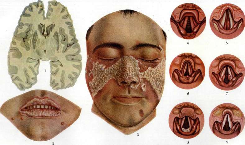
To the article Lupus erythematodes] Laryngitis, Laryngoscolin, Malaria. They occupy a smaller surface than in acute ones; mainly they are located on the true cords and in the interarytenoid space. Hyperemia is more of a venous character; tissue swelling occurs due to small-round-cell infiltration rather than serous imbibition; the flat epithelium of the true cords is thickened, on the posterior wall the ciliated epithelium is replaced by flat epithelium; the glands of the false cords are enlarged and secrete more mucus. Chronic catarrhs of the larynx are of two kinds: hyperplastic and atrophic. In the hyperplastic form, both the epithelial cover and the submucosal tissue undergo hyperplasia. The thickening of the true cords is either diffuse, uniform along the entire length (and then they have a fusiform shape with a rounded free edge), or partial, in places in the form of separate nodules, tubercles (l. nodosa). On the free edge of normal-appearing true cords, at symmetrical places, approximately in the middle of their length in individuals who use their voice intensively, point-like elevations sometimes appear [singer's nodes (see separate plate, figure 9)]; they consist of thickened epithelium and elastic tissue. More massive thickenings of the stratified squamous epithelium are sometimes observed in the region of the vocal process, where they have the appearance of mushroom-like elevations with a central depression [pachyder-mia circumscripta (see separate plate, figure 7)]; much more often, epithelial thickenings occur on the posterior wall of the larynx in the interarytenoid space, which acquires a grayish, bumpy surface [pachydermia diffusa (see separate plate, figure 8)]. In the same place, hyperplasia of the mucosa is observed in the form of a cushion (elevation) with a smooth, red surface (l. posterior hyperplastica). The hyperplastic process can also develop in the ventricles of Morgagni and lead to the formation of folds or rolls of the mucosa, which extend beyond the ventricles and cover the true cords (prolapsus ventriculi Morgagni); it can also develop in the subglottic space (see separate plate, figure 5), forming rolls parallel to the true vocal cords (l. hypoglottica chronica). The atrophic form of chronic laryngitis is much rarer than the hyperplastic; in it, the true cords are thinned and covered with dry crusts. Symptoms of chronic laryngitis are not as intense as in acute; they are expressed in rapid fatigability, weakness of the voice, in hoarseness: these voice defects can also be caused by pareses of the laryngeal muscles, mainly the vocal muscle and cricothyroid muscles. Diagnosis. Any chronic catarrh of the larynx, if there is no suitable etiological factor for it at hand (tobacco, alcohol, profession), is suspicious with respect to tuberculosis; suspicion is enhanced if there is doubt regarding the state of the lungs or if the laryngeal lesion is unilateral. Hyperplastic forms of chronic laryngitis bear great similarity to tuberculous infiltrates; however, the latter possess certain signs that distinguish them from tuberculosis; thus, hyperplasias on the posterior wall of the larynx in tuberculosis have the appearance of red wart-like conical elevations, while in laryngitis posterior hyperplastica these elevations (cushions) with a smooth, even surface are not so red; in pachydermia diffusa, thickenings with a bumpy surface have a grayish tint. Tuberculous infiltrates in the region of the false cords and Morgagni's ventricles can sometimes be confused with prolapse of the ventricle, but the latter are softer and more mobile upon probing. Decisive importance in all cases belongs to biopsy. Subglottic chronic laryngitis bears great similarity to scleroma of the larynx, but in the latter, characteristic changes in the nasopharynx usually exist simultaneously. Treatment. Preventive measures and general methods of treatment of diseases of the larynx - see Larynx. Specifically in acute laryngitis at the beginning of the disease, mustard plasters on the sternum, inhalation of water vapors, drinking warm alkaline liquid with milk and along with this complete vocal rest are prescribed; later - inhalation or atomization with alkaline liquids, instillation with a laryngeal syringe of menthol (1-2%) oil or cocaine (1-2%); for cough, codeine, morphine with apomorphine; pains upon swallowing in posterior laryngitis and inflammation of the epiglottis are alleviated by cold compresses on the neck (ice cravat) and swallowing small pieces of ice. In acute subglottic laryngitis (false croup) - a warming compress on the neck, inhalation of water vapors, a hot foot bath; internally apomorphine [for children 0.02 : 100 one teaspoon (dessert spoon) every hour]; if stenosis does not decrease, as a prophylactic anti-diphtheria serum 5,000 IU, an emetic and finally intubation. In erysipelas and phlegmon of the larynx - injection of polyvalent serum or vaccine, autovaccine, milk, Omnadin; swallowing small pieces of ice, scarification of edematous places, maintenance of heart activity; in asphyxia - tracheotomy. Treatment of chronic catarrh of the larynx presents a difficult task requiring patience and persistence on the part of both the patient and the physician. After eliminating etiological moments, inhalations or atomizations with alkaline (Ems, Borjomi) liquids - alone or with the addition of menthol, eucalyptus, turpentine - or painting the larynx with silver nitrate (1-5%), zinc chloride (1-3%), tannin-glycerin (3%), Gogol's solution (1/2 - 2%) are applied sequentially; the best results are given by systematic (3-4 times a decade) painting with solutions of silver nitrate of gradually increasing concentration (1-5%). In l. posterior hyperplastica and prolapsus ventriculi Morgagni - removal of infiltrates with a double laryngeal curette; singer's nodes disappear with prolonged vocal rest, more persistent ones are removed operatively.
Related articles
Mentioned in
Cite this page
“Laryngitis.” Soviet Medical Encyclopedia. English translation of Bolshaya Meditsinskaya Entsiklopediya, 1st ed. (Moscow, 1928–1936), ed. N. A. Semashko. https://sovietmedicalencyclopedia.pages.dev/article/laryngitis/